411 Original Article
Korean Circulation J 2005;35:411-414
ISSN 1738-5520
ⓒ 2005, The Korean Society of Circulation CASE REPORT
A Case of Complex Congenital Anomaly Combined with Congenital Pseudoarthrosis of the Left Clavicle: Is it a New Syndrome?
Wang-Soo Lee, M.D.1, Jung-Sun Kim, M.D.1, Kwang Ho Lee, M.D.2, Soo Hee Choi, M.D.2, Kyung Eun Lee, M.D.2, Sung Ho Lee, M.D.2, Kwang Je Lee, M.D.2, Sang Wook Kim, M.D.2, Tae Ho Kim, M.D.2, Chee Jeong Kim, M.D.2, Wang Seong Ryu, M.D.2 and Sin Weon Yun, M.D.3
1Department of Cardiology, Armed Forces Capital Hospital, Seongnam, 2Department of Internal Medicine,
3Pediatrics, College of Medicine, Chung-Ang University, Seoul, Korea
ABSTRACT
Congenital pseudoarthrosis of the clavicle is a rare disorder of the shoulder girdle, with only approximately 200 individual cases having been reported in the world literature to date. A persistent left superior vena cava (SVC) is the most common thoracic venous anomaly, and has been observed in 0.3% of the general population. Meso- cardia and an aneurysm of the main pulmonary artery, associated with bicuspid pulmonary valves, are both ex- tremely rare entities. We report the first case of a 23 year-old man with the above mentioned complex congenital anomaly. (Korean Circulation J 2005;35:411-414)
KEY WORDS:Pseudoarthrosis;Superior vena cava;Pulmonary valve;Aneurysm.
Introduction
Congenital pseudoarthrosis of the clavicle is a rare disorder, with only approximately 200 individual cases having been reported in the world literature to date. It should be differentiated from the more common form of pseudoarthrosis that occurs secondary to a fractured clavicle.1)2) The presence of a left superior vena cava (SVC) has been reported to occur in approximately 0.3% of the general population. They drain via the co- ronary sinus to the right atrium in more than 90% of patients, but rarely to the left atrium when associated with other congenital heart diseases.3-5) The prevalence of mesocardia is 2 in every 1000 deliveries(0.2%), so is very rare.6) Congenital abnormalities of the pulmonary valve are rare; 21 in 3600(0.58%) consecutive autopsies.
There are very few reports of bicuspid pulmonary valves, the majority of which are associated with congenital heart disease,7)8) combined with a pulmonary artery an- eurysm.9)
We report the first case of a 23 year-old man with the above mentioned complex congenital anomaly.
Case
A 23 year-old man, a soldier, presented with numbness of the left arm when he shouldered a pack. There was no history of trauma or other diseases. On examina- tion, he was of average build; with a blood pressure of 110/80 mmHg and pulse rate of 52 beats/minute. The breath sounds in both lung fields were clear, and re- gular heart beats, without murmur, were noted on au- scultation. Chest palpation revealed the absence of the distal 3/4 of the left clavicle(Fig. 1). There was no li- mitation of motion in the left shoulder. A chest X-ray showed mesocardia(Fig. 2). The 12-lead ECG revealed a normal sinus rhythm and right axis deviation. Tests of his serum VDRL, ANA, Anti-dsDNA and rheuma- toid factor were all negative, and the CRP was also within the normal range. Transthoracic and transesophageal echocardiography revealed bicuspid pulmonary valves, a normal pulmonary artery pressure, mild pulmonary regurgitation and aneurysmal dilatation of the main pulmonary artery, without stenosis(Fig. 3A). No other cardiac abnormality was observed, with the exception of a huge coronary sinus along the posterior side of the left atrium(Fig. 3B). A 3-dimensional CT scan showed a dilated main pulmonary artery, with a diameter of 4.4 cm(Fig. 4), and a persistent left SVC draining into the coronary sinus(Fig. 5). There were no stenotic le- sions of the subclavian artery and vein.
Received:January 28, 2005 Accepted:March 14, 2005
Correspondence:Kwang Je Lee, M.D.,Department of Internal Medicine, Chung-Ang University Medical Center, 224-1 Heukseok-dong, Dongjak- gu, Seoul 157-070, Korea
Tel: 82-2-6299-1404, Fax: 82-2-6299-1077 E-mail: [email protected]
412·Korean Circulation J 2005;35:411-414
Discussion
The etiology of congenital pseudoarthrosis is obscure, but some theories have been proposed. One, namely the vascular theory, proposes that it is due to the pres- sure exerted by the subclavian artery, but another suggests it is due to the separation of the two primary ossifi- cation centers.2)10) The abnormality occurs almost enti- rely on the right side, with involvement of the left side usually only occurs with dextrocardia and situs inversus.
Bilateral cases are typically reported with genetic pro- blems.1)10-12) Surgical management may be chosen due to their unaesthetic appearance and the development of thoracic outlet syndrome. Our approaches; however, varied in terms of the indications for surgery, the type of surgery and the timing of the reconstruction.1)2) 10)12)
In the early stages of embryological development, a left SVC is present as a counter part of a normal right-
sided SVC. However, it normally obliterates and forms the ‘Ligament of Marshall’ in adulthood.3)5) Residual persistence of the left SVC in adult life is normal in rabbits and some other mammals, but it is a rare abnor- mality in humans. Moreover, when associated with a congenital disease, its more relevant clinical implication
Fig. 1. The 3D-Chest CT: the apical lordotic and AP views, showing congenital pseudoarthrosis of the left clavicle. The distal 3/4 of the left clavicle is absent (arrow) and mesocardia is shown (arrow head).
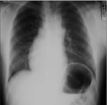
Fig. 2. Chest PA. Mesocardia with situs solitus.
Fig. 4. A 3D-Chest CT. Marked aneurysmal dilatation of the main pulmonary artery.
Fig. 3. The 2D-Echocardiogram. A: parasternal short axis view;
thickened, dysplastic partially bicuspid pulmonary valve (arrow) and marked dilatation of the main pulmonary artery (arrow head). B:
parasternal long axis view; huge coronary sinus (arrow) along the posterior side of the left atrium.
A B
Fig. 5. A 3D-Chest CT. Persistent left superior vena cava (arrow) draining into the coronary sinus.
Wang-Soo Lee, et al: A Case of Complex Congenital Anomaly·413
is its association with disturbances of cardiac impulse formation and conduction.3)13)14) Developmentally, the sinus node, atrioventricular(AV) node and the His bundle may be heavily influenced by the lack of reg- ression of the left SVC. The persistent presence of the left SVC alters the anatomic location and histology of the conducting system, paradoxically speaking. This pa- thologic substrate may predispose the patient to arrhy- thmias and sudden death.3)15) The prevalence of the left SVC in patient with congenital cardiac abnormalities is much higher than in the general population, ranging from 2.8 to 4.3%. In addition, about 10% of these sub- jects do not have a right SVC.3)13)16) Nsah et al.5) re- ported significantly more frequent associations between a persistent left SVC and AV canal defects, cor triatria- tum and mitral atresia. When not associated with other congenital cardiac anomalies, it is usually asymptoma- tic and hemodynamically insignificant.4) However, a persistent left SVC has important clinical implications under certain circumstances, such as the positioning of a left-sided pacemaker or implantable cardioverter-de- fibrillator(ICD), the placement of central venous lines for therapeutic purposes and hemodynamic monitoring, and cardiopulmonary bypass in patients undergoing cardiac surgery procedures.3)4)17)
Mesocardia means that the heart is in the middle of the thorax, with a prevalence of 4 in 1716 autopsied cases(0.2%); therefore, is a very infrequent diagnosis, and 10 different types of congenital mesocardia were found in 17 autopsied cases.6)18)
A pulmonary artery aneurysm is an uncommon vas- cular anomaly. Deterling and Clagett19) reviewed 109,571 autopsied cases, and only 8 cases of a pulmonary artery aneurysm were recorded. Its etiology and pathogenesis are not well known, but can be categorized as idiopa- thic and secondary types. More than 50% of pulmonary artery aneurysms are combined with congenital heart diseases, such as patent ductus arteriosus(PDA), ven- tricular septal defect(VSD) and atrial septal defect(A- SD). Other causes, such as infection, collagen vascular disease, bronchial tumor, iatrogenic(Swan-Ganz cathe- ter), and bicuspid pulmonary valves, may also be related to the pathogenesis of a pulmonary artery aneurysm.9)20) A pulmonary artery aneurysm is diagnosed with a mea- sured diameter more than 28 mm, which can be achieved by a combination of echocardiography, conventional CT scan, MRI and angiography. Recently, a 3-dimen- sional CT scan has also been widely used as an accurate, noninvasive diagnostic tool.20) The most common causes of death in the presence of an aneurysm of the pulmo- nary artery are congestive heart failure or aneurysm rupture.19) Correction is the most usual management of a pulmonary artery aneurysm of primary cause. Sur- gical intervention should be considered when in the presence of an aneurysmal progression in size, pul-
monary hypertension and dissection. Recently, the benign clinical courses have been reported, with une- ventful long-term follow-up. Thus, periodic monitoring of the pulmonary artery aneurysm diameter and pressure, and right ventricular dysfunction are recommended in the long-term follow-up.9)20)
Our patient had four rare ontogenic anomalies; con- genital pseudoarthrosis of the clavicle, a persistent left SVC, mesocardia and a pulmonary artery aneurysm combined with bicuspid pulmonary valves. We just observed and followed up, without specific treatment as the patient had no esthetic complaint, clinical symp- toms or hemodynamic instability. We report for the first time this new case of a complex congenital anom- aly. It is a new syndrome or not, that is question and needed observation.
REFERENCES
1) Lozano P, Doaz M, Riera R, Gomez FT. Venous thoracic outlet syndrome secondary to congenital pseudoarthrosis of the cla- vicle: presentation in the fourth decade of life. Eur J Vasc Endo- vasc Surg 2003;25:592-3.
2) Dzupa V, Bartonicek J, Zidka M. Fracture of the clavicle after surgical treatment for congenital pseudoarthrosis. Med Sci Mo- nit 2004;10:CS1-4.
3) Biffi M, Boriani G, Frabetti L, Bronzetti G, Branzi A. Left su- perior vena cava persistence in patients undergoing pacemaker or cardioverter-defibrillator implantation: a 10-year experience.
Chest 2001;120:139-44.
4) Sarodia BD, Stoller JK. Persistent left superior vena cava: case report and literature review. Respir Care 2000;45:411-6.
5) Nsah EN, Moore GW, Hutchins GM. Pathogenesis of persistent left superior vena cava with a coronary sinus connection. Pediatr Pathol 1991;11:261-9.
6) Geva T, Praagh SV. Abnormal systemic venous connections. In: Allen HD editor. Moss and Adams’ Heart Disease in Infant, Chil- dren, and Adolescents: include the fetus and young adult. 6th ed.
Philadelphia: LWW Company;2001. p.779-84.
7) Orrit J, Mestres CA, Agusti E, Pomar JL. Isolated bicuspid pul- monary valve: an unusual finding. J Heart Valve Dis 2004;13: 521-2.
8) Koletsky S. Congenital bicuspid pulmonary valves. Arch Pathol 1941;31:338-53.
9) Veldtman GR, Dearani JA, Warnes CA. Low pressure giant pul- monary artery aneurysm in the adult: natural history and ma- nagement strategies. Heart 2003;89:1067-70.
10) Lorente Molto FJ, Bonete Lluch DJ, Garrido IM. Congenital pseudoarthrosis of the clavicle: a proposal for early surgical treatment. J Pediatr Orthop 2001;21:689-93.
11) Karakurt L, Yilmaz E, Belhan O, Serin E. Pycnodysostosis asso- ciated with bilateral congenital pseudoarthrosis of the clavicle.
Arch Orthop Trauma Surg 2003;123:125-7.
12) Schnall SB, King JD, Marrero G. Congenital pseu-doarthrosis of the clavicle: a review of the literature and surgical results of six cases. J Pediatr Orthop 1988;8:316-21.
13) Campbell M, Deuchar DC. The left-sided superior vena cava. Br Heart J 1954;16:423-39.
14) Camm AJ, Dymond D, Spurrell RA. Sinus node dysfunction associated with the absence of right superior vena caca. Br Heart J 1979;41:504-7.
15) James TN, Marshall TK, Edwards JE. Cardiac electrical instabi-
414·Korean Circulation J 2005;35:411-414
lity in the presence of a left superior vena cava. Circulation 1976;54:689-97.
16) Karnegis JN, Wang Y, Winchell P, Edwards JE. Persistent left superior vena cava, fibrous remnant of the right superior vena cava and ventricular septal defect. Am J Cardiol 1964;14:573-7.
17) Mooney DP, Snyder CL, Holder TM. An absent right and per- sistent left superior vena cava in an infant requiring extracor- poreal membrane oxygenation therapy. J Pediatr Surg 1993;28: 1633-4.
18) Lev M, Liberthson RR, Golden JG, Eckner FA, Arcilla RA. The pathologic anatomy of mesocardia. Am J Cardiol 1971;28:428-35.
19) Deterling RA, Clagett OT. Aneurysm of the pulmonary artery: review of the literature and report of a case. Am Heart J 1947; 34:471-98.
20) Ahn JH, Lee KJ, Kim SW, et al. A case of asymptomatic patient with idiopathic pulmonary artery aneurysm. Korean Circ J 2003; 33:242-5.