Korean J Gastroenterol Vol. 67 No. 6, 337-340 http://dx.doi.org/10.4166/kjg.2016.67.6.337 pISSN 1598-9992 eISSN 2233-6869
CASE REPORT
Korean J Gastroenterol, Vol. 67 No. 6, June 2016 www.kjg.or.kr
소라페닙 치료 간세포암 환자에서 발생한 드레스 증후군 1예
김동균, 이성우, 남화성, 전동섭, 박나래, 남영희, 이수걸, 백양현, 한상영, 이성욱
동아대학교 의과대학 내과학교실
A Case of Sorafenib-induced DRESS Syndrome in Hepatocelluar Carcinoma
Dong Kyun Kim, Sung Woo Lee, Hwa Seong Nam, Dong Sub Jeon, Na Rae Park, Young Hee Nam, Soo Keol Lee, Yang Hyun Baek, Sang Young Han, and Sung Wook Lee
Department of Internal Medicine, Dong-A University College of Medicine, Busan, Korea
Sorafenib is currently the only targeted therapy available for advanced stage hepatocellular carcinoma (HCC). Cutaneous adverse events associated with sorafenib treatment include hand-foot skin reaction, but there has been no report of drug reaction (or rash) with eosinophilia and systemic symptoms (DRESS) syndrome. Here, we report a case of 72-year-old man with HCC and alcoholic liver cirrhosis who developed skin eruptions, fever, eosinophilia, and deteriorated hepatic and renal function under sorafenib treatment. He has since successfully recovered with conservative care. (Korean J Gastroenterol 2016;67:337-340) Key Words: DRESS syndrome; Sorafenib; Hepatocellular carcinoma
Received February 5, 2016. Revised March 28, 2016. Accepted April 24, 2016.
CC This is an open access article distributed under the terms of the Creative Commons Attribution Non-Commercial License (http://creativecommons.org/licenses/
by-nc/4.0) which permits unrestricted non-commercial use, distribution, and reproduction in any medium, provided the original work is properly cited.
Copyright © 2016. Korean Society of Gastroenterology.
교신저자: 이성욱, 49201, 부산시 서구 대신공원로 26, 동아대학교병원 소화기내과
Correspondence to: Sung Wook Lee, Department of Gastroenterology, Dong-A University Hospital, 26 Daesingongwon-ro, Seo-gu, Busan 49201, Korea. Tel: +82-51- 240-2983, Fax: +82-51-240-1510, E-mail: [email protected]
Financial support: None. Conflict of interest: None.
INTRODUCTION
Sorafenib (NexavarⓇ; Bayer Korea, Seoul, Korea) is a wide- ly-used oral tyrosine kinase inhibitor. It inhibits serine threo- nine kinases Raf-1, B-Raf; the receptor tyrosine kinase activ- ity of vascular endothelial growth factor receptors (VEGFRs) 1, 2, and 3; and platelet-derived growth factor receptor (PDGFR-).1 It is currently approved by the US Food and Drug Administration for the treatment of hepatocellular carcino- ma (HCC), metastatic renal cell carcinoma, and thyroid cancer.2,3
Sorafenib is associated with a number of adverse effects including fatigue, diarrhea, nausea, and weight loss.4 Cuta- neous manifestations (e.g., hand-foot skin reaction [HFSR], skin eruption, scalp dysesthesia, subungal splinter hemor- rhages, alopecia, and body hair loss), mostly mild to moder-
ate in severity, comprise the commonest side-effects.1,5 There are a few case reports of Stevens-Johnson syndrome (SJS) or drug reaction (or rash) with eosinophilia and sys- temic symptoms (DRESS) syndrome.
Here, we present a case of a patient with DRESS syndrome related to sorafenib prescribed to treat HCC.
CASE REPORT
A 72-year-old man was admitted through the emergency room (ER) with a whole body skin rash, onset a day before his visit. He was an alcoholic liver cirrhosis patient and had re- ceived transarterial chemoembolization (TACE), radiofre- quent ablation, and hepatic wedge resection for treatment of concomitant recurrent multifocal HCC. Because the HCC re- curred, he was prescribed sorafenib (NexavarⓇ) 400 mg
338 김동균 등. 소라페닙 치료 간세포암 환자에서 발생한 드레스 증후군
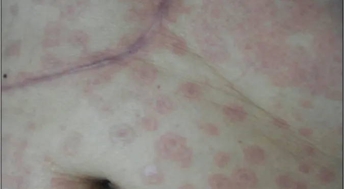
The Korean Journal of Gastroenterology Fig. 1. Abdominal skin lesion at admission. Multiple targetoid and
patch-like rash occurred in the lateral part of right thigh and spread to whole body.
Table 1. Serial Laboratory Results
BA 25 AD 1 AD 3 AD 5 AD 7 AD 8 AD 9 AD 11 AD 12 AD 14 AD 16 AD 19 DC 22 WBC (/L) 4,810 7,730 6,800 6,380 6,650 6,770 5,550 4,950 4,070 2,660 2,840 2,900 5,010 Seg. neutrophil (%) 42.7 80.5 76.2 66.9 48.1 39.3 52.0 51.5 48.2 43.2 37.8 34.8 50.9
Eosinophil (%) 6.4 9.7 12.9 18.0 22.9 27.9 17.0 18.0 12.5 13.9 16.5 14.5 7.2
Platelet (×103/L) 118 64 42 16 24 16 18 39 32 37 38 59 120
PT (INR) 1.05 1.15 1.97 1.99 2.03 1.82 1.67 1.43 1.95 1.50 1.02
Creatinine (mg/dL) 1.3 1.7 2.0 1.8 1.4 1.2 1.1 1.0 1.0 1.0 1.0 1.1
Serum albumin (g/dL) 4.1 3.1 2.8 2.7 2.7 2.8 2.8 2.8 3.1 2.8 2.9 3.6
AST (IU/L) 31 50 109 251 379 282 145 81 49 28 24 30 34
ALT (IU/L) 17 14 21 35 52 53 39 25 21 13 14 10 24
ALP (IU/L) 212 222 159 237 256 219 197 169 169 364
Total bilirubin (mg/dL) 0.9 2.8 4.3 8.6 10.9 13.5 15.6 15.0 12.1 8.9 5.7 4.3 1.6
Direct bilirubin (mg/dL) 0.3 1.4 2.5 5.9 7.8 9.6 10.8 10.4 8.7 6.5 4.5 3.6 1.4
CRP (mg/dL) 6.37 14.43 10.00 8.12 3.78 2.76 0.25
BA, before admission; AD, admission day; DC, discharge day; WBC, white blood cell; Seg., segmented.
twice daily. Five days after initiation of sorafenib, he had a mild sense of throat swelling and hoarseness. At 10 days af- ter starting treatment, he developed a whole body rash that started in the lateral part of the right thigh and spread to his whole body.
When he visited the ER, he complained of general weak- ness, whole body itching, headache, and feeling febrile. He had a fever of over 38oC that had not subsided for more than four days despite antipyretics. His other vital signs were stable.
The complete blood cell test revealed that the white blood cell (WBC) count was 7,730/L (segmented neutrophil 80.5%, eosinophil 9.7%, lymphocyte 5.0%), and platelet (PLT) count was 64,000/L (which was markedly decreased compared to the baseline level of 130,000/L). Serum chemistry findings showed increased levels of creatinine (1.7 mg/dL), total bilirubin (TB)/direct bilirubin (DB) (2.8/1.4 mg/dL), and CRP (6.37 mg/dL), all of which had been within
normal limits prior to the event. Serum AST/ ALT levels were not markedly increased (50/14 IU/L). Urinalysis was positive for protein, ketone, WBC, and urobilinogen.
We checked blood culture, ascitic fluid analysis and cul- ture, antibodies for cytomegalovirus, Epstein-Barr virus, her- pes simplex virus, Korean virus, leptospira and tsutsuga- mushi, and all were negative.
The skin rashes were multiple erythematous targetoid and patch-like appearance on the chest, abdomen, back, and lower extremities. Peculiar to the upper extremities was a rash with a small dot pattern (Fig. 1).
With the impression of DRESS syndrome, sorafenib was immediately discontinued. We performed conservative care with a topical steroid lotion, antihistamines, antipyretics (acetaminophen), empirical antibiotics, and intravenous flu- id therapy. Upon consultation with otolaryngology and oph- thalmology, there were not any other severe mucosal lesions in these systems.
The skin lesions, fever, and systemic symptoms such as itching, headache, and general weakness gradually sub- sided, and consequently, the patient did not need further medications for these discomforts. However, liver function decreased and eosinophilia worsened rapidly. Seven days af- ter the admission, WBC count was 6,770/L (segmented neutrophil 39.3%, eosinophil 27.9%, lymphocyte 20.8%), and PLT level was 16,000/L. Serum TB/DB levels increased to 13.5/9.6 mg/dL and AST/ALT was 282/53 IU/L. PT, which had been within normal limits upon admission, was 2.03 in INR, and serum albumin was 2.7 g/dL.
We continued supportive care of his deteriorated hepatic
Kim DK, et al. Sorafenib-induced DRESS Syndrome in Hepatocelluar Carcinoma 339
Vol. 67 No. 6, June 2016
function. On the 19th day of hospitalization, although the lab- oratory findings did not reach baseline, liver function and general condition noticeably improved (WBC 2,900/L [segmented neutrophil 34.8%, eosinophil 14.5%, lympho- cyte 27.6%]), PLT 59,000/L, serum creatinine 1.0 mg/dL, serum TB/DB 4.3/3.6 mg/dL, AST/ALT 30/10 IU/L, PT (INR) 1.50, serum albumin 2.9 g/dL, CRP 2.76 mg/dL). Table 1 lists the serial laboratory findings. The patient was transferred to a convalescent hospital for further supportive care. When he visited the outpatient department, almost all laboratory find- ings had normalized or returned to the baseline, and he was in a good physical condition state.
DISCUSSION
Cutaneous adverse events are common with sofafenib treatment. More than 90% of patients experience a skin re- action and 60-70% have HFSR.3,6 However, delayed cuta- neous hypersensitivity reactions, such as erythema multi- forme (EM), or severe cutaneous adverse reactions (SCARs), such as SJS, are very rare, although there are some recent reports.7,8 To the best of our knowledge, the literature does not include reports of toxic epidermal necrolysis (TEN) or DRESS syndrome.
The spectrum of SCARs comprises several disease enti- ties; acute generalized exanthematous pustulosis (AGEP), epidermal necrolysis (TEN, SJS), and DRESS.9 These con- ditions are defined mainly by clinical features of visceral organ involvement, hematologic abnormalities, and alterations of vital signs. Specific biological and histological findings are al- so important considerations.10
DRESS syndrome is a distinct, severe, and idiosyncratic re- action to a drug.11 The clinical manifestations usually appear two to eight weeks after initiation of the causative drug,12 and include skin lesions (e.g. rash, urticaria, maculopapular eruption, edema, vesicles, pustules, purpura, target lesion, erythroderma), fever, lymphadenopathy, hematologic abnor- malities, and deterioration of liver function.13
The regiSCAR scoring system is helpful for the diagnosis of DRESS syndrome.14 When this system was applied to this pa- tient, he scored 6 (fever of 38.9oC at admission, 0; no en- larged lymph nodes, 0; atypical lymphocytes, unknown, 0; eo- sinophilia, 2; skin involvement >50% of body surface and suggesting DRESS, 2; increased liver function tests and se-
rum creatinine, 2; resolution ≥15 days, 0). Therefore, this patient corresponds to a ‘definite case’.
Drugs that can cause DRESS syndrome include anti- convulsants, antidepressants, sulfonamides, antibiotics, an- ti-inflammatory drugs, anti-hypertensives.15 A recent system- atic review reported 44 drugs associated with 172 DRESS cases reports between January 1997 and May 2009, in PubMed and MEDLINE.16 Besides sorafenib, the medi- cations that our patient were taking were not in these drug categories. Moreover, he had been taking these medications for at least 20 months without any specific complications.
We did not check autoimmune markers or hepatitis viral markers during admission. Hepatitis B and C markers were checked about eight months before the admission, and were negative. We consider these to be limitations of this case report.
Early recognition and prompt discontinuation of the causa- tive drug is most important in the treatment of DRESS syndrome.17 Oral corticosteroids are often used in severe cases, although the effectiveness is not evident.10 Other therapies include cyclosporin, intravenous immunoglobulin, and plasmapheresis.18,19 In this case, we managed the pa- tient with supportive, symptomatic care, and topical steroid lotion. At the point when we considered oral steroids as the next step in treatment, the patient’s clinical manifestations improved. Therefore, we emphasize the importance of cessa- tion of the causative medication.
REFERENCES
1. Llovet JM, Ricci S, Mazzaferro V, et al; SHARP Investigators Study Group. Sorafenib in advanced hepatocellular carcinoma. N Engl J Med 2008;359:378-390.
2. Bruix J, Raoul JL, Sherman M, et al. Efficacy and safety of sor- afenib in patients with advanced hepatocellular carcinoma: sub- analyses of a phase III trial. J Hepatol 2012;57:821-829.
3. Sohn KH, Oh SY, Lim KW, Kim MY, Lee SY, Kang HR. Sorafenib induces delayed-onset cutaneous hypersensitivity: a case series. Allergy Asthma Immunol Res 2015;7:304-307.
4. Wood LS. Managing the side effects of sorafenib and sunitinib.
Community Oncol 2006;3:558-562.
5. Autier J, Escudier B, Wechsler J, Spatz A, Robert C. Prospective study of the cutaneous adverse effects of sorafenib, a novel mul- tikinase inhibitor. Arch Dermatol 2008;144:886-892.
6. Cohen PR. Sorafenib-associated facial acneiform eruption.
Dermatol Ther (Heidelb) 2015;5:77-86.
7. Namba M, Tsunemi Y, Kawashima M. Sorafenib-induced eryth- ema multiforme: three cases. Eur J Dermatol 2011;21:1015-
340 김동균 등. 소라페닙 치료 간세포암 환자에서 발생한 드레스 증후군
The Korean Journal of Gastroenterology 1016.
8. Ikeda M, Fujita T, Amoh Y, Mii S, Matsumoto K, Iwamura M.
Stevens-Johnson syndrome induced by sorafenib for metastatic renal cell carcinoma. Urol Int 2013;91:482-483.
9. Bouvresse S, Valeyrie-Allanore L, Ortonne N, et al. Toxic epi- dermal necrolysis, DRESS, AGEP: do overlap cases exist?
Orphanet J Rare Dis 2012;7:72.
10. Roujeau JC, Stern RS. Severe adverse cutaneous reactions to drugs. N Engl J Med 1994;331:1272-1285.
11. Choudhary S, McLeod M, Torchia D, Romanelli P. Drug reaction with eosinophilia and systemic symptoms (DRESS) syndrome. J Clin Aesthet Dermatol 2013;6:31-37.
12. Eshki M, Allanore L, Musette P, et al. Twelve-year analysis of se- vere cases of drug reaction with eosinophilia and systemic symp- toms: a cause of unpredictable multiorgan failure. Arch Dermatol 2009;145:67-72.
13. Peyrière H, Dereure O, Breton H, et al; Network of the French Pharmacovigilance Centers. Variability in the clinical pattern of cutaneous side-effects of drugs with systemic symptoms: does a DRESS syndrome really exist? Br J Dermatol 2006;155:422- 428.
14. Kardaun SH, Sidoroff A, Valeyrie-Allanore L, et al. Variability in the clinical pattern of cutaneous side-effects of drugs with sys- temic symptoms: does a DRESS syndrome really exist? Br J Dermatol 2007;156:609-611.
15. Wongkitisophon P, Chanprapaph K, Rattanakaemakorn P, Vachiramon V. Six-year retrospective review of drug reaction with eosinophilia and systemic symptoms. Acta Derm Venereol 2012;92:200-205.
16. Cacoub P, Musette P, Descamps V, et al. The DRESS syndrome:
a literature review. Am J Med 2011;124:588-597.
17. Fernando SL. Drug-reaction eosinophilia and systemic symp- toms and drug-induced hypersensitivity syndrome. Australas J Dermatol 2014;55:15-23.
18. Kirchhof MG, Miliszewski MA, Sikora S, Papp A, Dutz JP.
Retrospective review of Stevens-Johnson syndrome/toxic epi- dermal necrolysis treatment comparing intravenous im- munoglobulin with cyclosporine. J Am Acad Dermatol 2014;71:
941-947.
19. Koštál M, Bláha M, Lánská M, et al. Beneficial effect of plasma exchange in the treatment of toxic epidermal necrolysis: a series of four cases. J Clin Apher 2012;27:215-220.