www.krspine.org
Changes in Cervical Spine Range of Motion after Laminoplasty in Cervical Spondylotic Myelopathy
Jae-Sung Ahn, M.D., June-Kyu Lee, M.D., Woo-Wong Lee, M.D., Jung-Mo Hwang, M.D.
J Korean Soc Spine Surg 2012 Sep;19(3):85-89.
Originally published online September 30, 2012;
http://dx.doi.org/10.4184/jkss.2012.19.3.85
Korean Society of Spine Surgery
Department of Orthopedic Surgery, Inha University School of Medicine
#7-206, 3rd ST. Sinheung-Dong, Jung-Gu, Incheon, 400-711, Korea Tel: 82-32-890-3044 Fax: 82-32-890-3467
©Copyright 2011 Korean Society of Spine Surgery pISSN 2093-4378 eISSN 2093-4386
The online version of this article, along with updated information and services, is located on the World Wide Web at:
http://www.krspine.org/DOIx.php?id=10.4184/jkss.2012.19.3.85
This is an Open Access article distributed under the terms of the Creative Commons Attribution Non-Commercial License (http://
creativecommons.org/licenses/by-nc/3.0) which permits unrestricted non-commercial use, distribution, and reproduction in any medium, provided the original work is properly cited.
Journal of Korean Society of
Spine Surgery
Changes in Cervical Spine Range of Motion after Laminoplasty in Cervical Spondylotic Myelopathy
Jae-Sung Ahn, M.D., June-Kyu Lee, M.D., Woo-Wong Lee, M.D., Jung-Mo Hwang, M.D.
Department of Orthopedic Surgery, Chungnam National University, School of Medicine, Daejeon, Korea Study Design: A retrospective study.
Objectives: This study examined the cervical range of motion (ROM) of cervical spondylotic myelopathy patients, before and after open door laminoplasty.
Summary of Literature Review: Majority of the cases regarding the change of cervical range of motion after cervical laminoplasty showed decreased range of motion, and the results were diverse.
Materials and Methods: Of the 487 patients, who underwent open door laminoplasty at our hospital from March 1997 to March 2008, 98 had been followed for at least 2 years and had cervical flexion-extension lateral x-rays. In all patients, open door laminoplasty involved at least three segments: three, four, and five segments in 11, 52, and 35 patients, respectively. In previous cases, fixation involved sutures using suture anchors. The lordosis or kyphosis between C2 and C7 was analyzed using cervical flexion-extension lateral radiographs before and 2 years after the operation.
Results: The average patient age was 62.7 (range 32–82) years; 65 patients were male and 33 were female. From preoperatively to postoperatively, the average kyphosis of cervical flexion decreased from 10.7° to 7.8°, average lordosis decreased from 21.2° to 14.2°, and cervical ROM decreased from 31.9° to 22.0°, respectively (mean 9.9°, 31.0%).
Conclusions: We could observe decreased cervical range of motion after cervical laminoplasty for cervical spondylotic myelopathic patients. Thus, the treatment to prevent the postoperative decrease of cervical range of motion and further study to find a new treatment are thought to be essential.
Key Words: Cervical spondylotic myelopathy, Laminoplasty, Suture anchor, Range of motion(ROM)
Received: October 24, 2011 Revised: March 19, 2012 Accepted: August 6, 2012
Published Online: September 30, 2012 Corresponding author: Jae-Sung Ahn, M.D.
Department of Orthopaedic Surgery, Chungnam National University, College of medicine, 33, Munhwa-Ro, Jung-gu, Daejeon 301-721, Korea
TEL: 82-42-280-7353, FAX: 82-42-252-7098 E-mail: [email protected]
“This is an Open Access article distributed under the terms of the Creative Commons Attribution Non-Commercial License (http://
creativecommons.org/licenses/by-nc/3.0/) which permits unrestricted non-commercial use, distribution, and reproduction in any medium, provided the original work is properly cited.”
서 론
경추 후궁 성형술은 3분절 이상 다분절의 경추부 척수가 압박 을 받는 경우 척수관 확장에 효과적인 술식이다.1-5) 후방에서 경 추부의 감압을 하는 방법으로는 후궁절제술, 후궁절제술 후 유 합술 및 후궁성형술이 있다. 이러한 수술 방법들은 모두 경추부 의 감압에 효과적이라고 할 수 있으나 문제는 수술 후 후만 변 형과 운동 범위 감소가 발생할 수 있다는 것이다. 후궁 성형술은 후궁절제술 후 유합술에 비하여 감압을 시행하며 운동 분절을 유합하지 않음으로써 경추 운동범위를 보존할 수 있으며 후궁절 제술에 비하여 후만 변형의 발생 위험성이 적은 점이다. 이러한 후궁 성형술의 목적은 황색인대의 비후 등 후방에서의 척수 압 박을 직접 제거 할 수 있을 뿐만 아니라 척추관을 후방에서 확장 함으로서 척수를 후방으로 이동(posterior migration)시켜 전방 의 압박으로부터 간접적인 감압효과를 기대하는 것이다. 그러므 로 수술적 경추 만곡은 전만(lordosis)이어야 하며, 충분한 감압 을 얻기 위해서는 수술 후에도 전만이 유지되어야 한다. 또한 후
관절을 보존하고 후궁을 절제하지 않기 때문에 부분적인 섬유 성 강직과 더불어 수술 후 안정성을 유지할 수 있고 후궁을 보존 함으로서 척수에 대한 보호 역할도 기대할 수 있다. 그리고 다른 후궁 절제술 후 유합술에 비해 후궁 성형술은 유합을 하지 않아 경추 운동 범위를 보존하는 장점이 있다.6-8) 그러나 본 저자들은
Jae-Sung Ahn et al Volume 19 • Number 3 • September 2012
www.krspine.org
86
수술 전과 비교하여 경추 굴곡-신전 운동 범위가 감소하는 예를 경험하고 이러한 현상에 대한 고찰과 함께 경추 후궁 성형술 후 굴곡-신전 운동 범위의 관계에 대해 알아보고자 하였다.
대상 및 방법
1997년 3월부터 2008년 3월까지 본원에서 경추 후궁 성형술 을 시행한 487예 중 2년 이상 추시가 가능하였으며, 수술 전과 후에 경추부 굴곡-신전 측면 단순 방사선 촬영을 시행하였고 수 술 시 제 2 경추를 제외한 경추에 3분절, 4분절 또는 5분절의 경 추 후궁 성형술을 시행한 경추증성 척수증 환자 98예를 대상으 로 후향적 연구를 시행하였다. 모든 예에서 Hirabayashi 방법의 개문형(open door) 경추 후궁 성형술을 시행하였으며, 후궁 개 방을 유지하기 위하여 최근위와 최원위의 경추 측괴에 double knot suture anchor를 삽입하여 봉합사를 이용하여 고정하였고 수술 후 3개월간 필라델피아 경추 보조기(Philadelphia brace)를 착용하였고 보조기 제거 후에는 적극적인 경추부 재활 운동를 교육하였다. 또한 수술 전 진단에서 후종인대 골화증과 수술 후
천부 감염, 경첩부의 골절, 후궁간 유합 및 수술 후 일시적 신경 근병증의 합병증이 발생한 예는 제외되었다.
모든 환자들은 수술 전과 수술 후 2년에 경추 굴곡-신전 측면 단순 방사선 촬영을 시행하였고 제 2 경추 하단과 제 7 경추의 하단이 이루는 각도로 제 2-7 경추 간의 전만 또는 후만각을 측 정하였다(Fig. 1). 경추의 굴곡-신전 운동범위는 굴곡 시에 측정 한 각도와 신전 시에 측정한 각도의 차로 정의하였고, 수술 전과 수술 후 2년의 운동 범위를 비교하여 수술 전에 비하여 수술 후 2년의 운동범위가 얼마나 감소하였는지를 조사하였다. 1명의 연구자가 1주일 간격으로 2회씩 측정하였고, 통계분석은 paired sample T-test를 이용하였다.
결 과
1. 전만각 또는 후만각
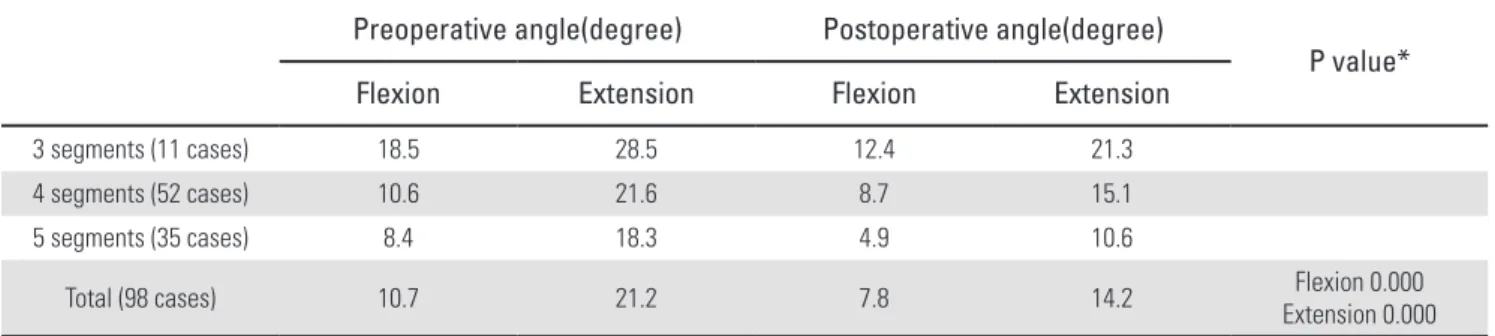
평균 연령은 62.7(32~82)세였고, 총 98예(남자 65예, 여자 33 예)였다. 경추부 굴곡 시 수술 전 후만각은 평균 10.7도에서 수 술 후 7.8도로 유의하게 감소하였고 경추부 신전 시 수술 전 전 만각은 평균 21.2도에서 수술 후 평균 14.2도로 유의하게 감소하 였다. 분절 범위에 따라서는 3분절의 경추 후궁 성형술을 받은 군에서는 경추부의 굴곡 및 신전시 후만각과 전만각이 수술 전 평균 18.5도와 28.5도에서 수술 후 평균 12.4도와 21.3도로 감소 하였고, 4분절의 경추 후궁 성형술을 받은 군에서는 경추부의 굴곡 및 신전시 후만각과 전만각이 수술 전 평균 10.6도와 21.6 도에서 수술 후 평균 8.7도와 15.1도로 감소하였다. 또한 5분절 의 경추 후궁 성형술을 받은 군에서는 경추부의 굴곡 및 신전시 후만각과 전만각이 수술 전 평균 8.4도와 18.3도에서 수술 후 평 균 4.9도와 10.6도로 감소하였다(P=0.000)(Table 1).
2. 운동 범위
경추부의 운동범위는 수술 전 평균 31.9도에서 수술 후 평균 22.0도로 9.9도(31.0%) 감소하였다. 분절 범위에 따라서는 3분 Fig 1. Flexion and extension angle of the cervical spine was measured by
cobb’s method from bottom of C2 to bottom of C7.
Table 1. Mean values of flexion and extension angle.
Preoperative angle(degree) Postoperative angle(degree)
P value*
Flexion Extension Flexion Extension
3 segments (11 cases) 18.5 28.5 12.4 21.3
4 segments (52 cases) 10.6 21.6 8.7 15.1
5 segments (35 cases) 8.4 18.3 4.9 10.6
Total (98 cases) 10.7 21.2 7.8 14.2 Flexion 0.000
Extension 0.000
*P-value by paired-sample T-test
절, 4분절, 5분절의 경추 후궁 성형술을 받은 군에서는 경추부 의 운동범위가 각각 수술 전 평균 47.0도, 32.2도, 26.7도에서 수 술 후 평균 33.7도, 23.8도, 15.5도로 각각 13.3도(28.3%), 8.4 도(26.1%), 11.2도(41.9%) 씩 유의하게 감소하였다(P=0.000) (Table 2).
고 찰
경추부 척수압박을 감압하기 위한 수술 방법으로는 전방 감압 및 추체유합술, 후궁 절제술, 후궁절제술 후 유합술, 후궁성형 술 등이 있다. 1950년도 후반부터 보편화된 전방감압 및 추체유 합술은 병소가 한두 분절에 국한되어 있을 때 효과적이나 3분절 이상에서 시행할 경우에는 인접 상하분절의 퇴행성 변화와 가 관절 형성 등이 문제가 된다.9,10) 후궁 절제술은 수술 후 불안정 성과 신경마비의 악화가 빈번하고 후궁 절제술 후 유합술은 수 술 후 운동범위 감소가 문제가 된다.11-13) 이와 같은 문제점을 해 결하기 위해 개발된 시술로서 후궁성형술은 수술 후 척추운동을 허용하면서도 수술로 인한 불안정성의 빈도가 낮고 비교적 안전 하게 척추관을 확장시킬 수 있다. Oyama 등14)은 척추관의 후궁 을 Z-성형술로 보존함으로서 수술 후 운동범위 감소를 예방할 수 있다고 하였고, Hirabayashi 등6-8)은 척추관의 편측을 개방하 는 후궁성형술(expansive open door laminoplasty)을 개발하여 좁은 척추관의 효과적인 감압과 운동범위 보존을 할 수 있다고 하였다.
Herkowitz1)는 45명의 다발성 경추증 환자에서 전방감압 및 유합술, 후궁절제술, 후궁성형술을 시행한 예를 비교하여 수술 후 임상평가 및 방사선학적 평가상 후궁성형술의 결과가 타수술 에 비해 우수하였다고 보고한 것과 같이 여러 저자들에 의해 후 궁 성형술이 경추의 분절 운동을 보존할 수 있는 방법으로 타수 술에 비해 우수하다고 알려져 있다.15-18)
하지만 Ratiff와 Cooper19)는 이제까지 보고된 다양한 후궁 성 형술에 대한 71편의 연구결과 2000명 이상의 환자를 분석하 여 후궁 성형술 수술 후 경추의 운동범위는 수술 전에 비하여
17~80% 감소하여 평균 50%정도 감소하였다고 보고하였고 현 재까지 대부분 보고된 바로는 수술 후 30-70% 정도의 경추 운 동 제한이 있으며, 보고자에 따른 운동범위 감소 정도에 차이가
많았다.20-24) 이에 본 저자들은 최근 장해 진단에서 경추부 운동
범위의 중요성이 대두되고 있는 점에 착안하여 후궁 성형술을 시행 후 운동범위 감소를 확인하고 경추 후궁 성형술의 장점인 운동범위의 보존의 정도를 알아보기 위하여 본 연구를 시행하였 다.
본 연구 결과 경추의 굴곡-신전 운동범위는 수술 전에 비하 여 수술 후에 31.0%가 유의하게 감소되었으며, 굴곡 및 신전 운 동범위가 모두 감소하였지만 신전의 감소가 뚜렷하였다. 본 연 구 결과가 Ratliff와 Cooper19)의 연구결과보다 경추 운동 제한보 다 경미하였지만, 운동 범위 감소가 발생하는 것을 확인할 수 있 었다. 이는 Ratliff와 Cooper19)의 연구 및 Tsuji 등25)이 보고한 바 와 같이 수술 전 경추의 후만, 불충분한 감압 및 수술 후 통증 때 문인 것으로 판단되나, 수술 중 근막과의 봉합 같은 수술 방법의 차이와 수술 후 3개월간의 필라델피아 경추 보조기 사용도 영향 을 미친 것으로 사료된다. 그러나 기존의 연구 결과보다 운동범 위의 감소가 경미했던 것은 보조기 제거 후 적극적 경추부 재활 운동의 시행과 밀접한 관계가 있을 것으로 사료된다. 비록 본 연 구가 후향적 연구이며, 타 수술 방법과 비교가 되지 않았고 단순 방사선 사진만으로 조사하였다는 한계가 있지만, 후궁 성형술 역시 수술 후 경추 운동 범위 감소가 온다는 것을 확인할 수 있 었다. 앞으로 다른 방법으로 시행한 수술의 결과와의 비교가 필 요할 것으로 사료된다.
결 론
경추증성 척수증 환자에서 경추 후궁 성형술 후 경추 운동범 위의 감소를 확인하였다. 따라서, 수술전 인자의 차이에 따른 경 추 운동범위 감소의 차이는 있겠지만, 수술 후 경추 운동범위 감 소를 최소화하는 예방이 중요하다고 사료 된다. 이에 저자는 수 술전·후 환자 및 보호자에게 경추 운동범위 감소 예방을 위한 재 Table 2. Mean values of change of ranges of motion(ROM) after the laminoplasty.
Preoperative ROM
(degree) Postoperative ROM
(degree) Change of ROM
(%) P value*
3 segments (11 cases) 47.0 33.7 13.3(28.3%)
4 segments (52 cases) 32.2 23.8 8.4(26.1%)
5 segments (35 cases) 26.7 15.5 11.2(41.9%)
Total (98 cases) 31.9 22.0 9.9(31%) 0.000
*P-value by paired-sample T-test
Jae-Sung Ahn et al Volume 19 • Number 3 • September 2012
www.krspine.org
88
활 운동에 대한 충분한 설명이 필요하며, 경추 운동범위 감소 예 방을 위한 수술법 등 추가적 방법에 대한 고찰이 필요하리라 사 료된다.
REFERENCES
1. Herkowitz HN. A comparison of anterior cervical fusion, cervical laminectomy, and cervical laminoplasty for the sur- gical management of multiple level spondylotic radiculopa- thy. Spine (Phila Pa 1976). 1988;13:774-80.
2. Herkowitz HN. The surgical treatment of cervical spondy- lotic radiculopathy and myelopathy. Clin Orthop Relat Res.
1989;(239):94-108.
3. Kamioka Y, Yamamoto H, Tani T, Ishida K, Sawamoto T. Postoperative instability of cervical OPLL and cervical radiculomyelopathy. Spine (Phila Pa 1976). 1989;14:1177- 83.
4. Kawai S, Sunago K, Doi K, Saika M, Taguchi T. Cervical laminoplasty (Hattori’s method). Procedure and follow-up results. Spine (Phila Pa 1976). 1988;13:1245-50.
5. Yoshida M, Otani K, Shibasaki K, Ueda S. Expansive lami- noplasty with reattachment of spinous process and extensor musculature for cervical myelopathy. Spine (Phila Pa 1976).
1992;17:491-7.
6. Hirabayashi K, Miyakawa J, Satomi K, Maruyama T, Wakano K. Operative results and postoperative progression of ossification among patients with ossification of cervi- cal posterior longitudinal ligament. Spine (Phila Pa 1976).
1981;6:354-64
7. Hirabayashi K, Watanabe K, Wakano K, Suzuki N, Sa- tomi K, Ishii Y. Expansive open-door laminoplasty for cervical spinal stenotic myelopathy. Spine (Phila Pa 1976).
1983;8:693-9.
8. Hirabayashi K, Satomi K. Operative procedure and results of expansive open-door laminoplassty. Spine (Phila Pa 1976). 1988;13:870-6.
9. Hilibrand AS, Carlson GD, Palumbo MA, Jones PK, Bohl- man HH. Radyculopathy and myelopathy at segments adjacent to the site of a previous anterior cervical artrodesis.
J Bone Joint Surg Am. 1999; 81:519-28.
10. Vitarbo E, Sheth RN, Levi AD. Open-door expansile cervi- cal laminoplasty. Neurosurgery. 2007;60(1 Suppl):S154-9.
11. Morimoto T, Ohtsuka H, Sakaki T, Kawaguchi M.
Postlaminectomy cervical spinal cord compression dem- onstrated by dynamic magnetic resonance imaging. Case
report. J Neurosurg. 1988;88:155-7.
12. Oiwa T, Hirabayashi K, Uzawa M, Ohira T. Experimental study on postlaminectomy deterioration of cervical spon- dylotic myelopathy. Influences of intradural surgery and persistent spinal block. Spine (Phila Pa 1976). 1985;10:717- 21.
13. Wang MY, Green BA. Open-door cervical expansile lami- noplasty. Neurosurgery. 2004;54:119-23.
14. Oyama M, Hattori S, Moriwaki N. A new method of cer- vical laminoplasty [in Japanese]. Cent Jpn J O rthop T rau- mat Surg. 1973;16:792-4.
15. Kaner T, Sasani M, Oktenoğlu T, Ozer AF. Clinical out- comes following cervical laminoplasty for 19 patients with cervical spondylotic myelopathy. Turkish Neurosurgery.
2009; 19: 121-6.
16. Baba H, Maezawa Y, Furusawa N, Imura S, Tomita K.
Flexibility and alignment of the cervical spine after lamino- plasty for spondylotic myelopathy. A radiographic study. Int Orthop. 1995;19:116-21
17. Kimura I, Shingu H, Nasu Y. Long-term follow-up of cervical spondylotic myelopathy treated by canal-expansive laminoplasty. J Bone Joint Surg Br. 1995;77:956-61.
18. Ogawa Y, Chiba K, Matsumoto M, et al. Long-term results after expansive open-door laminoplasty for the segmental-type of ossification of the posterior longitudinal ligament of the cervical spine: a comparison with nonseg- mental-type lesions. J Neurosurg Spine. 2005;3:198–204.
19. Ratliff JK, Cooper PR. Cervical laminoplasty: a critical re- view. Neurosurg. 2003;98:230-38.
20. Sakai Y, Matsuyama Y, Inoue K, Ishiguro N. Postoperative instability after lanimoplasty for cervical myelopathy with spondylolisthesis. J Spinal Disord Tech. 2005;18:1-5.
21. Vatsal DK, Husain M, Jha D, Chawla J. Square cervical laminoplasty incorporating spinous process: surgical tech- nique. Surg Neurol. 2003;60:131-5.
22. Ishibashi K. Expansive laminoplasty by sagittal splitting of the spinous process for cervical myelopathy: correlation of clinical results with morphological changes in the cervical spine. Kurume Med J. 2000;47:135-45.
23. Kamioka Y, Yamamoto H, Tani T, Ishida K, Sawamoto T. Postoperative instability of cervical OPLL and cervical radiculomyelopathy. Spine (Phila Pa 1976). 1989;14:1177- 83.
24. Suk KS, Kim KT, Lee SH, Lim YJ, Lee KW. Changes of Range of Motion and Sagittal Alignment of the Cervi-
cal Spine after Laminoplasty. J Korean Soc Spine Surg.
2005;12(4):247-54.
25. Tsuji T, Asazuma T, Masuoka K, et al. Retrospective cohort
study between selective and standard C3-7 laminoplasty.
Minimum 2-year follow-up study. Eur Spine J. 2007;16:
2072-7.
경추증성 척수증에서 경추 후궁 성형술 후 경추 운동범위의 변화
안재성 • 이준규 • 이우용 • 황정모 충남대학교 의학대학원 정형외과학교실
연구 계획: Suture anchor를 이용한 경추 후궁 성형술 후 경추 운동범위의 변화에 대한 후향적 연구
목적: 경추 후궁 성형술을 시행한 경추증성 척수증 환자에서 수술 전과 후의 경추 운동범위의 변화에 대해 살펴보고자 한다.
선행문헌의 요약: 경추 후궁 성형술 후 경추 운동범위의 변화에 대한 많은 연구에서 운동범위의 감소가 있었고, 다양한 결과를 보고하였다.
대상 및 방법: 1997년 3월부터 2008년 3월까지 본원에서 경추 후궁 성형술을 시행한 487예 중 2년 이상 추시가 가능하였으며, 수술 전과 후에 경추부 굴곡-신전 측면 단순 방사선 촬영을 시행하였던 경추증성 척수증 환자 98예를 대상으로 후향적 연구를 시행하였다. 모든 환자들은 3분절 이상의 경추 후궁 성형술을 시행 받았다. 전례에서 suture anchor를 삽입하여 봉합사를 이용하여 고정하였으며, 수술 전 및 수술 후 2년 추시 시에 시행한 경추부 굴 곡-신전 측면 단순 방사선 사진을 이용하여 제 2-7 경추 간의 전만 또는 후만각을 측정하였다.
결과: 평균 연령은 62.7(32~82)세였고, 총 98예(남자 65예, 여자 33예)였다. 경추부 굴곡 시 수술 전 후만각은 평균 10.7도에서 수술 후 7.8도로 유의하 게 감소하였고 경추부 신전 시 수술 전 전만각은 평균 21.2도에서 수술 후 평균 14.2도로 유의하게 감소하였다. 경추부의 운동범위는 수술 전 평균 31.9 도에서 수술 후 평균 22.0도로 9.9도(31.0%) 감소하였다. 분절 범위에 따라서는 3분절, 4분절, 5분절의 경추 후궁 성형술을 받은 군에서는 경추부의 운 동범위가 각각 수술 전 평균 47.0도, 32.2도, 26.7도에서 수술 후 평균 33.7도, 23.8도, 15.5도로 각각 13.3도(28.3%), 8.4도(26.1%), 11.2도(41.9%) 씩 유의하게 감소하였다.
결론: 경추증성 척수증 환자에서 경추 후궁 성형술 후 경추 운동범위의 감소를 확인하였다. 그러므로 수술 후 경추 운동범위 감소 예방을 위한 치료와 새로운 수술법에 대한 추가 연구가 필요할 것으로 사료된다.
색인 단어: 경추증성 척수증, 경추 후궁 성형술, Suture anchor, 운동범위 약칭 제목: 경추 후궁 성형술 후 경추 운동범위의 변화