ORIGINAL ARTICLE
Common Carotid Intima-media Thickness in Patients with Non-alcoholic Fatty Liver Disease: A Population-based Case-control Study
Kamran B. Lankarani1, Mojtaba Mahmoodi1, Mehrzad Lotfi2,3, Nima Zamiri1, Sayed Taghi Heydari4, Fariborz Ghaffarpasand5, Mohammad Kazem Fallahzadeh1, Meisam Babaeinejad2,3, Najmeh Maharlouei1, Omid Mirzaee1,6, Bita Geramizadeh7,8 and Payam Peymani1
Health Policy Research Center, Shiraz University of Medical Sciences, Shiraz1, Department of Radiology, Namazi Hospital, School of Medicine, Shiraz University of Medical Sciences, Shiraz2, Medical Imaging Research Center, Namazi Hospital, Shiraz University of Medical Sciences, Shiraz3, Department of Biostatistics, Jahrom University of Medical Sciences, Jahrom4, Trauma Research Center, Shiraz University of Medical Sciences, Shiraz5, Student Research Committee, Shiraz University of Medical Sciences, Shiraz6, Department of Pathology, School of Medicine, Shiraz University of Medical Sciences, Shiraz7, Transplant Research Center, Namazi Hospital, Shiraz University of Medical Sciences, Shiraz8, Iran
Background/Aims: Metabolic syndrome is a well-known risk factor for atherosclerosis. Non-alcoholic fatty liver disease (NAFLD) has features of metabolic syndromes. This study aimed to investigate the association between NAFLD and atherosclerosis.
Methods: In a population-based study in southern Iran, asymptomatic adult inhabitants aged more than 20 years were selected through cluster random sampling, and were screened for the presence of fatty liver and common carotid intima-media thickness (CIMT), with abdominal and cervical ultrasonography, respectively. Those with fatty liver were compared to the same number of individuals without fatty liver.
Results: Two hundred and ninety individuals were found to have fatty change on abdominal ultrasonography, and were labeled NAFLD. Compared to normal individuals, NAFLD patients had significantly higher prevalence of increased CIMT (OR, 1.66;
p<0.001). Those with hypertension (HTN), diabetes mellitus (DM), higher waist circumference (WC) and older ages had significantly higher prevalence of thick CIMT. Through adjusting the effects of different variables, we indicated that NAFLD could be an independent risk factor for thick common carotid intima-media (OR, 1.90; 95% CI, 1.17-3.09; p=0.009). It was also shown that age could be another independent risk factor for thick CIMT.
Conclusions: Individuals with risk factors such as HTN, DM, and high WC are prone to develop atherosclerosis of the carotid artery. The presence of NAFLD should be considered as another probable independent factor contributing to the development of carotid atherosclerosis. (Korean J Gastroenterol 2013;62:344-351)
Key Words: Thickness, intima-media; Artery, common carotid; Liver, fatty
Received April 24, 2013. Revised October 31, 2013. Accepted November 18, 2013.
CC This is an open access article distributed under the terms of the Creative Commons Attribution Non-Commercial License (http://creativecommons.org/licenses/
by-nc/3.0) which permits unrestricted non-commercial use, distribution, and reproduction in any medium, provided the original work is properly cited.
Correspondence to: Mojtaba Mahmoodi, Health Policy Research Center, Shiraz School of Medicine, Shiraz University of Medical Sciences, Zand Blvd., Shiraz, Iran.
Tel: +98-711-230-9615, Fax: +98-711-230-9615, E-mail: mahmoodimoj@gmail.com
Financial support: This project was financially supported by the Health Policy Research Center, Shiraz University of Medical Sciences, Shiraz, Iran. Conflict of interest:
None.
INTRODUCTION
Nonalcoholic fatty liver disease (NAFLD) encompasses a spectrum of pathologic conditions, ranging from simple stea- tosis, to nonalcoholic steatohepatitis (NASH) and cirrhosis.1 The disease is now the most common cause of elevated liver enzymes worldwide, including developing and developed
countries. Approximately 20-30% of adults in the general population of western countries have NAFLD,2,3 and its preva- lence increases to 70-90% among individuals who are obese or have diabetes.4 Recent study from Iran (as a developing country) indicated the prevalence of 21.5%.5 Regardless of its hepatic sequel, there is an increasing trend of evidence suggesting that NAFLD is the missing part of metabolic syn-
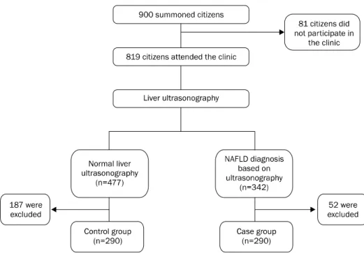
Fig. 1. A flow chart of the study.
drome (i.e. a sixth criterion). From this point of view, NAFLD strongly relates with diabetes mellitus, hypertension, and obesity,6 and consequently, with increased risk for car- diovascular events. The atherogenic effect of metabolic syn- drome was previously established, as well as its compo- nents; but the role of NAFLD in this regard is still being debated. There are evidences of increased rate of car- diovascular mortality in patients with NAFLD and/or cir- rhosis, but the role of NAFLD as the main etiology, or as just a co-morbid disease has remained unclear.7,8 Many pub- lished studies focusing on this issue were done in sympto- matic patients with NAFLD, or on patients in clinical follow up.
Few studies, if any, evaluated this relation in a pop- ulation-based setting, with the participation of asympto- matic individuals.
Intima-media thickness (IMT) of the common carotid ar- tery is a reliable predictor of atherosclerosis. Increased com- mon carotid intima-media thickness (CIMT) has been consid- ered as a marker of atherosclerosis and cardiovascular disease. This study was conducted to find the relation of NAFLD and atherosclerosis as the main predictor of future cardiovascular disease, in a population-based setting, through comparison of the IMT of carotid artery and other im- portant cardiovascular risk factors, between patients with NAFLD and controls.
SUBJECTS AND METHODS
1. Study population
This study was an extension of our previous study on the prevalence of fatty liver disease in the general population in Shiraz, southern Iran, which has recently been published.5 Based on ultrasonograhic finding, as described in the follow- ing parts, individuals with fatty liver disease were identified.
Flow chart of the study is demonstrated in Fig. 1. Among 819 individuals who agreed to participate in the study, 342 had evidence of fatty liver disease on ultrasonography in any form, from mild to severe (ultrasonographic criteria are de- scribed in the “study protocol” part).
We excluded patients with positive or suspicious results for HBsAg, anti-HCV and HIV. We also excluded subjects who had any history of liver disease, had suspicious hepatic-re- lated signs and symptoms during the physical exam or after history taking, had a history of pregnancy in the previous year, had a history of weight loss or weight gain in recent years, or who had had any kind of major organ failure. Exclusion of in- dividuals reporting alcohol consumption was ensured, by in- terviewing the participants at two different times. We ex- cluded pregnant women, or those who had delivered within the past six months. Due to different genetic and environ- mental background, which could have affected our results, non-Iranian residents were also excluded.
Fifty-two of these individuals were excluded, based on the
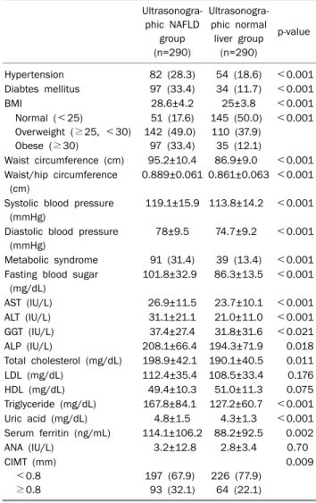
Fig. 2. Longitudinal ultrasonographic image of the common carotid artery.
Longitudinal ultrasonographic image of the common carotid artery, showing the carotid intima-media thickness in the posterior wall (diameter: 11 mm [A], 7 mm [B]).
above-mentioned exclusion criteria. One age matched con- trol group were selected from those who attended our clinic and had normal liver ultrasonography (n=477). Age matching was based on ten-year periods. The above-mentioned ex- clusion criteria were also applied for selecting the control group.
The approvals of the Institutional Review Board, as well as of the Ethics Committee of Shiraz University of Medical Sciences, were obtained prior to the start of the study (No.
HP-12-89). All participants gave their written informed con- sent.
2. Study protocol
All the individuals were invited to come to the Fatty Liver Special Clinic, Health Policy Research Center, Shiraz Univer- sity of Medical Sciences (Shiraz, Iran) for a comprehensive medical evaluation, including physical examination, and complete multi-disciplinary history taking (by physicians and nurses), and anthropometric assessments (by trained nurses). Venous blood samples were also taken, for related laboratory analysis.
Diabetes mellitus was defined according to the American Diabetes Association criteria, based on an fasting blood sug- ar (FBS) >126 mg/dL.9 Hypertension was defined according to the Seventh Report of the Joint National Committee on Prevention, Detection, Evaluation and Treatment of High Blood Pressure (JNC 7).10 Afterwards, liver ultrasonography and common carotid artery ultrasonography were performed for all participants.
The National Cholesterol Education Program - Adult Treatment Panel III (ATP III) published criteria11 were applied for the diagnosis of metabolic syndrome in our population, which defined metabolic syndrome as positivity for three or more of the following criteria: 1) FBS >110 mg/dL, or current use of hypoglycemic agents; 2) central obesity (waist circum- ference >102 cm in men, and >88 cm in women); 3) arterial blood pressure >130/85 mmHg, or current use of anti- hypertensive medications; 4) triglyceride levels >150 mg/dL, or current use of fibrates; 5) HDL cholesterol <40 mg/dL in men, and <50 mg/dL in women.
3. Liver ultrasonography
Participants underwent abdominal ultrasonography ac- cording to a standardized protocol, taking into consideration that ultrasonography can detect increased liver echogenicity, and confirm the diagnosis of NAFLD, particularly when hep- atic fat infiltration surpasses 33%.12 Real-time imaging of the liver was performed for each subject by an experienced radi- ologist, using a Shimadzu ultrasound machine (Shimadzu Inc., Tokyo, Japan) with a 5-MHz to 7-MHz transducer probe (curvilinear). The radiologist performing the ultrasonography was unaware of the clinical and laboratory results. The ultra- sonography diagnostic patterns of fatty liver disease were based on the presence of a ‘bright’ liver, with stronger echoes in the hepatic parenchyma than in the renal parenchyma, of- ten associated with unusually fine liver echotexture and ves- sel blurring, in the absence of findings suggestive of other chronic liver diseases.
Table 1. Clinical, Anthropometric and Laboratory Findings of Patients with NAFLD and Control Group (Ultrasonographic Normal Liver Group)
Ultrasonogra- phic NAFLD
group (n=290)
Ultrasonogra- phic normal
liver group (n=290)
p-value
Hypertension 82 (28.3) 54 (18.6) <0.001 Diabtes mellitus 97 (33.4) 34 (11.7) <0.001
BMI 28.6±4.2 25±3.8 <0.001
Normal (<25)
Overweight (≥25, <30) Obese (≥30)
51 (17.6) 142 (49.0)
97 (33.4)
145 (50.0) 110 (37.9) 35 (12.1)
<0.001
Waist circumference (cm) 95.2±10.4 86.9±9.0 <0.001 Waist/hip circumference
(cm)
0.889±0.061 0.861±0.063 <0.001
Systolic blood pressure (mmHg)
119.1±15.9 113.8±14.2 <0.001
Diastolic blood pressure (mmHg)
78±9.5 74.7±9.2 <0.001
Metabolic syndrome 91 (31.4) 39 (13.4) <0.001 Fasting blood sugar
(mg/dL)
101.8±32.9 86.3±13.5 <0.001
AST (IU/L) 26.9±11.5 23.7±10.1 <0.001
ALT (IU/L) 31.1±21.1 21.0±11.0 <0.001
GGT (IU/L) 37.4±27.4 31.8±31.6 <0.021
ALP (IU/L) 208.1±66.4 194.3±71.9 0.018
Total cholesterol (mg/dL) 198.9±42.1 190.1±40.5 0.011 LDL (mg/dL) 112.4±35.4 108.5±33.4 0.176
HDL (mg/dL) 49.4±10.3 51.0±11.3 0.075
Triglyceride (mg/dL) 167.8±84.1 127.2±60.7 <0.001 Uric acid (mg/dL) 4.8±1.5 4.3±1.3 <0.001 Serum ferritin (ng/mL) 114.1±106.2 88.2±92.5 0.002
ANA (IU/L) 3.2±12.8 2.8±3.4 0.70
CIMT (mm) <0.8 ≥0.8
197 (67.9) 93 (32.1)
226 (77.9) 64 (22.1)
0.009
Values are presented as the mean±SD or n (%).
For the comparison between patient and control data, student’s t-test were used for quantitative variables and the chi-square test was used to determine the statistical significance of differences in qualitative variables. p-value less than 0.05 is considered sta- tistically significant.
NAFLD, nonalcoholic fatty liver disease; CIMT, common carotid intima-media thickness.
4. Carotid artery ultrasonography
Carotid artery ultrasonography was performed using the same model of ultrasonography scanner (Sonoline G50;
Seimens AG, Munich, Germany). The protocol involved scan- ning of both common carotid, as well as internal and external carotid arteries. Subjects were examined by an expert radiol- ogist, following a standardized protocol. Briefly, IMT was as- sessed in the posterior wall of the artery as the distance be- tween the leading edge of the first and second echogenic lines (Fig. 2). All of the scans were recorded. The best-quality end diastolic image was captured in a longitudinal axis view that showed the bifurcation, and was analyzed off-line. From this image, three measurements of the common carotid ar- tery were performed at a distance of 0.5 cm, 1 cm, and 2 cm below the bifurcation of the common carotid, and the average measurement was used for further assessments. The cut-off value for the CIMT was set at 0.8 mm (CIMT ≥0.8 mm), in or- der to avoid missing any patients.13 All measurements were carried out by a single radiologist on previously recorded scans. At the time of reading, he was unaware of the patients’
identities, and clinical and laboratory characteristics.
5. Statistical analysis
Statistical analyses were performed using the SPSS soft- ware, version 17.0 (SPSS Inc., Chicago, IL, USA). Results are reported as the mean±SD for continuous variables, and as number (%) for categorical variables. For the comparison be- tween patient and control data, student’s t-test were used for quantitative variables and the chi-square test was used to de- termine the statistical significance of differences in qual- itative variables. Multiple logistic regression analysis (adjust- ed OR with multiple logistic regression) was performed for the adjustment of different risk factors and variables associated with the development of thick CIMT.
RESULTS
1. Comparison of clinical characteristics between NAFLD and the control group
Table 1 shows the demographical, clinical and laboratory characteristics of patients and controls. The 290 patients with NAFLD and the same number of controls were matched in term of age. In the NAFLD group, 160 (55.2%) participants
were females, while in the controls, 174 (60%) individuals were females (p=0.27). The mean age of patients with NAFLD was 46.4±11.9 years, with a value of 45.4±12.4 years in the controls (p=0.90). Patients with NAFLD had a higher blood pressure (both systolic and diastolic) than the controls, with statistical significance (p<0.001). Mean FBS was sig- nificantly higher in patients with NAFLD, than in the controls (101.8±32.9 vs. 86.3±13.5 mg/dL, p<0.001). Levels of liv- er aminotransferases were higher in patients with NAFLD, than in the controls (p<0.001). We also noticed that the level
Table 2. Comparison between CIMT and Metabolic Syndrome- related Factors
CIMT
≥0.8 mm (n=157)
CIMT
<0.8 mm (n=423)
p-value
Age (yr) Hypertension Diabetes mellitus Triglyceride (mg/dL) HDL (mg/dL) Waist circumference (cm)
NAFLD Serum ferritin (ng/mL)
54.4±11.4 58 (36.9) 52 (33.1) 153.7±79.7
50.7±9.9 93.3±9.6
93 (59.2) 110.3±107.7
42.8±10.8 78 (18.4) 79 (18.7) 145.1±74.5
50.1±11.2 90.3±10.9
197 (46.6) 98±97.5
<0.001
<0.001
<0.001 0.251 0.506 0.002
0.007 0.222
Values are presented as the mean±SD or n (%).
For the comparison between those with thick CIMT and subjects with normal CIMT, student’s t-test were used for quantitative variables, and the chi-square test was used to determine the statistical significance of differences in qualitative variables.
p-value less than 0.05 is considered statistically significant.
CIMT, common carotid intima-media thickness; NAFLD, nonalco- holicfatty liver disease.
Table 3. Multivariate Analysis for the Risk Factors of CIMT ≥0.8 mma
Variable p-value
OR (CIMT ≥0.8 mm/
<0.8 mm)
95% CI
NAFLD
No 1 -
Yes 0.009 1.91 1.17-3.10
Hypertension
No 1 -
Yes 0.238 1.34 0.82-2.19
Diabetes mellitus
No 1 -
Yes 0.611 1.14 0.68-1.89
Waist circumferenceb
Normal 1 -
High 0.054 0.62 0.38-1.01
Age (yr)
18-29 1 -
30-39 0.166 0.27 0.04-1.710
40-49 0.048 3.50 1.01-12.18
50-59 <0.001 10.83 3.07-38.20
≥60 <0.001 22.76 6.08-85.21
CIMT, common carotid intima-media thickness; NAFLD, nonalco- holic fatty liver disease.
aAdjusted OR with multiple logistic regression.
bWaist circumference >102 cm in men and >88 cm in women was considered high.
of serum ferritin was significantly higher in patients with NAFLD, than in the controls (p=0.002). Those with NAFLD had a significantly higher prevalence of increased CIMT, in comparison with normal individuals (OR, 1.66; 95% CI, 1.15-2.41; p<0.001). We also observed that patients with NAFLD had a higher prevalence of metabolic syndrome (OR, 2.94; 95% CI, 1.93-4.47; p<0.001), than the controls.
However, in the NAFLD group, no correlation was found be- tween the presence of metabolic syndrome and the higher occurrence of increased CIMT (38.5% in the NAFLD patients with metabolic syndrome, in comparison with 29.1% in the NAFLD patients without metabolic syndrome, p=0.136).
2. Association between CIMT and metabolic syndrome A comparison between CIMT and metabolic syndrome- related risk factors is depicted in Table 2. The mean age of subjects with CIMT equal to or greater than 0.8 mm was 54.4±11.4 years, with a value of 42.8±10.8 years in those with CIMT less than 0.8 mm (p<0.001). Subjects with thick CIMT had a higher prevalence of HTN, in comparison to those with normal CIMT (36.9% vs. 18.4%, p<0.001). This is also true for those with NAFLD, in contrast to subjects without NAFLD (59.2 vs. 46.6, p=0.007).
Individuals with thick CIMT had a significantly higher waist circumference, than those with normal CIMT (93.3±9.6 vs.
90.3±10.9 cm, p=0.002).
3. Multivariate analysis of risk factors for CIMT ≥0.8 mm
Table 3 indicates the results of the multiple logistic re- gression analysis (with adjusted OR) of five criteria of meta- bolic syndrome (diabetes mellitus, hypertension, trigly- ceride, HDL, and waist circumference), as well as NAFLD for predicting thick common carotid intima-media. Age and se- rum ferritin level were also mentioned in our analysis, in order to adjust their impact on the CIMT. The table shows that NAFLD could be an independent risk factor for thick common carotid intima-media (OR, 1.9; 95% CI, 1.17-3.09; p=0.009).
Statistical analysis showed that patients between 40 to 49 years old were more prone to develop thick CIMT, in- dependent of the presence of other risk factors for thick CIMT (OR, 3.5; 95% CI, 1.01-12.18; p=0.048). This would be more prominent for patients who were between 50 to 59 years old, as well as those above age 60, for detecting thick CIMT (OR, 10.83; 95% CI, 3.07-38.2; p<0.001 and OR, 22.76; 95% CI, 6.08-85.21; p<0.001, respectively).
DISCUSSION
The prevalence of NAFLD is growing globally, as a result of the increase of type II diabetes mellitus and obesity.14 The NAFLD is closely related to insulin resistance, and to the pres- ence of markers of oxidative stress in plasma and endothelial dysfunction. The NAFLD is actually mentioned as the hepatic manifestation of the metabolic syndrome.15 The increase in risk of cardiovascular disease in patients with metabolic syn- drome is considerable.16
The importance of NAFLD is highlighted by new data sug- gesting that NAFLD can be an independent risk factor for car- diovascular disease.17,18 As a matter of fact, the hypothesis that NAFLD is not only a marker of cardiovascular disease, but may also be involved in its pathogenesis,19 has started to gain more evidence. This situation is in concordance with the new guidelines for the diagnosis and treatment of patients with NAFLD, which emphasize an early and more accurate di- agnosis of this pathologic condition, in order to better define the disease progression from simple fatty changes without inflammation, to NASH, cirrhosis, and end-stage liver disease.20
Our population-based case-control study suggests that cardiovascular risk factors, such as increased IMT, occur more frequently among NAFLD patients, when compared to healthy individuals, confirming previous findings in patients’
NAFLD.8,18,21 Although some of these studies included only NAFLD patients with elevated liver aminotransferases, most surveys stated that NAFLD is associated with increased CIMT, regardless of the rise of liver enzymes plasma levels.21 Recent studies concluded that the same process occurs in cerebral arteries, which causes increased cerebrovascular resistance.22 This leads to a higher risk of morbidity and mor- tality related to non-hepatic consequences of NAFLD.23 Through adjusting the effects of other variables, such as DM and age, we also indicated that NAFLD could be an in- dependent risk factor for thick common carotid intima-media (OR, 1.91; 95% CI, 1.17-3.01; p=0.009). Randomized pro- spective clinical trials are needed, to sufficiently evaluate the direct and indirect impacts of NAFLD on the atherosclerotic process.
In our study, we observed that the age of 50 years is a suit- able cut-off value for predicting CIMT ≥0.8 mm. This finding is applicable in practice, because fatty changes in the liver
are usually incidental findings in abdominal ultrasono- graphy, and there is a permanent controversy regarding which patients should undergo further laboratory and para- clinical work-up. Recent surveys support the argument that patients with non-alcoholic fatty changes of liver have a high- er death probability due to cardiovascular complications, in comparison to the complications of liver dysfunction.24 Therefore, it is crucial to perform cardiovascular evaluations in patients with NAFLD, even if no other cardiovascular risk factor is observed.25 Overwhelming evidence suggests that cardiovascular disease determines the outcomes in patients with NAFLD more frequently, and to a greater extent, than the progression of liver disease.24,26,27
Serum ferritin was increased in NAFLD patients, in com- parison with the controls. The role of ferritin in the human body is to store and deliver iron to the cells, and the levels of ferritin in plasma reflect the total body iron supply. Ferritin is also an acute-phase reactant that may have elevated plasma levels, in the case of infection, inflammation, and other stimuli.28 Because there are no proofs at present to advocate for a role of excess iron in the pathogenesis of NAFLD,29 it can be hypothesized that a higher ferritin level in these patients could be linked to the presence of an inflammatory back- ground associated with steatosis in hepatocytes, and even necrosis. This concept is supported by our findings, which in- dicate that the serum ferritin level was strongly correlated with markers of hepatocyte injury (alanine amino- transferase, aspartate aminotransferase and gamma-glu- tamyl transpeptidase correlated with ferritin levels, p
<0.001). Although growing evidence suggests a link be- tween serum ferritin, insulin resistance, and NAFLD,30 the in- creased serum ferritin levels may just be in response to, and along with the atherogenic course undergoing in vessel walls.
This can be explained on the basis of a mechanism that states that ferritin genes are up-regulated by inflammatory cytokines, and are prone to induction in the process of plaque formation.31
The gold standard for the diagnosis of NAFLD is liver biop- sy, but because of ethical reasons, the procedure was not ap- plied in our study. As ultrasonography is the most common and readily available method for the diagnosis of fatty changes in liver, we used it, in addition to excluding known eti- ologies of liver disease. Studies have proven that the optimal hepatic steatosis threshold for radiologic detection is 33% of
liver texture, and this is the range of moderate to severe NAFLD. Therefore, regardless of changes in liver enzymes, abdominal ultrasonography is sensitive enough for the de- tection of fatty changes in the liver.12
Several studies have evaluated the relationship between NAFLD and CIMT, and support the idea of independent asso- ciations between NAFLD and atherosclerosis.8,20,32-35
One of the important defects of many of these studies is that their cases groups are selected from those who were re- ferred to clinics or private physicians. Consequently, these NAFLD subjects do not actually represent the general population. The causes for referral of these NAFLD patients were the development of signs and symptoms that made ei- ther them, or their family physicians, initiate diagnostic workups. On the other hand, in our study, the cases group was randomly selected from the general population. The cases did not complain of any symptoms, and their physical exam and history taking by trained physicians revealed no sign and symptoms, NAFLD being detected by ultrasonography.
In summary, we suggest that NAFLD could be a risk factor for carotid atherosclerosis, independently of its association with the metabolic syndrome. The clinical corollary of our findings suggests that the detection of fatty changes in liver on abdominal ultrasonography should provide an alert on the probability of the presence of multiple underlying car- diovascular risk factors, warranting evaluation and treat- ment, as cardiovascular atherosclerosis-related morbidity and mortality are elevated by the pathological course of fatty liver disease.
ACKNOWLEDGEMENTS
We sincerely thank all the subjects who participated in our study, as well as their families. We also express our gratitude to all the staff of the Motahari Polyclinic laboratory for their cooperation.
REFERENCES
1. Ko JS. Nonalcoholic fatty liver disease. Korean J Gastroenterol 2010;56:6-14.
2. Levene AP, Goldin RD. The epidemiology, pathogenesis and his- topathology of fatty liver disease. Histopathology 2012;61:141- 152.
3. Vernon G, Baranova A, Younossi ZM. Systematic review: the epi- demiology and natural history of non-alcoholic fatty liver disease
and non-alcoholic steatohepatitis in adults. Aliment Pharmacol Ther 2011;34:274-285.
4. Ahmed MH, Abu EO, Byrne CD. Non-alcoholic fatty liver disease (NAFLD): new challenge for general practitioners and important burden for health authorities? Prim Care Diabetes 2010;4:
129-137.
5. Lankarani KB, Ghaffarpasand F, Mahmoodi M, et al. Non alco- holic fatty liver disease in southern Iran: a population based study. Hepat Mon 2013;13:e9248.
6. Lee SY, Kim SK, Kwon CI, et al. Clinical characteristics of health screen examinees with nonalcoholic fatty liver and normal liver function test. Korean J Gastroenterol 2008;52:161-170.
7. Margariti E, Deutsch M, Manolakopoulos S, Papatheodoridis GV.
Non-alcoholic fatty liver disease may develop in individuals with normal body mass index. Annals of Gastroenterology 2012;25:
45-51.
8. Sookoian S, Pirola CJ. Non-alcoholic fatty liver disease is strongly associated with carotid atherosclerosis: a systematic review. J Hepatol 2008;49:600-607.
9. Basevi V, Di Mario S, Morciano C, Nonino F, Magrini N. Comment on: American Diabetes Association. Standards of medical care in diabetes--2011. Diabetes Care 2011;34(Suppl. 1):S11-S61.
Diabetes Care 2011;34:e53, author reply e54.
10. Lenfant C, Chobanian AV, Jones DW, Roccella EJ; Joint National Committee on the Prevention, Detection, Evaluation, and Treat- ment of High Blood Pressure. Seventh report of the Joint National Committee on the Prevention, Detection, Evaluation, and Treatment of High Blood Pressure (JNC 7): resetting the hy- pertension sails. Hypertension 2003;41:1178-1179.
11. Grundy SM, Cleeman JI, Daniels SR, et al; American Heart Association; National Heart, Lung, and Blood Institute.
Diagnosis and management of the metabolic syndrome: an American Heart Association/National Heart, Lung, and Blood Institute Scientific Statement. Circulation 2005;112:2735- 2752.
12. Saadeh S, Younossi ZM, Remer EM, et al. The utility of radio- logical imaging in nonalcoholic fatty liver disease. Gastroenter- ology 2002;123:745-750.
13. Dougherty RS, Brant WE. Vascular ultrasound. In: Brant WE, Helms CA, eds. Fundamentals of diagnostic radiology. 3rd ed.
Philadelphia: Lippincott, Williams & Wilkins, 2007:1027.
14. Yki-Järvinen H. Nutritional modulation of nonalcoholic fatty liver disease and insulin resistance: human data. Curr Opin Clin Nutr Metab Care 2010;13:709-714.
15. Kotronen A, Westerbacka J, Bergholm R, Pietiläinen KH, Yki-Järvinen H. Liver fat in the metabolic syndrome. J Clin Endocrinol Metab 2007;92:3490-3497.
16. Musso G, Gambino R, Bo S, et al. Should nonalcoholic fatty liver disease be included in the definition of metabolic syndrome? A cross-sectional comparison with Adult Treatment Panel III cri- teria in nonobese nondiabetic subjects. Diabetes Care 2008;
31:562-568.
17. Targher G, Day CP, Bonora E. Risk of cardiovascular disease in patients with nonalcoholic fatty liver disease. N Engl J Med 2010;
363:1341-1350.
18. Targher G, Bertolini L, Rodella S, et al. Nonalcoholic fatty liver dis-
ease is independently associated with an increased incidence of cardiovascular events in type 2 diabetic patients. Diabetes Care 2007;30:2119-2121.
19. Maurantonio M, Ballestri S, Odoardi MR, Lonardo A, Loria P.
Treatment of atherogenic liver based on the pathogenesis of nonalcoholic fatty liver disease: a novel approach to reduce car- diovascular risk? Arch Med Res 2011;42:337-353.
20. Loria P, Adinolfi LE, Bellentani S, et al; NAFLD Expert Committee of the Associazione Italiana per lo studio del Fegato. Practice guidelines for the diagnosis and management of nonalcoholic fatty liver disease. A decalogue from the Italian Association for the Study of the Liver (AISF) Expert Committee. Dig Liver Dis 2010;42:272-282.
21. Targher G, Bertolini L, Padovani R, et al. Relations between car- otid artery wall thickness and liver histology in subjects with non- alcoholic fatty liver disease. Diabetes Care 2006;29:1325- 1330.
22. Karakurt F, Carlioglu A, Koktener A, et al. Relationship between cerebral arterial pulsatility and carotid intima media thickness in diabetic and non-diabetic patients with non-alcoholic fatty liv- er disease. J Endocrinol Invest 2009;32:63-68.
23. Ying I, Saposnik G, Vermeulen MJ, Leung A, Ray JG. Nonalcoholic fatty liver disease and acute ischemic stroke. Epidemiology 2011;22:129-130.
24. Targher G, Day CP, Bonora E. Risk of cardiovascular disease in patients with nonalcoholic fatty liver disease. N Engl J Med 2010;363:1341-1350.
25. Thakur ML, Sharma S, Kumar A, et al. Nonalcoholic fatty liver dis- ease is associated with subclinical atherosclerosis independent of obesity and metabolic syndrome in Asian Indians. Atheroscle- rosis 2012;223:507-511.
26. Assy N, Djibre A, Farah R, Grosovski M, Marmor A. Presence of coronary plaques in patients with nonalcoholic fatty liver
disease. Radiology 2010;254:393-400.
27. Szollár L. Fatty liver and global cardiometabolic risk. Orv Hetil 2010;151:1946-1950.
28. Manousou P, Kalambokis G, Grillo F, et al. Serum ferritin is a dis- criminant marker for both fibrosis and inflammation in histologi- cally proven non-alcoholic fatty liver disease patients. Liver Int 2011;31:730-739.
29. Marchesini G, Bugianesi E, Forlani G, et al. Nonalcoholic fatty liv- er, steatohepatitis, and the metabolic syndrome. Hepatology 2003;37:917-923.
30. Zelber-Sagi S, Nitzan-Kaluski D, Halpern Z, Oren R. NAFLD and hyperinsulinemia are major determinants of serum ferritin levels. J Hepatol 2007;46:700-707.
31. Pang JH, Jiang MJ, Chen YL, et al. Increased ferritin gene ex- pression in atherosclerotic lesions. J Clin Invest 1996;97:
2204-2212.
32. Brea A, Mosquera D, Martín E, Arizti A, Cordero JL, Ros E.
Nonalcoholic fatty liver disease is associated with carotid athe- rosclerosis: a case-control study. Arterioscler Thromb Vasc Biol 2005;25:1045-1050.
33. Fracanzani AL, Burdick L, Raselli S, et al. Carotid artery in- tima-media thickness in nonalcoholic fatty liver disease. Am J Med 2008;121:72-78.
34. Kim HC, Kim DJ, Huh KB. Association between nonalcoholic fatty liver disease and carotid intima-media thickness according to the presence of metabolic syndrome. Atherosclerosis 2009;
204:521-525.
35. Nambi V, Chambless L, He M, et al. Common carotid artery in- tima-media thickness is as good as carotid intima-media thick- ness of all carotid artery segments in improving prediction of cor- onary heart disease risk in the Atherosclerosis Risk in Communities (ARIC) study. Eur Heart J 2012;33:183-190.