http://dx.doi.org/10.12671/jkfs.2014.27.1.29
29
Copyright ⓒ 2014 The Korean Fracture Society. All rights reserved.
This is an Open Access article distributed under the terms of the Creative Commons Attribution Non-Commercial License (http://creativecommons.org/licenses/
by-nc/3.0) which permits unrestricted non-commercial use, distribution, and reproduction in any medium, provided the original work is properly cited.
Received February 20, 2013 Revised July 6, 2013 Accepted October 12, 2013
Address reprint requests to: Seoung Joon Lee, M.D., Ph.D.
Department of Orthopedic Surgery, Konkuk University Medical Center, 120-1 Neungdong-ro, Gwangjin-gu, Seoul 143-729, Korea Tel: 82-2-2030-7360ㆍFax: 82-2-2030-7369
E-mail: lsjmd@naver.com
전방 부정유합된 원위 요골에서 개방성 쐐기형 절골술의 임상 결과
이승준 ⋅최진호
건국대학교 의학전문대학원 정형외과학교실
The Clinical Results of Opening Wedge Osteotomy in the Volarly Malunited Distal Radius
Seoung Joon Lee, M.D., Ph.D. , Jin Ho Choi, M.D.
Department of Orthopedic Surgery, Konkuk University School of Medicine, Seoul, Korea
Purpose: To report the clinical results of opening wedge osteotomy graft in the volarly malunited distal radius.
Materials and Methods: Ten patients with volarly malunited distal radius fractures treated by opening wedge osteotomy were included in this study. Grip power, range of motion of the wrist, radiographic parameter and Mayo wrist scores were retrospectively evaluated.
Results: At the final follow-up, the rotation of the forearm, the range of motion of wrist, and the grip power were improved.
The average radial inclination improved to 22.2o, the average volar tilting improved to 5.6o, and the average ulnar variance improved to 0.8 mm. The average Mayo wrist score was improved to 85.6.
Conclusion: Opening wedge osteotomy for volarly malunited distal radius was considered as one of the good treatments to restore anatomy of the distal radius and distal radioulnar joint and also to improve the function of the wrist joint.
Key Words: Distal radius, Malunion, Opening wedge osteotomy
서 론
원위 요골 골절은 상지에서 발생하는 가장 흔한 골절로, 쉽게 치료될 수 있는 골절로 인식되고 있으나 실제로 원위 요골 골절 후 합병증은 6%-80%로 적지 않게 보고되고 있 으며, 증상을 유발하는 부정유합은 5%로 보고되고 있다5). 원위 요골의 부정유합은 원위 요척골 관절면의 불일치와 요 수근 관절의 생역학적 변화를 유발하여 완관절의 통증, 수
부 악력 감소, 전완부의 회전 제한 및 완관절 운동 범위 감 소 등의 증상이 나타날 수 있다6,9,11,12,14,17)
. 여러 저자들이 후방으로 부정유합된 원위 요골에 대하여 교정 절골술을 이 용하여 좋은 치료 결과를 보고하였으나3,4,7,10), 전방으로 부 정 유합된 요골 골절에 대한 교정 절골술의 치료 결과를 보고하는 문헌은 많지 않다. 이에 저자들은 원위 요골의 부 정유합 중 전방으로 부정유합된 원위 요골에 대하여 시행한 개방성 쐐기형 절골술의 치료 결과를 보고하고자 한다.
대상 및 방법
1. 연구 대상
2001년 3월부터 2011년 3월까지 건국대학교병원 정형외 과에서 원위 요골 골절 후 부정유합으로 진단받은 환자 중
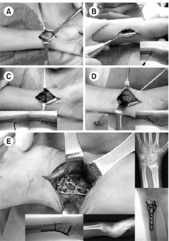
Fig. 1. Intraoperative photographs show surgical approach (A), wedge osteotomy and wedge shaped iliac bone (B, C, D), and plate fixation (E).
전방으로 부정유합된 10예를 대상으로 하였다. 본 저자들 은 전완부의 회전 운동 범위에 제한이 있거나 통증이 있는 경우를 수술의 적응증으로 고려하였으며, 방사선 계측에서 요골의 전방 경사각 20o 이상, 요골 단축 6 mm 이상일 경 우도 수술의 대상으로 고려하였다. 원위 요골 관절과 원위 요척골 관절면 내 골절이 있거나 관절염이 진행된 환자, 수부에 다른 동반 손상이 있는 환자는 연구 대상에서 제외 하였다. 10예의 평균 연령은 45세(범위: 25-65세)였으며 남 성이 8예, 여성이 2예였다. 요골 골절 후 처음 치료 방법 은 도수 정복 후 석고 고정술 6예, 도수 정복 후 K-강선 고정술 2예, 도수 정복 후 외고정 장치 및 K-강선 고정술 2예였다. 수상 후 절골술을 받기까지 기간은 평균 16주(범 위: 14-20주)였고 평균 추시 기간은 21개월(범위: 12-30개 월)이었다. 수술 전 완관절의 신전은 평균 39.5o (범위:
20o-60o), 굴곡은 평균 43.2o (범위: 20o-70o)였으며, 전완부 의 회외전은 평균 49o (범위: 30o-70o), 회내전은 73o (범위:
60o-90o)였다. 수술 전 이환된 수부의 악력은 평균 16 kg (범위: 5-20 kg), 정상인 반대측 수부의 평균 악력 35 kg
(범위: 20-50 kg)이었다.
2. 수술 방법 및 수술 후 처치
수술은 10예 모두에서 Henry의 전방 도달법을 이용하여 요골의 부정유합된 부위를 노출시킨 후 C-arm으로 완관절 의 측면을 투시하면서 K-강선이 원위 요골의 관절면에 수 평이 되게 삽입한 후 이 K-강선을 따라 관절면에 평행이 되게 얇은 절골기를 이용하여 절골술을 시행하였다. 절골 된 부위를 lamina spreader로 벌리고 C-arm을 보면서 요골 의 전방 경사각과 길이, 사면각을 정상 범위로 회복시켰다.
K-강선을 이용하여 절골된 부위를 일시적으로 고정한 후 자가 장골 피질골을 절골된 부위에 삽입 후 잠금 금속판으 로 고정하였다(Fig. 1). 수술 후 4주 동안 단상지 석고 고 정을 하였으며, 이 기간 동안에도 전완부의 능동적 회전 운동은 시작하였으며 수술 후 4주째 석고를 제거하고 완관 절의 능동적, 수동적 관절 운동을 시작하였다.
3. 수술 전, 후 평가
수술 전에 컴퓨터 단층 촬영을 하여 관절 내 골절 유무 여부와 원위 요척골 관절면의 상태를 확인하였으며, 완관 절의 진성 전후방 및 측면 단순 방사선 사진을 촬영하여 요골의 전방 경사각(volar tilting), 요골 사면각(radial in- clination), 척골 변위(ulnar variance)값을 수술 전과 최종 추시에 비교하였다. 또한, 수술 전과 최종 추시 시에 수부 의 악력 검사, Mayo wrist 점수, 완관절의 운동 범위 측정 을 하여 임상적 결과를 분석하였으며, Wilcoxon signed rank test을 이용하여 통계적 유의성을 평가하였다. 연구분 석에는 IBM SPSS ver. 22.0 (IBM Co., Armonk, NY, USA) 을 사용하였다.
결 과
1. 완관절 운동 범위 및 악력 측정
수술 후 최종 추시 시 완관절의 운동 범위 측정 결과, 완관절의 신전은 평균 62o (범위: 45o-80o), 굴곡은 평균 53.5o (범위: 40o-80o)였으며, 전완부의 회외전은 평균 82o (범위: 70o-90o), 회내전은 평균 85o (범위: 80o-90o)로 수술 전에 비하여 향상되었다(p<0.05) (Table 1).
2. 방사선 계측
요골의 전방 경사각은 수술 전 평균 24.4o (범위: 18o-
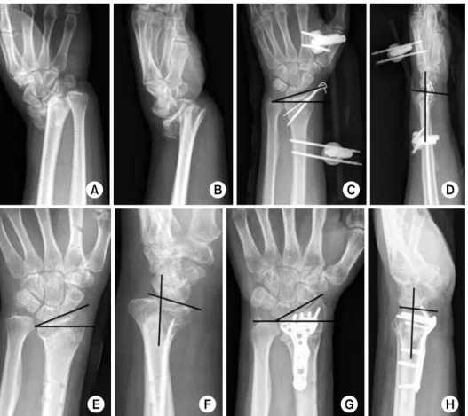
Fig. 2. (A, B) Initial simple radiographs show unstable di- stal radius fracture. (C, D) Si- mple radiographs after external fixation and K-wire fixation show radial inclination of 20o, volar tilt 5o, and neutral ulnar variance. (E, F) Simple radiographs after removal of external fixation and K-wire show the volarly malunited distal radius with volar tilt of 25o, 12o radial inclination, 3 mm ulnar positive variance.
(G, H) Simple radiographs after open wedge osteotomy show a good alignment of distal radius with volar tilt of 5o, 25o radial inclination, 0 mm ulnar variance.
Table 1. The Preoperative and Postoperative Range of Motion and Grip Power
Case No.
Range of motion (o)
Grip power (kg)
Extension Flexion Supination Pronation
Preoperative Final Preoperative Final Preoperative Final Preoperative Final Preoperative Final Normal 1
2 3 4 5 6 7 8 9 10
20 60 30 50 35 60 40 30 40 30
75 70 55 80 55 80 60 50 50 45
20 70 40 60 30 50 50 40 30 30
60 80 50 50 45 60 50 60 40 40
30 60 50 70 30 30 40 50 70 60
70 80 80 90 80 80 80 80 90 90
70 80 70 90 70 70 60 70 80 70
80 90 80 90 80 80 90 80 90 90
5 15 20 30 20 15 10 10 20 15
15 40 40 40 35 35 30 45 35 20
20 40 35 50 40 30 40 40 35 20
p-value 0.001134 0.003727 0.000187 0.006603 0.000587
30o)에서 최종 추시 평균 5.6o (범위: 0o-10o)로 측정되었고, 요골 사면각은 수술 전 평균 14.7o (범위: 10o-20o)에서 최 종 추시 평균 22.2o (범위: 18o-25o)로 증가하였다. 척골의 변위는 수술 전 평균 +4.6 mm (범위: 3-8 mm)에서 최종 추시 평균 +0.8 mm (범위: 0-2 mm)로 감소하였다(Fig.
2, 3, Table 2).
3. Mayo wrist 점수 및 수부 악력 검사
최종 추시 시 악력은 평균 35.6 kg (범위: 18-44 kg)으로
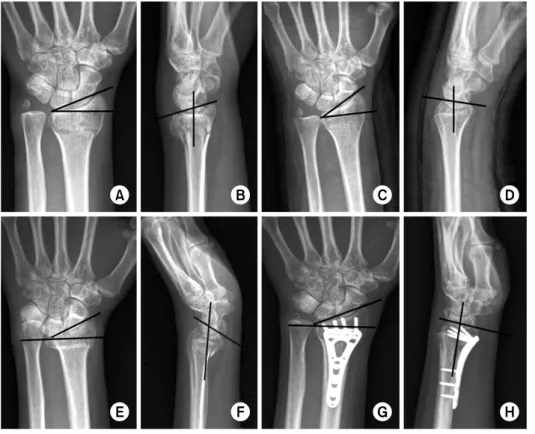
Fig. 3. (A, B) Initial simple radiographs show Colles’ fra- cture with ulnar styloid frac- ture. (C, D) Simple radiogra- phs after closed reduction and splinting show a good align- ment of distal radius. (E, F) At 2 months after trauma, simple radiographs show vo- larly displaced distal radius fracture with volar tilt of 30o, 15o radial inclination, 5 mm ulnar positive variance. (G, H) Simple radiographs after open wedge osteotomy show a good alignment of distal radius with volar tilt of 10o, 23o radial inclination, 0 mm ulnar variance.
Table 2. Preoperative and Postoperative Radiological Values
Case No. Volar tilting (o) Radial inclination (o) Ulnar variance (mm)
Preoperative Final Preoperative Final Preoperative Final
1 2 3 4 5 6 7 8 9 10
30 20 30 26 25 18 24 28 20 23
10 0 10 3 5 0 5 10 8 5
15 17 16 14 12 20 10 15 12 16
23 22 23 25 25 24 22 20 18 20
5 4 3 5 3 4 5 8 5 4
0 2 0 1 0 0 1 2 1 1
p-value <0.05 <0.05 <0.05
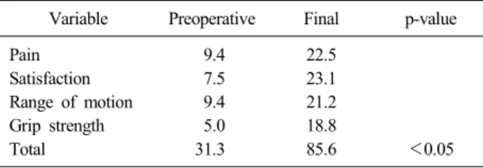
수술 전 12.6 kg에 비하여 호전되었으며(p<0.05) Mayo wrist 점수는 수술 전 평균 31.3점(범위: 30-50점)에서 최종 추시 시 평균 85.6점(범위: 70-90점)으로 향상되어 환자의 통증과 만족도에서 좋은 결과를 보였다(p<0.05) (Table 3).
고 찰
원위 요골의 전방 부정유합은 시상 축, 축성 축, 그리고 수평 축에서 복합적으로 원위 요척골 관절과 요수근 관절
에 해부적 및 생역학적 변화를 유발하여 통증, 관절 운동 범위 감소, 악력 감소, 미용 문제 그리고 드물게 건 파열 등을 일으킨다5,11,17).
증가한 요골의 전방 경사각은 척골 두의 후방 아탈구, 굴곡건의 장력 감소, 완관절의 신전 운동 범위 감소를 일 으킬 수 있는데, 척골 두의 후방 아탈구는 삼각섬유연골 복합체의 장력 증가와 원위 요척골 관절면의 불일치를 유 발하여 전완부의 회전 운동, 특히 회외전 운동 범위의 감 소와 통증을 야기하며, 또한 요골의 단축은 척골 충돌 증
Table 3. Mayo Wrist Score
Variable Preoperative Final p-value Pain
Satisfaction Range of motion Grip strength Total
9.4 7.5 9.4 5.0 31.3
22.5 23.1 21.2 18.8
85.6 <0.05
후군을 유발하여 통증, 악력 감소 그리고 완관절 운동 범 위 감소의 원인이 될 수 있다9,12,14).
사체를 이용한 생역학 실험에서도 20o 이상의 전방 또는 후방 경사각의 증가는 요수근 관절의 생역학에 문제를 일 으킬 수 있다고 보고되었으며, 4-6 mm 이상의 요골 단축 은 척골 양성 변이를 일으켜 척골 충돌 증후군 및 삼각섬 유연골 복합체에 문제를 일으킬 수 있다고 보고되었다1,13). Adams1)는 생역학 실험을 통해 요골의 단축이 요골 경 사각 감소와 후방 각변형보다 원위 요척 관절의 동역학과 삼각 섬유연골 복합체에 더 큰 영향을 미친다고 보고하였 고, Ambrose와 Posner2), Taleisnik와 Watson18)은 부정유합 중 정상 전방 경사각의 소실이 가장 큰 문제라고 하였다.
따라서 전방 부정유합된 원위 요골 골절의 치료 목표는 요 골 경사각, 요골 사면각, 요골의 길이를 정상으로 복원시켜 정상 생역학의 회복과 요수근 관절과 원위 요척골 관절의 기능 회복에 있다.
교정 절골술의 적응증으로 Sato 등16)은 통증, 완관절의 운동 제한 등 임상적 증상이 중요하다고 하였으며, Shea 등17)도 해부적 변형 정도보다 통증과 기능적 장애가 중요 하다고 하면서 원위 요척골 관절에 관절염이 있는 경우에 는 교정 절골술만으로는 증상의 호전을 기대할 수 없다고 하였다. 본 저자들도 전완부의 회전 운동 범위 제한이 있 거나 통증이 있는 경우를 우선적인 수술 적응증으로 고려 하였으며, 보고된 사체 생역학 실험 결과를 기준으로 하여 방사선 계측에서 요골의 전방 경사각 20o 이상, 요골 단축 6 mm 이상일 경우도 수술의 대상으로 판단하였다.
절골술의 방법으로는 여러 저자들이 개방성 쐐기형 절골 술 후 금속판 고정과 골 이식술의 좋은 결과를 보고하고 있는데11,14,15,17,19)
, Thivaios와 McKee19)는 sliding 절골술로 골 이식술 없이 정상적인 완관절과 원위 요척골 관절의 해 부학적 회복과 좋은 임상 결과를 얻었다고 하였으며, Wada 등20)은 요골의 쐐기형 절골술과 동시에 척골 단축술을 같 이 시행하여 골 이식술 없이 쉽게 요골의 길이를 회복하여 좋은 결과를 얻을 수 있었다고 보고하였다. 본 연구에서는 개방성 쐐기형 절골술을 하였으며 본 저자들의 경험상 수 술 중 요골의 전방 경사각이 교정되면 생각보다 요골의 길
이를 회복시키는 것이 어렵지 않았지만 요골 단축이 심한 경우에는 수술을 쉽게 하기 위하여 척골 단축술이 필요할 수 있다고 생각한다.
수술 후 합병증으로 Wada 등20)과 Kanterewicz 등8)은 고령의 환자에서는 골밀도의 감소로 인한 금속판의 고정력 소실이 발생할 수 있으므로 수술의 대상 범위에서 제외하 는 것이 좋다고 하였으나 Sato 등16)은 절골술 후 잠금 금 속판을 이용하여 45세 이상의 고령의 환자에서도 좋은 결 과를 얻었다고 보고하였다. 많지는 않지만 본 연구의 증례 중 4예가 50세 이상이었으며 절골된 부위에 고정력 실패나 교정 각도의 소실 등의 합병증이 보이지 않았다. 이는 비 록 이들의 골밀도를 측정하지 않았다는 제한점이 있으나 잠금 금속판을 이용한 고정과 자가 장골 피지골 이식으로 절골된 부위에 안정성을 얻을 수 있었기 때문이라 생각한다.
또 다른 합병증으로 교정 절골술 후에도 원위 요척골 관 절의 통증 또는 전완부의 회전 제한이 30%에서 보고되고 있으며17), 이러한 합병증을 예방하기 위하여는 수술 전에 원위 요척골 관절에 관절염 또는 관절면의 손상이 있는지 를 세심히 확인해야 하며 원위 요척골 관절에 관절염 병변 이 있을 경우에는 절골술과 동시에 Darrach, 척골의 인공 관절술 등의 원위 요척골 관절에 대한 수술적 치료가 필요 하다17). 본 연구에서는 이런 점을 고려하여 수술의 대상을 선정할 때 원위 요골 관절과 원위 요척골 관절면 내 골절 이 있거나 관절염이 진행된 환자는 제외하였으며, 수술 중 에도 항상 원위 요척골 관절의 불안정성이 있는지를 확인 하였다. 비록 본 연구는 연구 대상의 수가 충분하지 않다 는 제한점이 있으나 본 저자들은 전방 부정유합된 원위 요 골에서 개방성 쐐기형 절골술 후 금속판 고정 및 골 이식 술로 좋은 결과를 기대할 수 있을 것으로 생각한다.
결 론
전방으로 부정유합된 원위 요골의 골절에서 개방성 쐐기 형 절골술은 원위 요골의 해부적 형태를 정상으로 회복시 켜 통증의 감소, 운동 범위와 악력의 향상시킬 수 있는 수 술적 방법으로 생각된다.
References
1) Adams BD: Effects of radial deformity on distal radio- ulnar joint mechanics. J Hand Surg Am, 18: 492-498, 1993.
2) Ambrose L, Posner MA: Biplanar osteotomy for the treatment of malunited colles’ fractures. Presented at the 43rd Annual Meeting of the American Society for Surgery
of the Hand (Baltimore, Sep. 14-17, 1998). Baltimore, American Society for Surgery of the Hand: 1998.
3) Bronstein AJ, Trumble TE, Tencer AF: The effects of distal radius fracture malalignment on forearm rotation: a cadaveric study. J Hand Surg Am, 22: 258-262, 1997.
4) Brown JN, Bell MJ: Distal radial osteotomy for mal- union of wrist fractures in young patients. J Hand Surg Br, 19: 589-593, 1994.
5) Cooney WP 3rd, Dobyns JH, Linscheid RL: Complica- tions of Colles' fractures. J Bone Joint Surg Am, 62:
613-619, 1980.
6) Fernandez DL: Radial osteotomy and Bowers arthro- plasty for malunited fractures of the distal end of the radius. J Bone Joint Surg Am, 70: 1538-1551, 1988.
7) Jupiter JB, Ruder J, Roth DA: Computer-generated bone models in the planning of osteotomy of multidirec- tional distal radius malunions. J Hand Surg Am, 17: 406- 415, 1992.
8) Kanterewicz E, Yañez A, Pérez-Pons A, Codony I, Del Rio L, Díez-Pérez A: Association between Colles' frac- ture and low bone mass: age-based differences in post- menopausal women. Osteoporos Int, 13: 824-888, 2002.
9) Kazuki K, Kusunoki M, Shimazu A: Pressure dis- tribution in the radiocarpal joint measured with a densi- tometer designed for pressure-sensitive film. J Hand Surg Am, 16: 401-408, 1991.
10) Ladd AL, Huene DS: Reconstructive osteotomy for mal- union of the distal radius. Clin Orthop Relat Res, (327):
158-171, 1996.
11) Linder L, Stattin J: Malunited fractures of the distal ra- dius with volar angulation: corrective osteotomy in 6 cases using the volar approach. Acta Orthop Scand, 67: 179-181, 1996.
12) McQueen M, Caspers J: Colles fracture: does the ana- tomical result affect the final function? J Bone Joint Surg Br, 70: 649-651, 1988.
13) Pogue DJ, Viegas SF, Patterson RM, et al: Effects of distal radius fracture malunion on wrist joint mechanics. J Hand Surg Am, 15: 721-727, 1990.
14) Prommersberger KJ, Froehner SC, Schmitt RR, Lanz UB: Rotational deformity in malunited fractures of the distal radius. J Hand Surg Am, 29: 110-115, 2004.
15) Prommersberger KJ, Van Schoonhoven J, Lanz UB:
Outcome after corrective osteotomy for malunited fractures of the distal end of the radius. J Hand Surg Br, 27:
55-60, 2002.
16) Sato K, Nakamura T, Iwamoto T, Toyama Y, Ikegami H, Takayama S: Corrective osteotomy for volarly mal- united distal radius fracture. J Hand Surg Am, 34: 27-33, 2009.
17) Shea K, Fernandez DL, Jupiter JB, Martin C Jr:
Corrective osteotomy for malunited, volarly displaced frac- tures of the distal end of the radius. J Bone Joint Surg Am, 79: 1816-1826, 1997.
18) Taleisnik J, Watson HK: Midcarpal instability caused by malunited fractures of the distal radius. J Hand Surg Am, 9: 350-357, 1984.
19) Thivaios GC, McKee MD: Sliding osteotomy for de- formity correction following malunion of volarly displaced distal radial fractures. J Orthop Trauma, 17: 326-333, 2003.
20) Wada T, Isogai S, Kanaya K, Tsukahara T, Yamashita T: Simultaneous radial closing wedge and ulnar shortening osteotomies for distal radius malunion. J Hand Surg Am, 29: 264-272, 2004.
Copyright ⓒ 2014 The Korean Fracture Society. All rights reserved.
This is an Open Access article distributed under the terms of the Creative Commons Attribution Non-Commercial License (http://creativecommons.org/licenses/
by-nc/3.0) which permits unrestricted non-commercial use, distribution, and reproduction in any medium, provided the original work is properly cited.
http://dx.doi.org/10.12671/jkfs.2014.27.1.29
전방 부정유합된 원위 요골에서 개방성 쐐기형 절골술의 임상 결과
이승준 ⋅최진호
건국대학교 의학전문대학원 정형외과학교실
목 적: 전방으로 부정유합된 원위 요골에서 개방성 쐐기형 절골술의 임상 결과를 보고하고자 한다.
대상 및 방법: 전방 부정유합된 원위 요골에 대하여 개방성 쐐기형 절골술로 치료를 받은 10예를 대상으로 하였다. 수부의 악력, 완관절의 운동 범위, 방사선 계측(요골 전방 경사각, 요골 사면각, 척골 변이)을 후향적으로 조사하였으며, Mayo wrist 점수를 측정하였다.
결 과: 최종 추시 시 전완부의 회외전과 완관절의 운동 범위, 수부의 악력은 향상되었다. 요골의 사면각은 평균 22.2o, 전방 경사각은 평균 5.6o로, 척골 변이는 평균 0.8 mm로 향상되었고 Mayo wrist 점수는 평균 85.6점으로 향상되었다.
결 론: 전방으로 부정 유합된 원위 요골에서 개방성 쐐기형 절골술은 원위 요골과 원위 요척골 관절의 해부학을 정상으로 복원시키고 완관절의 기능을 향상시킬 수 있는 좋은 치료 방법 중 하나로 생각된다.
색인 단어: 원위 요골, 부정유합, 개방성 쐐기형 절골술
접수일 2013. 2. 20 수정일 2013. 7. 6 게재확정 2013. 10. 12 교신저자 이 승 준
서울시 광진구 능동로 120-1, 건국대학교병원 정형외과
Tel 02-2030-7360, Fax 02-2030-7369, E-mail lsjmd@naver.com
35