I. 서론
치주치료의 궁극적인 목표는 치주질환으로 인하 여 파괴된 치주조직의 재생에 있으며, 이는 염증에 이환된 치근면의 신생 백악질의 형성, 신생 골의 침 착, 이 두 조직간에 기능적으로 배열되는 신생 결합 조직섬유 및 상부 치은조직의 재형성을 의미한다.
그러나 치주조직의 치유시 치근백악질, 치주인대, 치 조골 및 치은조직으로부터 유래되는 다양한 세포들 의 영향을 받아 치유되는 양태가 매우 다른데1,2), 치 근면 활택술과 같은 통법의 치주치료시 주로 긴 접 합상피로 치유되나 치주조직의 재생을 도모하기 위 해서는 혈관이 없고 고도로 석회화된 백악질이나 상 아질 면에 새로운 결합조직의 부착, 골형성의 시작과 증진 및 치은상피세포의 치근단이동의 방지 등이 필 요하다3).
치은상피세포의 치근단이동을 기계적으로 막기위 하여 다양한 차폐막을 이용하는 조직유도재생술이 이용되고 있고4,5), 생화학적인 방법으로 구연산과 염 산테트라싸이클린같은 약제로 치근면을 탈회시켜 일형 교원섬유를 노출시키든가 섬유아세포의 이주, 부 착 , 교 원 질 합 성 을 증 가 시 킨 다 고 알 려 진 fibronectin을 치근면에 도포하여 신생 결합조직의 부착을 향상시키는 방법이 있으며6-8), fibroblast growth factors(FGFs), platelet-derived growth fac-
tors(PDGFs), insulin-like growth factors(IGFs) 및 transforming growth factor-β(TGF-β)와 같은 성장인 자를 사용하여 세포의 화학주성, 증식, 분화 및 세포 기질형성을 자극하여 치주조직의 재생을 도모하는 방법이 있다9-11). 또한 강력한 골형성 유도물질로 알 려진 골형태형성단백질(bone morphogenetic pro- teins, BMPs)이 치주조직의 재생에 미치는 영향에 대 해서도 연구되고 있다12-19).
1965년 Urist가 수종의 산으로 탈회한 골기질을 근 육과 피하에 이식한 후 이소성 연골이나 골의 분화 가 일어난다는 사실을 밝힌 이래20), 태생기 및 태생 기 이후에도 미분화 골형성 전구세포를 분화시키는 물질을 발견하여 골형태형성단백질이라 명명하였다
21). 기능적 생물검정을 통하여 천연의 골형태형성단 백질이 정제되었고 분자 클로닝과 재조합된 인간의 골형태형성단백질의 발현으로 수종의 골형태형성단 백질을 발견, 분류가 이루어지게 되었다22-24). 인간의 골형태형성단백질은 BMP-1, BMP-2(BMP-2a), BMP- 3(Osteogenin), BMP-4(BMP-2b), BMP-5, BMP - 6(human homologue of the murine, Vgr-1), BMP- 7(Osteogenic protein-1), BMP-8(Osteogenic protein- 2), BMP-9, BMP-10, Dpp(Decapentaplegic protein), GDP-10(BMP-3b), BMP- 12(GDF-7), BMP-13(GDF- 6) 등으로 분류되었으며 BMP-1을 제외한 이들은 일 반적인 특성 뿐 아니라 아미노산 서열과 삼차원적인
골형태형성단백질이 백서치주인대세포와 두개관세포에 미치는 영향
최진근ㆍ이만섭ㆍ권영혁ㆍ허 익 경희대학교 치과대학 치주과학교실
대한치주과학회지 : Vol. 29, No. 4, 1999
구조가 유사하고 카르복시말단에 cystein을 공유하 는 등 TGF-β와 유사성을 가지는 TGF-β의 상과 (superfamily)로 분류된다13,15,18,19). 또한 이들은 아미 노산서열의 유사성에 따라 다시 BMP-2/-4군, BMP- 5/-6/-7/-8군, BMP-3/-12/-13군의 3개의 소군으로 나 누어지며, 이들은 생체내에서 여러세포에 의해 합성 되어 dimerizing, glycosylation과정을 거쳐 dimeric, glycosylated protein의 형태로 존재한다15).
골형태형성단백질의 알려진 기능으로는 태생기 분화 및 구강악안면조직을 포함한 신체 골격형성과 기관형성에 관여하며 태생기 이후에도 치유될 골조 직으로 세포들의 이주와 부착, 세포들의 증식, 세포 들의 생합성및 연골아세포와 골아세포의 분화를 촉 진시켜 골형성을 유도하게 된다18,25,26). 특히 TGF-β, IGFs, FGFs와 같은 성장인자들은 골에 존재하는 분 화된 골형성세포에만 작용하므로 골조직 형성에는 한계가 있는 반면 골형태형성단백질은 미성숙 연골 아세포와 골아세포의 분화촉진 뿐아니라 이미 성숙 된 골아세포에도 영향을 미치므로 골결손부의 근접 도에 상관없이 거의 무제한적인 골형성 능력을 가진 다고 할 수 있다13,15,27).
치주조직의 재생에는 치조골과 치주인대에서 유 래되는 세포가 주로 관여함은 이미 밝혀진 사실이다
1,2,28,29). Melcher 등 (1986)30)은 in vitro에서 골세포가 백악질 내지는 골과 비슷한 조직을 형성함을 보고하 였고, Nojima 등 (1990)31)과 Arceo 등(1991)32)은 각 각 in vitro에서 치주인대세포가 높은 alkaline phos- phatase 활성도를 보이는 골아세포의 표현형을 나타 낸다고 하였고, 적절한 배지환경에서는 골과 비슷한 석회화결절(mineralized nodule)을 형성한다고 하였 는데 이는 치주인대에서 나타나는 골아세포, 백악아 세포 및 이 들의 전구세포들이 대부분 치조돌기의 골수강에서 유래됐을 가능성을 나타낸다. 따라서 in vitro에서 어떤 세포의 골아세포 표현형을 나타나게 하는 골형태형성단백질의 작용이 치주조직을 구성 하는 여러 세포들에도 비슷한 효과를 준다는 사실은 치주조직재생에 있어서 골형태형성단백질의 국소송 달의 타당성을 갖게한다.
다양한 실험실연구와 동물실험을 통해 골형태형
성단백질의 골화능력이 검증되었는데, 여러 가지 골 형태형성단백질 중 BMP-2/4군이 낮은 농도에서 골 형성 능력이 뛰어나 많은 실험에서 자주 이용되고
있다33,34). 특히 BMP-2/4군은 뇌, 심장, 폐, 신경조직,
눈, 신장, 간, 췌장 뿐 아니라 치배, 상아모세포층, 구 개궁 및 두개안면부조직의 발생에 기여하는 등 구강 악안면영역의 발생에 많은 영향을 미친다13,35). 시험 관에서 BMP- 2/4군을 골형성전구세포, 골아세포 및 연골세포에 적용한 결과 alkaline phosphatase (ALPase)의 활성 증가, 화학주성의 향상, 부갑상선호 르몬 자극에 의한 cAMP의 생산 증가, 교원질의 합 성, DNA의 합성, 단백질의 축적 및 석회화결절의 형 성을 관찰하였고27,36-43) , 이를 토대로 여러가지 실험 동물에서 BMP-2/4가 신생골의 형성, 신생결합조직 부착부의 증가 및 신생백악질의 형성을 촉진하는 등 치주조직의 재생에 영향을 미치는 것이 확인되었다
44-50). 특히 implant 주변의 신생골 형성시 골형태형
성단백질에 의해 골화가 촉진되므로 implant 치료시 골형태형성단백질의 활용은 implant의 성공률을 높 이고 환자의 무치악기간을 줄일 수 있는 장점이 있 다 하겠다51-53).
이상의 연구에서와 같이 골형태형성단백질 특히 BMP-2/4군은 태생기의 발생 및 분화에 관여하고 태 생기이후에도 골의 정상적인 대사과정, 골절부위의 치유, 구강악안면 영역의 수복, 손상된 치주조직의 재생 및 implant 주위골의 생성에 이용될 수 있다. 그 러나 치주질환으로 인하여 손상된 치주조직의 재생 시 골형태형성 단백질의 효과를 최대한 발휘할 수 있는 운반체에 대한 연구, 치주조직의 재생에 관여하 는 전구세포에 효과적인 골형태형성단백질의 검증 및 이의 효과적인 사용농도와 골형태형성 단백질간 또는 골형태형성단백질과 다른 성장인자와의 길항 작용에 대한 연구가 더 필요할 것으로 생각된다.
이에 본 연구는 백서의 치주인대세포와 두개관세 포를 배양하고 이에 BMP-2/4군을 적용하여, 세포증 식속도, 총단백질양, alkaline phosphatase 활성도 및 시험관적 상태에서 석회화 형성 등의 성상변화를 비 교 관찰하여 치주조직유도재생술과 골재생유도술시 골형태형성단백질의 임상적 사용근거를 확립하고자
하였다.
II. 실험재료 및 방법
1. 실험재료본 연구에서 사용된 골형태형성단백질은 미국의 R&D Systems에서 구입한 것으로 Human Bone mor- phogenetic protein-2(Met 1-Arg 282)과 Human Bone morphogenetic protein-4(Ser 293 - Arg 408)가 혼합되어진 것이다. 실험에 사용하기 위하여 Phosphate Buffered Saline(PBS)와 Bovine Serum Albumin을 희석하여 혼합한 후 사용하였고, 시료는 사용하는 동안 -20℃ 냉장고에서 보관하였다.
2. 세포의 배양
(1) 백서두개관세포(Rat Calvaria Cell, RCV)의 배양 백서두개관세포를 배양하기 위해서 무게100g의 백 서 (Sprague Dawley. SD)를 Pentobarbital Sodium(Tokyo Industrial Chem., Japan)으로 복강마 취한 후 70%알코올로 두피를 세척하고 경추탈락법 을 이용하여 희생하였다. 외과용 가위로 하악골을 상악골에서 분리시킨 후 두피를 박리하고 두개관을 분리하고 연조직을 완전하게 제거하였다. 순수 두개 관을 1 x 1mm 정도로 얇게 세절한 후 Fungizone과 Penicillin Steptomycin이 섞인 Dulbeco's Modified Eagle's Medium(DMEM, Gibco, U.S.A.)에 배양접시 에 부착시킨 다음 20% Fetal Bovine Serum(FBS, Gibco, U.S.A)와 100㎕/ml Penicillin 100㎕/ml Streptomycin이 포함된 DMEM을 넣고 온도 37℃,습 도 100%, 5% CO2 공기가 혼합된 배양기(Vision Scientific Co., Korea)에서 배양하고 2세대가 경과한 다음 계대배양액인 10% FBS와 100㎕/ml Penicillin과 100㎕/ml Strptomycin이 포함된 DMEM으로 3일에 한번씩 배양액을 교환하면서 밀생까지 배양하였다.
(2) 백서치주인대세포(Rat Periodontal Ligament cell, RPDL )의 배양
100g짜리 백서를 5일동안 0.4% β-aminopropioni- trile을 5일 동안 섭취시킨 다음 상악골의 대구치를 뽑아낸 다음 이틀 후에 Pentobarbital Sodium로 마취 시킨 후 상악골의 대구치 부위를 소독된 해부실험용 Set를 이용하여 잘라낸다. 잘라낸 부위에서 세포를 채취한 다음 Culture dish plate에 담고 20% FBS DMEM과 1배의 Fungizone과 100㎕/ml Penicillin 100㎕/ml Strptomycin이 포함된 DMEM으로 배양한 다음 2세대가 되면 계대배양액인 10% FBS DMEM과 100㎕/ml Penicillin과 100㎕/ml Streptomycin이 포함 된 DMEM으로 3일에 한번씩 배양액을 바꾸면서 밀 생이 될 때까지 배양하였다.
3. 실험방법
(1) 세포 증식속도 측정
6well의 배양접시(Corning Co., USA)에 백서두개 관세포와 백서치주인대세포 공히 배양 5-8세대의 세 포를 각각 1×10⁴/ml 씩 넣은 후 세포배양기에서 24 시간 배양했다. 배양 24시간 후 세포가 배양접시 바 닥에 완전히 부착된 상태를 확인한 후 상층액을 제 거하고 실험군에는 BMP- 2/4을 배양액 1ml당 25ng, 100ng, 250ng을 산정하여 주입하였고 대조군에는 계 대배양액으로 배양하였다. 배양 1, 2, 3, 5, 7일 후 Phosphate Buffered Saline(PBS)으로 3회 세척하고 0.05% Trypsin/0.02% EDTA(Gibco, USA)로 처리한 다음 세포를 배양기의 바닥으로부터 완전히 박리한 다음 Trypan Blue로 염색하고 도립현미경(Olympus Co., Japan)하에서 염색되지 않은 흰색의 세포만 혈 구세포측정기 (Haemacytometer)를 이용하여 세포 수를 측정하였다.
(2) 총단백질량 측정
초기 밀생상태의 백서두개관세포와 백서치주인대 세포를 Trypsin으로 처리하여 그 수를 측정한 다음 6well tissue culture plates(Corning Co., USA)에 각 well당 1×10⁴개씩 세포를 분주하였다. 대조군에서 는 BMP-2/4가 함유되지 않은 상태에서 접종하였고, 실험군에는 nodule medium 1ml당 25ng, 100ng,
250ng의 BMP-2/4을 혼합하여 접종한 다음 2, 5, 7일 후 배양액을 버리고 PBS로 3번 세척한 다음 Lysis완 충용액(0.02% Nonodent F-40)을 0.5ml첨가하여 30 초간 초음파분쇄기(Ultrsonic Dismembrator Model- 300, Fisher Co., USA)로 분쇄한 다음 200㎕를 취한 후 Protein assay kit시약(BIO-RAD, U.S.A) 5ml에 넣 고 Vortex Mixer(Vision Scientific Co., Korea)을 이용 하여 혼합한 후 UV-VIS Spectrophotometer(Shimatsu Co., Japan)로 595nm에서 흡광도를 측정하고 정규 곡선을 이용하여 세포내의 단백질함량을 측정하였 다.
(3) Alkaline phosphatase 활성도 측정
초기 밀생상태의 백서두개관세포와 백서치주인대 세포를 Trypsin으로 처리하여 세포의 수를 측정한 후에 6well tissue culture plates에 각 well당 1×10⁴ 개씩 세포를 분주하였다. 대조군에서는 BMP-2/4가 함유되지 않은 상태에서 접종하였고 실험군에서는 nodule medium 1ml당 25ng, 100ng, 250ng의 BMP- 2/4을 혼합하여 접종한 다음 2, 5, 7일 후 배양액을 버리고 PBS로 3번 세척한 다음 세포층에 1ml의 Lysis 완충용액을 넣고 30초간 초음파분쇄기로 분쇄한 다 음 300㎕를 취하여 기질액 2ml에 넣어주고 15분간 Wather bath(Vision Scientific Co., Korea)에 넣어준 다음 발색액 2ml을 넣어주고 Vortex Mixer을 이용하 여 혼합 후 UV-VIS Spectrophotometer로 500nm에서 흡광도를 측정하고 정규곡선을 이용하여 alkaline phosphatase의 활성도를 측정하였다.
(4) 석회화 과정 관찰
백서두개관세포와 치주인대세포를 6-well tissue culture plates에 각 well당 1x10⁵개씩 접종한 후 BMP-2/4가 포함되지 않은 일반배양액으로 배양한 BMP-2/4 비처리군과 배양액 1ml당 100ng의 BMP- 2/4을 혼합하여 배양초기 1일 동안만 적용한 BMP- 2/4 초기처리군과 배양 14일동안 계속 적용한 BMP- 2/4 장기처리군으로 세분하였다.
모든 세포는 각각 배양 7일, 14일에 배양액을 버리 고 PBS로 세척하여 10% Neutral formalin solution에
30분 간 고 정 하 고 증 류 수 로 2회 세 척 한 다 음 Hematoxylin solution으로 10초 염색한 다음 흐르는 물에 20분간 세척하고 Eosin solution에 1분간 재염 색한 후 30분 동안 재세척하고 계열알코홀을 이용하 여 건조하여 봉입하고 도립 현미경하에서 관찰촬영 하여 세포의 형태변화를 비교한다.
4. 통계 분석
각 실험군에 대해서 3번씩 측정하여 평균하였고, 각 시간별 실험군의 농도에 따른 변화와, 각 농도의 실험군의 시간에 따른 변화를 unpaird Student's t- test를 이용하여 검증하였다.
III. 실험 성적
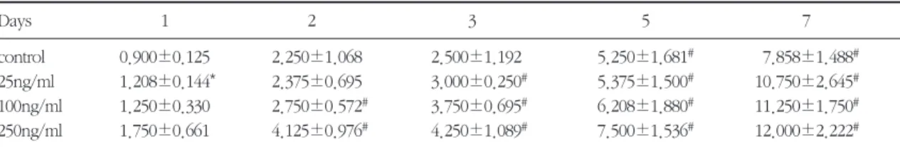
1. 세포의 증식속도배양한 백서두개관세포와 백서치주인대세포를 BMP-2/4군을 넣지 않은 대조군과 배양액 1ml당 25, 100, 250 ng/ml을 접종 배양한 실험군의 세포증식율 을 배양 1, 2, 3, 5, 7일에 측정하여 비교하였다(Table 1, Fig. 1, Table 2, Fig. 2). 백서두개관세포의 경우 대 조군에서는 배양 3일 까지는 세포수의 증가가 완만 하였으나 배양 5일부터는 증가폭이 컸으며, 25 ng/ml 투여군에서는 배양 3일부터, 100 ng/ml 투여 군과 250 ng/ml 투여군에서는 배양 2일부터 유의성 있게 증가하였다(p<0.05). 각 배양일별 대조군과 각 실험군간의 농도에 따른 세포수의 유의한 차이는 발 견할 수 없었다. 백서치주인대세포의 경우 대조군에 서는 배양 3일 까지는 세포수의 증가가 완만하였으 나 배양 5일부터는 증가폭이 컸으며, 100ng/ml 투여 군에서는 배양 3일부터, 25ng/ml 투여군과 250 ng/ml 투여군에서는 배양 5일부터 유의성있게 증가 하였다(p<0.05). 각 배양일별 대조군과 각 실험군간 의 농도에 따른 세포수의 유의한 차이는 발견할 수 없었다. 전체적으로 볼 때 두개관세포보다 치주인대 세포의 증식율이 더 높았다.
2. 총단백질양
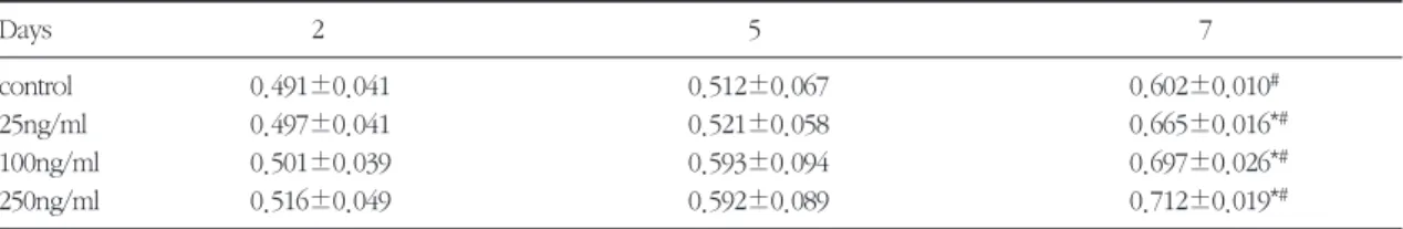
배양한 백서두개관세포와 백서치주인대세포를 BMP-2/4를 넣지 않은 대조군과 배양액 1ml당 25, 100, 250 ng/ml을 접종 배양한 실험군의 총단백질양 을 배양 2, 5, 7일에 측정하여 비교하였다(Table 3, Fig. 3, Table 4, Fig. 4). 두 세포 모두 배양 5일까지는 큰 변화가 없었으나 배양 7일 후에는 대조군, 실험군 모두 유의한 차이를 보였다(p<0.05). 농도에 따른 변 화는 두 세포 모두 배양 7일에서 대조군에 비해 각 실험군이 유의한 차이를 보였다(p<0.05).
3. Alkaline phosphatase 활성도
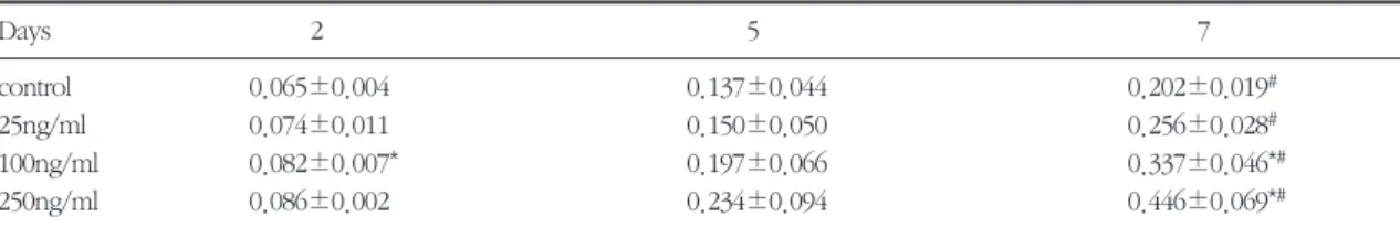
배양한 백서두개관세포와 백서치주인대세포를 BMP-2/4를 넣지 않은 대조군과 배양액 1ml당 25, 100, 250 ng/ml을 접종 배양한 실험군의 alkaline phosphatase의 활성도를 배양 2, 5, 7일에 측정하여 비교하였다(Table 5, Fig. 5, Table 6, Fig. 6). 백서두
개관세포는 배양 5일까지는 큰 변화가 없었으나 배 양 7일 후에는 대조군, 실험군 모두 유의한 차이를 보였다(p<0.05). 농도에 따른 변화는 배양 7일에서 100 ng/ml 투여군과 250 ng/ml 투여군이 대조군에 비해 유의한 차이를 보였다(p<0.05). 백서치주인대 세포는 배양 5일부터 대조군, 실험군 모두 유의한 차 이를 보였다(p<0.05). 농도에 따른 변화를 보면 배양 5일에서 100 ng/ml 투여군과 250 ng/ml 투여군이 대 조군에 비해 유의한 차이를 보였고 (p<0.05), 배양 7 일에서 모든 실험군이 대조군에 비해 유의한 차이를 보였다 (p<0.05).
4. 석회화 과정 관찰
(1) 백서치주인대세포의 배양
BMP-2/4 비처리군에서 배양 7일째보다 14일째 소 견에서 단층배열 세포의 분포가 더 밀집된 소견외에 는 커다란 변화가 관찰되지 않았다.(사진 1, 2 참조) BMP-2/4 초기 처리군에서는 배양 7일째에 세포가
Table 1. Proliferation of rat calvaria cell with bone morphogenetic protein-2/4(cell No.: 1×10⁴/ml).(mean±SD. n=3).
Days 1 2 3 5 7
control 0.900±0.125 2.250±1.068 2.500±1.192 5.250±1.681# 7.858±1.488#
25ng/ml 1.208±0.144* 2.375±0.695 3.000±0.250# 5.375±1.500# 10.750±2.645# 100ng/ml 1.250±0.330 2.750±0.572# 3.750±0.695# 6.208±1.880# 11.250±1.750# 250ng/ml 1.750±0.661 4.125±0.976# 4.250±1.089# 7.500±1.536# 12.000±2.222#
*significantly different from the control on dose-dependent manner, p<0.05
#significantly different from the 1st. day on time-dependent manner, p<0.05
Fig. 1. Proliferation of rat calvaria cell with bone morpho- genetic protein-2/4(cell No.: 1×10⁴/ml). (mean±
SD. n=3)
Fig. 2. Proliferation of rat periodontal ligament cell with bone morphogenetic protein-2/4(cell No.: 1×
104/ml). (mean±SD. n=3) 14
12 10 8 6 4 2
01 2 3 5 7
25 20 15 10 5
01 2 3 5 7
Day in culture Day in culture
control 25ng/ml 100ng/ml 250ng/ml
control 25ng/ml 100ng/ml 250ng/ml
Cell No.(1×10⁴/ml) Cell No.(1×10⁴/ml)
Table 2. Proliferation of rat periodontal ligament cell with bone morphogenetic protein-2/4(cell No.: 1×10⁴/ml).(mean±SD.
n=3).
Days 1 2 3 5 7
control 0.708±0.190 1.708±0.629 3.208±1.214 4.375±0.450# 16.666±1.880#
25ng/ml 1.000±0.433 1.044±0.301 1.801±0.581 4.291±0.563# 19.208±7.269#
100ng/ml 1.416±0.314* 1.916±0.438 4.000±0.875# 4.583±0.629# 19.500±11.103
250ng/ml 1.583±0.288* 2.000±0.753 3.208±1.134 5.000±1.205# 21.916±4.947#
*significantly different from the control on dose-dependent manner, p<0.05
#significantly different from the 1st. day on time-dependent manner, p<0.05
Table 3. Protein determination of rat calvaria cells treated with bone morphogenetic protein-2/4(mean±SD. n=3).
Days 2 5 7
control 0.491±0.041 0.512±0.067 0.602±0.010#
25ng/ml 0.497±0.041 0.521±0.058 0.665±0.016*#
100ng/ml 0.501±0.039 0.593±0.094 0.697±0.026*#
250ng/ml 0.516±0.049 0.592±0.089 0.712±0.019*#
*significantly different from the control on dose-dependent manner, p<0.05
#significantly different from the 1st. day on time-dependent manner, p<0.05
Table 4. Protein determination of rat periodontal ligament cells treated with bone morphogenetic protein-2/4(mean±SD.
n=3).
Days 2 5 7
control 0.526±0.027 0.556±0.035 0.873±0.006#
25ng/ml 0.506±0.049 0.549±0.015 0.909±0.007*#
100ng/ml 0.537±0.018 0.559±0.019 1.029±0.015*#
250ng/ml 0.532±0.031 0.586±0.046 1.086±0.008*#
*significantly different from the control on dose-dependent manner, p<0.05
#significantly different from the 1st. day on time-dependent manner, p<0.05
Fig. 3. Protein determination of rat calvaria cells treated with bone morphogenetic protein-2/4(mean±SD, n=3).
Fig. 4. Protein determination of rat periodontal ligament cells treated with bone morphogenetic protein- 2/4(mean±SD, n=3).
2 Days 5 Days 7 7 Days 0.8
0.7 0.6 0.5 0.4 0.3 0.2 0.1
0 0 25 100 250
BMP-2/4 conc.(ng/ml)
1.2 1 0.8 0.6 0.4 0.2
0 0 25 100 250
2 Days 5 Days 7 Days
BMP-2/4 conc.(ng/ml)
Protein(㎍/ml) Protein(㎍/ml)
일부에서는 세포가 특정방향으로 극성을 띄며 (점 선) 증식되어 전체적으로 농염되게 나타나 응집상태 로 배양되고, 14일째는 부분적으로 농염된 부위(▶
◀)가 관찰되었다. (사진 3, 4 참조) BMP-2/4 장기처 리군은 배양 7일째 이미 농염된 세포응집상태가 여 러부위(*)에서 나타났고, 14일째에는 세포극성이 현 저하여 40배의 배율상에서 대부분의 배양접시에 농 염된 세포응집상(▶◀)이 관찰되었으며 응집인접부 에는 공동현상 (▷◁)이 보였다. (사진 5, 6 참조)
(2) 백서두개관세포의 배양
BMP-2/4 비처리군에서는 배양초기에 일부 세포 의 밀집상(*)이 나타났으나 시간이 지남에 따라 세포 가 특정방향으로 극성을 띄며 (점선) 증식되었다.
(사진 7, 8 참조) BMP-2/4 초기처리군에서는 배양 7 일에 세포증식에 따른 밀집현상과 극성상 (점선) 이 관찰되었으며 14일 후에는 심화되어 일부에서는 세 포상이 응집농염(▶◀)되고 공동현상(▷◁))도 심화 되었다(사진 9, 10 참조). BMP-2/4 장기처리군에서 Table 5. Alkaline phosphatese activity of rat calvaria cells treated with bone morphogenetic protein-2/4(mean±SD. n=3).
Days 2 5 7
control 0.065±0.004 0.137±0.044 0.202±0.019#
25ng/ml 0.074±0.011 0.150±0.050 0.256±0.028#
100ng/ml 0.082±0.007* 0.197±0.066 0.337±0.046*#
250ng/ml 0.086±0.002 0.234±0.094 0.446±0.069*#
*significantly different from the control on dose-dependent manner, p<0.05
#significantly different from the 1st. day on time-dependent manner, p<0.05
Table 6. Alkaline phosphatese activity of rat periodontal ligament cells treated bone morphogenetic protein-2/4(mean±
SD. n=3).
Days 2 5 7
control 0.076±0.002 0.097±0.006# 0.420±0.022#
25ng/ml 0.069±0.001* 0.089±0.005# 0.565±0.007*#
100ng/ml 0.083±0.006 0.197±0.014*# 0.583±0.004*#
250ng/ml 0.077±0.006 0.202±0.005*# 0.725±0.010*#
*significantly different from the control on dose-dependent manner, p<0.05
#significantly different from the 1st. day on time-dependent manner, p<0.05
Fig. 5. Alkaline phosphatese activity of rat calvaria cells treated with bone morphogenetic protein- 2/4(mean±SD, n=3).
Fig. 6. Alkaline phosphatese activity of rat periodontal lig- ament cells treated with bone morphogenetic pro- tein-2/4(mean±SD, n=3).
0.6 0.5 0.4 0.3 0.2 0.1
0 0 25 100 250
2 Day 5 Day 7 Day
BMP-2/4 conc.(ng/ml)
ALPase activity(IU) 2 Days
5 Days 7 Days 0.8
0.7 0.6 0.5 0.4 0.3 0.2 0.1
0 0 25 100 250
BMP-2/4 conc.(ng/ml)
ALPase activity(IU)
는 배양 7일에서 세포극성의 심화로 세포공동현상이 보였고 14일에는 크고 강한 농염상태(▶◀)가 관찰 되었다 (사진 11, 12 참조).
IV. 총괄 및 고찰
치주질환은 치태 세균에 의한 만성 염증성 질환으 로 치조골을 포함한 치주조직의 파괴를 보이는데, 치 주조직의 특성상 고도로 석회화된 백악질과 주위의 치조골 및 이 둘을 연결하는 치주인대 등의 구조로 이루어져 조직파괴의 기전은 물론 치주조직의 재생 에 있어서도 다양한 세포들에 의한 복잡한 치유과정 을 거치게 된다1-3).
치주조직의 재생을 위한 다양한 치료방법이 사용 되고 있는데, 세포부착능 향상을 위한 치근면 처치제 의 사용, 골결손부의 재생을 위한 골이식술, 특정 세 포의 선별적 이주를 이용한 조직유도재생술, 질환에 이환되기 이전의 조직위치를 재건하기 위한 치관변 위판막술과 함께 골형태형성단백질을 포함하는 다 양한 polypeptide growth factors의 임상적용이 연구 되고 있다4,6,9,12,54).
골형태형성단백질은 골의 세포외기질의 유기 및 무기성분에 긴밀히 부착되어 있는 단백질로 골의 분 화과정의 기시제로 작용하여 태생기 분화 및 구강악 안면조직을 포함한 신체골격형성과 기관형성에 관 여하며 태생기 이후에도 치유될 골조직으로 세포들 의 이주와 부착, 세포들의 증식, 세포들의 생합성 및 연골아세포와 골아세포의 분화를 촉진시켜 골형성 을 유도하게 된다18,25,26).
1994년 Urist의 가설에 의하면 골형태형성단백질은 미분화간엽세포를 자극하여 정상적인 골대사과정, 골 절부위의 치유 및 이식골의 생착 등에 관여한다고 하 였고 골형태형성단백질에 의한 골형성과정을 세 과정 으로 설명하였다. 첫 단계인 화학주성시 골형태형성 단백질을 이식한 조직으로 다형핵백혈구에 이어 섬유 아세포가 이주하여 기질에 세포부착이 발생한다. 두 번째 분열과정에서는 이식 후 3일 째 간엽세포가 증식 되어 서로 응집하고, 마지막 분화과정에서는 이식 후 5일 째 간엽세포가 연골아세포로 분화되고 이식 후 9
일 째 혈관이 형성되고 이어서 섬유아세포의 성숙과 연골의 석회화가 일어난다. 이식 후 10 내지 12일 째 골아세포가 나타나 신생 골기질을 형성하고 이 때 연 골아세포는 석회화된 연골을 제거하기 시작한다. 이 시기에 골아세포와 연골아세포에 의한 alkaline phos- phatase의 활성이 최고를 이룬다. 이식 후 12 내지 18 일 째 골아세포가 신생골을 remodel시킨다. 이식 후 21일 째 골수강을 가진 신생 골이 형성된다18).
본 연구에 사용된 골형태형성단백질-2/4군은 다른 군의 골형태형성단백질과 성장인자 등에 비하여 유 전자 서열이 가장 유사하고 낮은 농도에서 골형성 능력이 뛰어나 많은 실험에 이용되고 있다27,33,34). 또 한 골형태형성단백질-2/4군은 치배, 상아모세포층, 구개궁 및 두개안면부조직의 발생에 기여하며 손상 된 치주조직의 치유시 치근흡수를 방지하고 치아의 유착을 줄여주며 치주인대와 백악질 및 치조골의 생 성을 촉진하는 등 치주조직의 재생에 영향을 미치는 것이 확인되었다13,33,44,45).
이러한 골형태형성단백질의 골형성능을 입증하기 위하여 많은 시험관적 실험이 시행되었는데 골형성 과정을 나타내는 다양한 specific marker의 발현을 측정한다. 이 specific marker에는 alkaline phos- phatase의 활성도, 부갑상선 호르몬 자극에 의한 cAMP의 생산, osteocalcin의 합성, osteonectin의 합 성, osteopontin의 합성, matrix Gla protein의 합성, proteoglycan의 합성, 교원질의 합성, DNA 합성 및 석회화결절의 형성 등이 있다.
골형성세포의 경우 분화과정에 따라 나타나는 specific marker들이 다른데 간엽세포에서 전골아세 포로 분화될 때는 I형 교원질의 생산이 증가하고, 전 골아세포에서 골아세포로 분화될 때는 alkaline phosphatase의 활성도, osteonectin의 합성, matrix Gla protein의 합성이 증가하며, 골아세포에서 골세 포로 분화될 때는 osteopontin과 osteocalcin의 합성 이 증가한다55).
본 연구에서도 골형태형성단백질-2/4군에 의한 두 개관세포와 치주인대세포의 성상변화를 관찰하기 위하여 세포증식속도, 총단백질양, alkaline phos- phatase의 활성도 및 석회화결절형성을 측정하였다.
세포의 증식속도를 보면 두 세포 모두 각 배양일별 대조군과 각 실험군간의 농도에 따른 세포수의 유의 한 차이는 발견할 수 없었으나 골형태형성단백질의 농도가 증가할수록 세포의 증식율이 높게 나타났다.
이는 Chaudhari 등 (1997)37)과 Yamaguchi 등 (1991)39)의 결과와도 일치한다. 전체적으로 볼 때 두 개관세포보다 치주인대세포의 증식율이 더 높았다.
세포의 총단백질양을 보면 두 세포 모두 배양 5일 까지는 큰 변화가 없었으나 배양 7일 후에는 대조군, 실험군 모두 유의한 차이를 보였다. 농도에 따른 변 화는 두 세포 모두 배양 7일에서 대조군에 비해 각 실험군이 유의한 차이를 보였다.
Alkaline phosphatase는 유기인산 에스테르를 가 수분해하여 석회화가 이루어지는 부위에서 국소적 으로 인산이온의 농도를 증가시키는 효소로서, 세포 외 기질에 calcium phosphate를 침착시킴으로써 석 회화를 유도하는 기능을 갖는다. 미분화간엽세포가 골아세포로 분화되어 신생골을 형성하는 과정 중, 초 기 세포분화 시기에 많이 관찰되며 spectrophotome- try나 BCA protein assay 방법으로 확인할 수 있다.
본 연구의 alkaline phosphatase의 활성도를 보면 두 개관세포는 배양 5일까지는 큰 변화가 없었으나 배 양 7일 후에는 대조군, 실험군 모두 유의한 차이를 보였다. 농도에 따른 변화는 배양 7일에서 100ng/ml 투여군과 250ng/ml 투여군이 대조군에 비해 유의한 차이를 보였다. 치주인대세포는 배양 5일부터 대조 군, 실험군 모두 유의한 차이를 보였다. 농도에 따른 변화를 보면 배양 5일에서 100ng/ml 투여군과 250ng/ml 투여군이 대조군에 비해 유의한 차이를 보 였고, 배양 7일에서 모든 실험군이 대조군에 비해 유 의한 차이를 보였다. 본 실험에서는 골형태형성단백 질의 농도가 증가할수록 alkaline phosphatase의 활 성도도 계속 증가하였는데 Chaudhari 등 (1997)37)의 연구에서는 골형태형성단백질-2군의 농도가 21.5 ng/ml에서 태생두개관세포의 alkaline phosphatase 의 활성도가 최대치를 보였고, Takuwa 등 (1991)40) 의 연구에서는 골형태형성단백질-2군의 농도가 200 ng/ml이 될 때까지 MC3T3-E1 골아세포의 alkaline phosphatase의 활성도가 계속 증가하였고, Thies 등
(1992)27)의 연구에서는 골형태형성단백질-2군의 농 도가 1㎍/ml가 될 때까지도 W-20-17 stromal cell의 alkaline phosphatase이 농도가 최대치를 기록하지 못 하였다. 각 실험들 간의 alkaline phosphatase 활 성도의 차이는 배양한 세포의 종류와 배양조건이 다 르기 때문인 것으로 생각된다.
석회화결절의 형성은 골조직재생의 기본이 되는 석회화능력을 암시하는 중요한 지표로 glucocor- ticoids, β-Glycerophosphate 등을 배양액에 첨가시 켰을 때 석회화결절의 형성이 증가한다고 보고하고
있다57,58). 본 연구에서 두 세포 모두 100ng/ml의
BMP-2/4 농도에서 배양 7일 후 석회화결절 형성을 보였는데 결절은 원형으로 주위에 세포가 밀집되어 있었으며 결절 중앙부위가 염색액에 농염되고 주위 로 연하게 염색되었다.
본 연구에서는 백서의 두개관세포와 치주인대세포 를 사용하였는데 두개관세포는 골아세포로서 많은 실험에 이용되고 있는데 골아세포의 각 분화단계에 따른 세포종류에 따라 골형태형성단백질에 의한 성 상변화가 다르게 나타날 수 있으며 같은 종류의 골아 세포라도 원시배양(primary culture)한 세포와 후속배 양(subculture)한 세포의 성질이 다를 수 있다. 후속배 양시 trypsin과 같은 분해효소에 의해 정상세포의 성 질이 변할 수 있고37), 원시배양시 배양시간이 오래 걸 리고 적절한 배지환경을 형성하기 힘든 단점이 있으 나 세포의 고유한 성질을 유지할 수 있어 좀더 믿을만 한 실험결과를 얻을 수 있다56). 본 연구에서도 백서의 두개관세포를 원시배양하여 사용하였다.
치주인대세포는 치주조직의 주 구성세포로 다양 한 성질을 나타내는데 섬유아세포이면서도 골이나 백악질을 형성할 수 있어 골원성섬유아세포 또는 osteoblastic fibroblast로 명명되고 있다31,32). Melcher 등 (1986)30)은 치주인대세포를 골기질상 및 상아질 기질상에 배양하면 신생 골양조직이 형성된다고 보 고하였고, Nojima 등 (1990)31)과 Arceo 등 (1991)32) 은 in vitro에서 치주인대세포가 높은 alkaline phos- phatase 활성도를 보이는 골아세포의 표현형을 나타 낸다고 하였고, 적절한 배지환경에서는 골과 비슷한 석회화결절을 형성한다고 하였다. 본 연구에서 골형
태형성단백질-2/4이 배양세포의 석회화에 미치는 영 향을 규명하기 위하여 배양액에 골형태형성단백질- 2/4을 함유하여 배양한 경우의 비교실험에서 백서치 주인대세포와 백서두개관세포 모두에서 공히 골형 태형성단백질을 투여한 경우에서 세포들의 증식속 도가 빠르고 세포의 응집을 위한 특정 방향으로의 극성화와 응집현상이 빠르게 나타난다는 사실을 확 인할 수 있었다. 따라서 응집된 세포들의 염색농도 도 시간이 갈수록 농염되는 현상이 나타났고 응집범 위도 더 넓게 확산되어 석회화를 촉진하는 것을 알 수 있었다. 본 실험과정에 골형태형성단백질-2/4의 투여기간의 변화를 관찰하기 위하여 배양세포에 전 혀 투여하지 않은 경우와 1일만 첨가한 경우의 비교 에서 괄목할 만한 속도로 세포응집에 빠르게 나타난 다는 사실을 알 수 있었다. 그러나 1일만 첨가한 경 우와 실험기간 동안 계속적으로 투여한 경우의 비교 에서는 전실험기간 동안 투여한 경우가 더 신속하게 응집이 된다는 사실을 알 수 있었다. 세포의 특정방 향으로의 극성화와 응집현상은 세포외기질형성의 전단계 현상이라는 사실을 미루어 석회화과정의 일 부분이라 유추되며 중요한 점은 특정세포가 1회라도 골형태형성단백질-2/4에 노출되면 세포의 변화는 당 분간 지속된다는 사실을 확인할 수 있어 임상응용에 기준이 되리라 생각된다.
본 연구결과를 종합하여 보건데 골형태형성단백 질-2/4군이 백서의 두개관세포와 치주인대세포의 증 식속도, 총단백질 형성, alkaline phosphatase의 활성 도 및 석회화 과정에 많은 영향을 미치는 것으로 나 타나 치주조직의 재생시 골형태형성단백질-2/4군의 임상적용의 근거를 확인할 수 있었다. 차후 다른 골 형태형성단백질과 또는 다른 성장인자와의 길항작 용에 대한 연구가 더 필요하며, 임상적용시 유용한 운반체에 대한 연구와 함께 적절한 사용농도에 대한 연구가 계속되어야 할 것으로 사료된다.
V. 결론
골형태형성단백질-2/4군을 백서의 두개관세포와 치주인대세포에 적용하여 세포증식속도, 총단백질
양, alkaline phosphatase 활성도 및 석회화결절 형성 능을 분석하여 다음과 같은 결과를 얻었다.
1. 세포증식율은 양 세포 모두 실험군, 대조군에서 시간이 지남에 따라 증가하였으나(p<0.05), 골 형태형성단백질-2/4군의 농도에 따른 유의한 차이는 없었다. 백서두개관세포보다 백서치주 인대세포가 더 많은 세포증식을 보였다.
2. 총단백질양은 양 세포 모두 실험군, 대조군에서 시간이 지남에 따라 증가하였으나 큰 차이는 없었고, 배양 7일 후 모든 실험군이 대조군에 비해 유의한 차이를 보였다 (p<0.05).
3. Alkaline phosphatase 활성도는 양 세포 모두 실험군, 대조군에서 시간이 지남에 따라 증가하 였다 (p<0.05). 또한 백서두개관세포에서는 배 양 7일 후 실험 2군, 실험 3군이 대조군에 비해 유의한 차이를 보였고(p<0.05), 백서치주인대 세포에서는 배양 5일 후 실험 2군, 실험 3군이, 배양 7일 후 모든 실험군이 대조군에 비해 유의 한 차이를 보였다 (p<0.05).
4. 백서치주인대세포와 두개관세포 배양 모두에 서 BMP-2/4를 처리한 군이 대조군에 비해 빠른 극성화, 세포응집현상 및 농염상을 보였고, BMP-2/4를 백서치주인대세포와 두개관세포 배 양과정에서 1일동안만 처리하여도 세포의 극성 화와 응집현상이 대조군보다 빠르게 관찰되었 다. 전체적으로 백서치주인대세포보다 두개관 세포가 BMP-2/4에 더 민감한 영향이 있는 것으 로 나타났다.
상기 결과를 근거로 골형태형성단백질-2/4군은 특 히 100ng/ml이상 농도에서 백서치주인대세포와 백 서두개관세포의 골형성 과정을 의미하는 여러 지표 가 농도의존형으로 나타나게 하는 것으로 미루어 골 형성 과정에 관여하는 것을 알 수 있었고, 이를 근거 로 차후 장기간의 시험관적 실험과 생체실험을 통한 임상적용 가능성을 확인할 수 있었다.
VI. 참고문헌
1. Iglhaut, J., Aukhil, I., Simpson, D.M., Johnston,
M.C., and Kock, G. : Progenitor cell kinetics during tissue regeneration in experimental peri- odontal wounds. J. Periodont. Res., 23:107-117, 1988.
2. McCulloch, C.A.G. and Bordin, S. : Role of fibroblast subpopulations in periodontal physiol- ogy and pathology. J. Periodont. Res., 26:144- 154, 1991.
3. Wikesjö U.M.E., Nilveus, R.E., and Selvig, K.A. : Significance of early healing events on periodon- tal repair : a review. J. Periodontol., 63:158-165, 1992.
4. Nyman, S., Lindhe, J., Karring, T., and Rylander, H. : New attachment following surgical treat- ment of human periodontal disease. J. Clin.
Periodontol., 9:290-296, 1982.
5. Gottlow, J., Nyman, S., Karring, T., and Lindhe, J. : New attachment formation as a result of con- trolled tissue regeneration. J. Clin. Periodontol., 11:494-503, 1984.
6. Ripamonti, U., Petit, J-C., Lemmer, J., and Austin, J.C. : Regeneration of the connective tis- sue attachment on surgically exposed roots using a fibrin-fibronectin adhesive system. An experimental study on the baboon(Papio ursi- nus). J. Periodont. Res., 22:320-326, 1987.
7. Wikesjö U.M.E., Claffey, N., and Chirstersson, L.A. : Repair of periodontal furcation defects in beagle dogs following reconstructive surgery including root surface demineralization with tetracycline hydrochloride and topical fibronectin application. J. Clin. Periodontol., 15:73-80, 1988.
8. Caffesse, T.G., Nasjleti, C.E., and Anderson, G.B. : Periodontal healing following guided tis- sue regeneration with citric acid and fibronectin application. J. Periodontol., 62:21-29, 1991.
9. Lynch, S.E., Williams, R.C., and Polson, A.M. : A combination of platelet-derived and insulin-like
growth factors enhances periodontal regen- eraion. J. Clin. Periodontol., 16:545-548, 1989.
10. Lynch, S.E., Ruiz de, C.G., and Williams, R.C. : The effects of short-term application of a combi- nation of platelet-derived and insulin-like growth factors on periodontal wound healing. J.
Periodontol., 62:458-467, 1991.
11. Rutherford, R.B., Niedrash, C.E., Kennedey, J.E., and Charette, M.F. : Platelet-derived and insulin-like growth factors stimulate regeneration of periodontal attachment in monkeys. J.
Periodont. Res., 27:285-290, 1992.
12. Reddi, A.H. and Cunningham, N.S. : Initiation and promotion of bone differentiation by bonemorphogenetic proteins. J. Bone Min. Res., 8(2):s499-s502, 1993.
13. Ripamonti, U. and Reddi, A.H. : Periodontal regeneration: potential role of bone morpho- genetic proteins. J. Periodont. Res., 29:225-235, 1994.
14. Shigeyama, Y., D'Errico, J.A., Stone, R., and Somerman, M.J. : Commercially-prepared allo- graft material has biological activity in vitro. J.
Periodontol., 66:478-487, 1995.
15. Wozney, J.M. : The potential role of bone mor- phogenetic proteins in periodontal reconstruc- tion. J. Periodontol., 66:506-510, 1995.
16. Riley, E.H., Lane, J.M., Urist, M.R., Lyons, K.M., and Lieberman, J.R. : Bone morphogenetic pro- tein-2: biology and application. Clin. Ortho. Rel.
Res., 324:39-46, 1996.
17. Ripamonti, U. and Reddi, A.H. : Tissue engi- neering, morphogenesis, and regeneration of the periodontal tissues by bone morphogenetic pro- teins. Crit. Rev. Oral. Biol. Med., 8(2):154- 163, 1997.
18. Lee, M.B. : Bone morphogenetic proteins: back- ground and implications for oral reconstruc- tionn. A review. J. Clin. Periodontol., 24:355-
365, 1997.
19. Wozney, J.M. and Rosen, V. : Bone morpho- genetic protein and bone morphogenetic pro- tein gene family in bone formation and repair.
Clin. Ortho. Rel. Res., 346:26-37, 1998.
20. Urist, M.R. : Bone formation by autoinduction.
Science, 150:893-899, 1965.
21. Urist, M.R. and Strates, B.S. : Bone morpho- genetic protein. J. Dent. Res., 1392-1406, 1971.
22. Wang, E.A., Rosen, V., and Cordes, P. : Purificationn and characterization of other dis- tinct bone inducing facters. Proc. Natl. Acad.
Sci. USA., 85:9484-9488, 1988.
23. Luyten, F.P., Cunningham, N.S., and Ma, S. : Purification and partial amino acid sequence of osteogenin. a protein in initiating bone differen- tiation. J. Biol. Chem., 264:13377-13380, 1989.
24. Wozney, J.M., Rosen, V., and Celeste, A.J. : Novel regulatiors of bone formation: Molecular clones and activities. Science, 242:1528-1534, 1988.
25. Reddi, A.H. : Regulation of local differentiation of cartilage and bone by extracellular matrix: A cascade type mechanism. Prog. Clin. Biol. Res., 110:261-268, 1982.
26. Ripamonti, U. and Reddi, A.H. : Growth and morphogenetic factors in bone induction: Role of osteogenin and related bone morphogenetic proteins in craniofacial and periodontal repair.
Crit. Rev. Oral. Biol. Med., 3:1-14, 1992.
27. Thies, R.S., Bauduy, M., Ashton, B.A., Kurtzberg, L., wozney, J.M., and Rosen, V. : Recombinant human bone morphogenetic pro- tein-2 induces osteoblastic differentiation in W- 20-17 stromal cell. Endocrinology, 130:1318- 1324, 1992.
28. Gould, T.R.L., Melcher, A.H., and Brunette, D.M. : Migration and division of progenitor cell populations in the periodontal ligament after
wounding. J. Periodont. Res., 15:20-42, 1980.
29. Bowers, G.M.,Schallhorn, R.G., and Mellonig, J.T. : Histologic evaluation of new attachment in human intrabony defects. A literature review. J.
Periodontol., 53:509-514, 1982.
30. Melcher, A.H., McCulloch, C.A.G., Cheong, T., Nemeth, E., and Shiga, A. : Synthesis of cemen- tum-like tissue in vitro by cells cultured from bone - a light and electron microscope study. J.
Periodont. Res., 21:592-612, 1986.
31. Nojima, N., Kobayashi, M., Sionome, M., Takahashi, N., Suda, T., and Hasegawa, K. : Fibroblastic cells derived from bovine periodon- tal ligament have the phenotypes of osteoblasts.
J. Peridont. Res., 25:179-185, 1990.
32. Arceo, N., Sauk, J.J., Moehring, J., Foster, R.A.
and Somerman, M.J. : Human periodontal cells initiate mineral-like nodules in vitro. J.
Periodontol., 62:499-503, 1991.
33. King, G.N., King, N., and Hughes, F.J. : The effects of two delivery systems for recombinant human bone morphogenetic protein-2 on peri- odontal regeneration in vivo. J. Periodont. Res., 33:226-236, 1998.
34. Hughes, F.J., Clooyer, J., Stanfield, S., and Goodman, S. : The effedts of bone morphogenic protein-2,4 and 6 on differentiation of rat osteoblastic cells in vitro. Endo., 136:2671-2677, 1995.
35. Reddi, A.H. : Initiation and promotion of bone differentiation by bone morphogenetic proteins.
J. Bone Min. Res., 8(sup. 2):s499-s502, 1993.
36. Chen, D., Harris, M.A.., Rossini, G., Dunstan, C.R., Dallas, S.L., Feng, J.Q., Mundy, G.R., and Harris, S.E. : Bone morphogenetic protein 2(BMP-2) enhances BMP-3, BMP-4, and bone cell differentiation marker gene expression dur- ing the induction of mineralized bone matrix for- mation in cultures of fetal rat calvarial
osteoblasts. Calcif. Tissue Int., 60:283-290, 1997.
37. Chaudhari, A., Ron, E., and Rethman, M.P. : Recombinant human bone morphogenetic pro- tein-2 stimulates differentiation in primary cul- tures of fetal rat calvarial osteoblasts. Mol. Cell.
Biochem., 167:31-39, 1997.
38. Ong, J.L., Carnes, D.L., Cardenas, H.L., and Cavin, R. : Surface roughness of titanium on bone morphogenetic protein-2 treated osteoblast cells in vitro. Implant Dent., 6:19-24, 1997.
39. Yamaguchi, A., katagiri, T., Ikeda, T., Wozney, J.M., Rosen, V., Wang, E.A., Kahn, A.J., Suda, T., and Yoshiki, S. : Recombinant human bone morphogenetic protein-2 stimulates osteoblastic maturation and inhibits myogenic differentiation in vitro. J. Cell Biol., 113:681-687, 1991.
40. Takuwa, Y., Ohse, C., Wang, E.A., Wozney, J.M., and Yamashita, K. : Bone morphogenetic protein-2 stimulates alkaline phosphatase activity and collagen synthesis in cultured osteoblasts cells, MC3T3-E1. Biochem. Biophys. Res.
Commun., 174:96-101, 1991.
41. Hiraki, Y., Inoue, H., Shigeno, C., Sanma, Y., Bentz, H., Rosen, D.M., Asada, A., and Suzuki, F. : Bone morphogenetic proteins(BMP-2 and BMP-3) promote growth and expression of the differentiated phenotypeof rabbit chondrocytes and osteoblastic MC3T3-E1 cells in vitro. J. Bone Mine. Res., 6:1373-1384, 1991.
42. Kubler, N. and Urist, M.R. : Allogenic bone and cartilage morphogenesis: Rat BMP in vivo and in vitro. J. Cranio-Max.-Fac. Surg., 19:283-288, 1991.
43. Puleo, D.A. : Dependence of mesenchymal cell responses on duration of exposure to bone mor- phogenetic protein-2 in vitro. J. Cell Physiol., 173:93-101, 1997.
44. Ripamonti, U., Heliotis, M., van den Heever, B., and Reddi, A.H. : Bone morphogenetic proteins
induce periodontal regeneration in the bab- bon(Papio ursinus). J. Periodont. Res., 29:439- 445, 1994.
45. Sigurdsson, T.J., Lee, M.B., Kubota, K., Turek, T.J., Wozney, J.M., and Wikesjö, U.M.E. : Periodontal repair in dogs: Recombinant human bone morphogenetic protein-2 significantly enhances periodontal regeneration. J.
Periodontol., 66:131-138, 1995.
46. Kinoshita, A., Oda, S., takahashi, K., Yokota, S., and Ishikawa, I. : Periodontal regeneration by application of recombinant human bone mor- phogenetic protein-2 to horizontal circumferen- tial defects created by experimental periodontitis in beagle dofgs. J. Periodontol., 68:103-109, 1997.
47. King, G.N., King, N., Cruchley, A.T., Wozney, J.M., and Hughes, F.J. : Recombinant human bone morphogenetic protein-2 promotes wound healing in rat periodontal fenestration defects. J.
Dent. Res., 76:1460-1470, 1997.
48. King, G.N., King, N., and Hughes, F.J. : The effect of root surface demineralization on bone morphogenetic protein-2-induced healing of rat periodontal fenestration defects. J. Periodontol., 69:561-570, 1998.
49. Giannobile, W.V., Ryan, S., Shih, M.S., Su, D.L., Kaplan, P.L., and Chan, T.C.K. : Recombinant human osteogenic protein-1(OP-1) stimulates periodontal wound healing in class III furcation defects. J. Periodontol., 69:129-137, 1998.
50. Sigurdsson, T.J., Tatakis, D.N., Lee, M.B., and Wikesjö U.M.E. : Periodontal regenerative potential of space-providing expanded polytetra- fluoroethylene membranes and recombinant human bone morphogenetic proteins. J.
Periodontol., 66:511-521, 1995.
51. Cook, S.D., Salkeld, S.L., and Rueger, D.C. : Evaluation of recombinant human osteogenic
protein-1(rhOP-1) placed with dental implants in fresh extraction sites. J. Oral Implat., 21:281-289, 1995.
52. Hanisch, O., Tatakis, D.N., Boskovic, M.M., Rohrer, M.D.,and Wikesjö U.M.E. : Bone forma- tion and reosseointegration in peri- implantitis defects following surgical implantation of rhBMP-2. Int. J. Oral Maxillofac. Implants, 12:604-610, 1997.
53. Sigurdsson, T.J., Fu, E., Tatakis, D.N., Rohrer, M.D., and Wikesjö U.M.E. : Bone morpho- genetic protein-2 for peri-implant bone regener- ation and osseointegration. Clin. Oral. Impl.
Res., 8:367-374, 1997.
54. Howell, T.H., Martuscelli, G., and Oringer, R.J. : Polypeptide growth factors for periodontal
regeneration. Cur. Opin. In Periodontol., 3:149- 156, 1996.
55. Hall, B.K. : Bone. Vol. 1 : The osteoblast and osteocyte. CRC press, pp. 171-192, 1992.
56. Wong, G. : Bone, Vol. 1 : Isolation and behavior of isolated bone-forming cells. CRC press, pp.
172-192. 1992.
57. Bellows, C.G., Aubin, J.E., Heersche, J.N.M., and Antoz, M.E. : Mineralized bone nodules formed in vitro from enzymatically released rat calvaria cell population. Calcif. Tissue Int., 38:143-154, 1993.
58. Nakamura, O. and Caplan, A.I. : Noncollagenous matrix protein-enhanced mineral deposition in osteoblast-like cell culture. J. Bone Miner. Res., 12:17-25, 1994.
사진부도 설명
Figure 1. Photomicrographs of Rat Periodontal ligament cell(RPDL) on 7 days culture without BMP-2/4 appli- cation(x 40).
Confluence state of Monolayer of RPDL populations on 7 days after culture without any remakable phenomenon on cell.
Figure 2. Photomicrographs of Rat Periodontal ligament cell(RPDL) on 14 days culture without BMP-2/4 application(x 40).
RPDL cell showed more density than one week before but there is no polarity.
Figure 3. Photomicrographs of Rat Periodontal ligament cell(RPDL) on 7 days culture with 1 day application of BMP-2/4 on culture medium(x 40).
Confluence state of polarity Monolayer of RPDL cell were divided with 2 different density by aggre- gation on 7 days after culture(dot line).
Figure 4. Photomicrographs of Rat Periodontal ligament cell(RPDL) on 14 days culture with day application of BMP-2/4 on culture medium(x 40).
Highly aggregated and stained cells were observed in the center of culture dish partly(▶◀).
Figure 5. Photomicrographs of Rat Periodontal ligament cell(RPDL) on 7 days culture with total application of BMP-2/4 on culture medium during experiment period(x 40).
The number of high stained and aggregated cell were more than other group(*).
Figure 6. Photomicrographs of Rat Periodontal ligament cell(RPDL) on 14 days culture with total application of BMP-2/4 on culture medium during experiment period(x 40).
Wide spreaded high stained and aggregated cell were found in this slide(▶◀).
There were some vacancy area between aggregated cell which had multidirected polarity(▷◁).
Figure 7. Photomicrographs of Rat calvaria cell(RCV) on 7 days culture without BMP-2/4 application(x 40).
Slight cell aggregation and polarity of Monolayer of RCV were revealed.(*)
Figure 8. Photomicrographs of Rat calvaria cell(RCV) on 14 days culture without BMP-2/4 application(x 40).
Remarkable RCV aggregation divided 2 different density and stain by aggregation(dot line). All the cell showed more poalrity to specipic direction.
Figure 9. Photomicrographs of Rat calvaria cell(RCV) on 7 days culture with first 1 day application of BMP- 2/4 on culture medium.(x 40)
Cultured RCV showed more remarkable aggregation, and high stained phenomenon which made 2 distinguished area.(dot line)
The shape of cell poalrity to specipic direction were more obvious.
Figure 10. Photomicrographs of Rat calvaria cell(RCV) on 14 days culture with first 1 day application of BMP- 2/4 on culture medium.(x 40)
Distinguished 2 area(dot line) with high stained and aggregated cell were observed more remark- able(▶◀). There were many vacancy area between aggregated cell which had multidirected polarity(▷◁).
Figure 11. Photomicrographs of Rat calvaria cell(RCV) on 7 days culture with total application of BMP-2/4 on culture medium during experiment period(x 40).
The portion of cell aggregated area extended more widely compared with other groups(dot line), and more large vacancy area prominent(▷◁).
Figure 12. Photomicrographs of Rat calvaria cell(RCV) on 14 days culture with total application of BMP-2/4 on culture medium during experiment period.(x 40)
Large portion of vacancy area were prominent(▷◁), and aggregated cell were severe stained in partly(▶◀).
사진부도 ( I )
사진부도( II )
-Abstract-
A Study of the Effects of Bone Morphogenetic Protein
on the Characteristics of Rat Periodontal Ligament and Calvaria Cells
Jin-Keun Choi, Man-Sup Lee, Young-Hyuk Kwon, Yeek Herr
Department of Periodontology, Division of Dentistry, Graduate school, Kyung-Hee University
Bone morphogenetic protein-2/4 (BMP-2/4) are members of Transforming Growth Factor-β (TGF-β) super- family and they may differentiate the osteoprogenitor cell and induce formation of cartilage and bone in vivo.
This study was performed to investigate the effects of bone morphogenetic protein-2/4 on the characteristics of rat periodontal ligament cells(RPDL) and rat calvaria cells(RCV). In the control group, the cells were cultured alone with Dulbeco's Modified Eagle's Medium contained with 20% fetal bovine serum, 100㎕/ml penicillin, 100㎕/ml streptomycin. In the experimental groups, recombinant human bone morphogenetic protein-2/4 (25ng, 100ng, 250ng/ml) were added into the above culture condition. And then each group was characterized by examing the cell proliferation at 1, 2, 3, 5, 7th day, the amount of total protein synthesis and alkaline phos- phatase activity at 2, 5, 7th day. And also, the calcified nodule was examed.
The results were as follows ;
1. Both RCV and RPDL cells in both control and experimental groups proliferated during the entire experi- mental period, but there is no stastically significant difference according to the BMP-2/4 concentration.
2. Amount of total protein synthesis of both cells in both groups was steadily increased until 5th day, but all experimental groups were significantly different from the control group at 7th day.
3. Alkaline phosphatase activity of both cells in both groups was increased during the entire experiment period. In RCV cells, the experimental group treated with 100ng/ml and 250ng/ml BMP-2/4 were signifi- cantly different from the control group at 7th day. In RPDL cells, the experimental group treated with 100ng/ml and 250ng/ml BMP-2/4 were significantly different from the control group at 5th day, and all experimental groups were significantly different from the control group at 7th day.
4. In the both of the cultured Rat Periodontal ligament and calvaria cell treated with BMP-2/4 to compared with control group, it revealed more rapid cell polarization, cell aggregation and hyperchromatic stained on HE agent, and even though only 1 day treated with BMP-2/4 both RPDL and RCV showed more rapid cell reaction than control group. More sensivitve cell reaction of RCV were observed than RPDL in this experiment.
From the above results, we could conclude that BMP-2/4 influenced the induction, proliferation and differen- tiation of bone forming cells