The Journal이Medicineand Ufe Scien
∞
Vol. 7. NO. 2,2010제주산 홍해삼에서 분리한 collagen 의 human
fibroblast
cell growth
에
미치는 영향
임 희경,조문제
제주대학교 의과대학 생화학교싣
l_~_~
→Ab
은땐Pt
→→-」
Effect of collagen purified from red sea cucumber (Stichopus japonicus) in Jeju on
growth of human fibroblast cells
HeeKyung
Lim;
MoonjaeCho
Department01 Biochemislry.Sch
∞
1이Medicine and Instilute 0' Medical Science.Jeju. KoreaPepsin-solubilized collagen (PSC) from red sea cucumber (Stichopus japanicus) was puri
’
ied 10 study the elfecls on human libroblasl cell line. PSC (1.929) was puriled oul 01 10여。
f sea cucumber body. SDS-PAGE analysis 01 puri’
ied PSC indicated that collagen from sea cucumber was a1ß type collagen type 1. The PSC did not have cy1otoxicity on various cell Iines. The treatmenl 01 PSC to human fibroblast cells increase the mRNA 01 MMP-1 gene not that of MMP-2 compared to untreated control. However,in case of MMP-2 ,mRNA is decreased upon PSC trealment. Trealment 01 PSC suppressed the synlhesis 01 collagen in vivo. Unlike 10 fibroblast,kera1inocyte reduced the MMP-2 produclion upon PSC treatment. (J Med Ufe Sci 2010;7:22-25)Key Words : red sea cucumber: Stichopus japanicus; collagen; MMP--1,MMP-2; Fibroblast 서
론
해상 (Echinodermata: Holothuroidea}은 동양에서 예로부터 식용과 약용으로 넘리 사용되어 오고 있으며 현재에는 그 수요가 증가추세이다 이는 특히 여러 내상과 외상의 치료제로 사용되었 고 또한 강장제로서 이용되기도 하였다 최근 해상으로부터 분리 한 다당체는"J'
암농력이 있다고 얄려졌으며 1). frondan이 A5는 화학요법 부작용 경감능력2l 홍띨 함유하는 다당체는 세포의 증식에 영향월 준다고 얄려지고 있다3) 그러나 해상 롤라겐의 fibrobiast 세포의 성장에 이치는 영향에 관한 연구는 보고되지 않았다 해삼의 약 70%는 콸라겐이라는 환용성 단백질로 이푸어져 있다4) 롤라겐은 홍물 피부조직과 뼈의 연콜조직을 이루는 대표 적인 구조 단백질이다-5) 현재 많은 피부미용, 의료,제약,피복「분 야에 이르기 까지 다양한 상엽적인 용도로 사용되고 있다.)특히 피부 재생 분야에서 콜라겐 용풍의 용도가 급증하고 있다.7,8) 그러나 광우병에 의한 소유래 콜라겐에 대한 사용거부와 모슐람Address lor correspandence : MoonjaeCh
。
Departmenl이 Biochemistry,Sch。이 이 Medicine and lns1ituteof Medical Science,Cheju National Universily,66 jeju daehakno, 690-756,Jeju,Korea E예13i1:[email protected] 국가의 종교적인 돼지 휠라겐의 사용거부는 콜라겐의 다른 출처 룹 요구하는 실정이다‘ 최근 거대 흥해상 (Parastichopus @뼈rnicus}2.로부터 콜라겐응 분리 풀리화학적 성질을 규명한 바있다9) 본 논문에서는 제주산 흥해상으로 부터 분리한 맹신 7)-용성 콜라겐 (pepsin-s이ubilized collagen. PSCs)의 인간 상며세포 (fibrobfast ceU)의 성장에 미치는 영향을 연구하였다
L
실험‘및방법 1.롤라겐의 분획 콜라젠 추출을 위하여 5배의 냉수로 흥해상을 잘 씻은 후 disaggregating solution을 이용하여 균질화한 후 3 일 동안 41"; 에서 S미π19 하연서 collagen fibril 융 조성하였다 3일 후 원싱 분리 하여 비한라겐성 단액진의 제거와 잔존 효소의 불환성화웰 위하여 잔λ}에 대하여 20배에 해당하는 0.1 N NaOH륜 가하여 2 일 동안 교반한 다음 원심분리 (7500rpm,lhr) 하여 상총액융 제거하였다 이어서 알랑리 처리 샌에 대하여 산가용성 혈라겐 의 제조흉 위하여 10배 (w/v)에 해당히는 0.5 M acetic acid 만올,pepsm 가용성 혈라겐의 제조블 위하여 10배 (w/v) 에 해당 하는 0.5 M acetic acid 이외에 단백칠의 1%(w/v)에 해당하는 pepsin (EC 3.4.23.1. Sigma-Afdrich"!nc .. St. 1ρuis,MO}융-Effect of collagen purified from red sea cucumber(St이JOPU8japomcus) in Jeju on growth of human fibroblast cells 가하여 3일 동안 교반한 후 원심분리 (750α-pm ,lhr) 하여 콜라 겐이 가용화되어 있는 산가용성 분획 및 pepsm 가용성 분획을 회수하였다 이어서 산가용성 콜라겐 추출용액 및 pepsm 기용성 콜라겐 추풀용액에 NaCI 용액 (최종 0.8 M)을 가하여 염석 및 원심분리 (7500rpm ,lhr) 하여 정제를 시도하였다 침전물은 다시 0.5 M acetic acìd에 가용화 시킨 후 염을 제거하기 위하여 0.02 M NfuHP04 buffel에 투석 및 원심분리 (7500rpm ,lhr) 하였다 분리된 산기용성 콜라겐 및 pepsm 가용성 콜라겐은 동결건조하 여 실험을 위한 산가용성 콜라겐 시료 및 pepsm 가용성 콜라겐 시료로사용하였다 2,서|포배앙 Fîbroblast (사람 섬유아세포)는 제주대학병원에서 얻은 7살 남아의 포경수술로 인해 적출된 피부조직을 이용하여 분려하였 다 적출한 조직을 collagenase 를 이용하여 표피와 진피로 분리 한 후, Trypsîn 을 이용하여 진피에서 섬유아세포를 분리하여 형태를 확인하였다 확인된 섬유아세포는 DMEM애지에서 10% FBS (fetal bo띠ne serum) ,1% penicî1lin-sìreptomycîn을 가하 여 100 φ dish 에서 배양기 내부 공기 5% CO,농도에서 37'C 에 서 배$봐였다 HaCaT (Human keratinocyte) ‘HeLa (Human cervical cancer) 세포의 경우 DMEM배지에서 10 % FBS ,1% penìcillin- sìreptornycin을 가하여 배$봐였고, MCF-7 (Human breast cancer) 세포의 경우 RPM1-1640 배지에서 10% FBS, 1% perricil1in-streptomycîn 을 가하여 배양하였다
3
서|포독성확인96 well plate에 lx1CJ‘cellsl마의 세포를 180띠 씩 seeding 하여 16시간 동안 배양한 후,흥해삼 엠신 가용성 콜라겐 (PSC) 을 농도별록 처리하여 2일간 배양하였다 MI'1‘용액 (5,땅111&) 은 20.,2 씩 첨가하고 4시간 동안 배
%i
한 후,상등액을 제거하였고, DMSO를 15C때 씩 첨가하여 570mn에서 Sunrise mìcroplate reader (Tecan,Sa1zburg. Aus미a)로 흡광도를 측정하였다4 흥해삼 콜라겐 생리활성 4.1 RNA분리
세포 (1 X 1O'cellsl뼈)를 60 • dish 에 분주하고 16 시간 배양 한 후,PSC 를 처리하여 24시간 동안 배양하였다 후에 맴I-zol (Invììrogen)을 이용하여 total RNA를 분리하였다 세포어
I
TRI-zol을 첨가하여 균질화한 후, 콜로로포릎을 첨가하여 원심분리 하였다 상층액에 동량의 이소프로판올을 첨가하여 원심 분리시 켜 뻐A를 침전시카고 75%의 DEPC 처리된 에탄올로 세척한 후,건조시켜 DEPC 처리된 증류수에 녹였다 260mn의 흡광도를 측정하여 RNA를 정량하였고,A2601A280nm의 비율이 1.7-1.9 범위 내의 값을 갖는 RNA를 실험에 사용하였다 모든 실험은 RNase-free 한 조건하에서 이루어졌다4.2 RT-PCR
23
1""의 total RNA를 olìg이dT)I8 primer,dNTP (0.5마Æ),lunit RNase inhìbitor 그리고 M-MuLV reverse transcriptase (2U)로 70'C 5min,25'C 5min ,42'C 60min ,그리고 70'C 에서 15min heatìng 시킴으로서 반응을 중지시켰다 Polymerase Chain Reaction (PCRl은 합성된 cDNA로부터 MMP-l ,MMP-2 , Collagen 빼e 1,GAPDH를 증폭시커가 위하여 1μQ cDNA,4따A 의 5'과 3↑ primer,10Xbuffer (IOr마~Tris-HCI ,pH 8.3 ,50
mM KCI,0.1% 맘iton X-100),250마~dNTP ,25mM MgClι l unìt Taq polymerase (Promega ,USA)를 섞고 며S며led water_로 전체를 25μE로 맞춘 다음 Perkin-Elmer Therrnal Cycler를 이용 하여 PCR을 설시하였다 PCR 수행에 사용한 pnmer sequences 는 각각 뻐1!P-1 forward pηmer ,5’GATCATCGGGACAACT CTCCT-3\ MMP-l reverse primer,5‘TCCGGGTAGAAGGGA 'ITT GTG-3’,MMP-2 forward primer,5'-TG GCAAGTACGG CT!CTG-3 ’.MMP-2 reverse primer,5'-TICTIGTCGC GGT CGTAG-3' ,Type 1 collagen forward primer,5'-CTGGCAAA GAAG GCGGCAAA-3',Type 1 collagen reverse primer,5'-CT CACCACGATCAC CACTCT-3',GAPDH forward primer ,5'-G AAGGTGAAGGTCGGAG TC-3' ,GAPDH reverse primer ,5'-G AAGATGGT GATG쉰GA'πTC-3 ’였다 이때 PCR 조건은 94'C1
45초,60'C/45초,72'C/60 초,30 회이며,PCR 에 의하여 생성된 산물은 1.5% agarose gel에서 전기영동을 실시하고 ethìdium bromìde로 염색하여 특정 band 를 확인하였다
5. CeJl prolifera1ion assay
Cell proliferation 효과논 roche (11 647 229 001) 사의 제품 을 시용하여 측정하였다 0.5 X 1O'celll뼈 의 fibr얘last 세포를 96 well p녀%에 180μU씩 seeding 하고 16 시간 동안 배òJ'한 후
PSC를 농도별로 처리하였다 그리고 BrdU를 20 때Iwell로 처리 하고 48 시간 동안 배양하였다 48시간 후 배지를 제거한 후 FixDenant 200μQlwell 넣고 30분간 상온에서 incubation 하고 FixDenant solution 제거한 후 anti-BrdU POD solution 100μQ Iwell 넣고 상온에서 100분간 incubation 하였다 100 분 후 an디body solution 제거한 후 washìng solution 200μI/well 로 washìng을 3회 하고,substrate solution 100μI/well 처리 한 후 발색반응을 한 후 stop solution (lM ffiSO~}을 25씨Iwell로 처리 하고 450mn 와 690mn 에서 흡광도를 측정하였다
결과및고찰
1,흥해삼콜라겐분리 산가용성 콜라겐과 산불용성 콜라겐, 웹신가용성 콜라겐과 렘신불용성 콜라겐을 분리하였을 때 산기용성 콜라겐 (0.24g) 은 거의 분리가 되지 않았다 웹신을 이용한 콜라겐 분리에서는 웹신가용성에 많은 양의 콜라겐 (1.92g) 이 분리된 것을 확인 할 수 있어 해삼에서의 콜라겐 분리에는 웹신의 시용이 중요한 것으 로 사료되어졌다 (Table 1)Hee Kyung Lim,Moon,îae Cho
Table 1. Pepsin effect in purification of col1agen from sea cucumber 100 Acid soluble Acid Ìnsoluble Pepsin soluble Pepsin insoluble
Collagen I Sea cucumber ↑00 9 0.24 g 2.208 g 1.92 g 0.2352 g ” s
、
.i:' i 므 > 80 60 40.•4>-
‘~활호냉
、、
o”
o 50 100 250 500 1000 PSC concentration (쩌1m
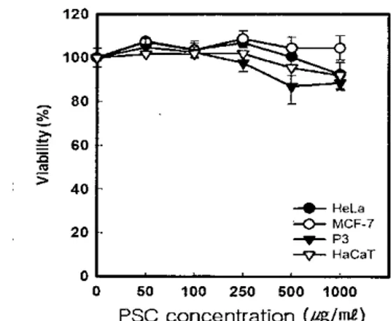
ι)Figure-2.-CytotoxÌcÌty- → of.-P-SC-from ←sea---cucumber. ~ Cells
were seeded in 96 well plate with concentratîon of 1x 10' cell/me and cultured 16 hr then treated with indicated concentration of PSC. After 48 hr incubation ,check the viability 、꺼th MIT assay
2
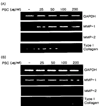
콜라겐 패턴분석흥해삼으로부터 분리된 콜라겐을 전기영동으로 패턴 분석한 결과 ra 얘↓ cali 의 collagen type 1과 비교하였을 때 거의 유사한 패턴을 보였으냐,pepsm 에 의해 말단잔기가 잘려면서 사이즈가 약간 작아진 것을 확인 할 수 있었고,type 1의 a,ß,Y 밴드가 전반적으로 사이즈가 약간 작게 나타난 것을 확인할 수 있었다 (Fìg.ll 이는 Lìu 등이 발표한 결과와 유사하며 이들은 해삼의 PSC는 alß 의 제1헝 콜라겐 (collagen type 1) 으로 정의하였다1이
20
-“-HeLa '-c←
MCF-7 •..•..•. P3-‘
7- HaCaT 4. 흥해상 콜라겐 생리활성 3. 세포독성확인Figure 1. SDS-PAGE analysis of purified sea cucumber PSC. Purified PSC in indîcated time points were analyzed by 6.5% SDS-PAGE and visiblized by coomassÌe blue staining
겐 처리가 세포의 콜라겐 합성을 억제히는 것으로 나타났다 섬유아세포와는 달리 각질세포 (keratìnocyte) 는 MMP-2 는 PSC 처리에 따라 놓도 의존적으로 줄어드는 반면 MMP-l 의 발현에는 변화가 없었다 각질세포가 피부 재생과정 중 이동에 관여하는 것으로 알려졌으며 MMP-l 은 이통에 관여합으로 섬유 아세포처럼 줄지 않는 것으로 사료되어진다 흥해삼 PSC 가 섬유아세포의 성장에 미치는 효과를 알아보기 위하여 BrdU 를 이용하여 세포의 증식을 측정하였다 PSC를 일부는 세포 배양액에 처리하고 일부는 세포 배양 접시에 코팅을 하여 48 시간동안 배양하여 세포의 증식을 DNA에 끼어틀어간 BrdU의 양을 측정하여 조사하였다 그 결과 Fig1니re 4에서 보는 바와 같이 낮은 농도를 배양액에 처리한 결과 성장이 증가하는 현상을 보이나 고놓도에서는 영헝
F
을 주지 못하였다세포외 기질 (Exlracell띠ar malrix. ECM) 의 주요 구성성분인 롤라겐은 피부의 섬유아세포에서 생성되는 주요 기질 단백질이 다 콜라겐은 피부,건 (tendon)‘뼈 및 지아의 유기 물질의 대부 분을 형생하는데,특히 뼈와 피부 (진피)에그 함유량이 놓다 콜라겐의 주된 기능으로는 피부의 기계적 견고성,결합조직의 저 항력과 조직의 결합력,세포의 접착의 지탱,세포 분할과 분화 (유기체의성장 혹은 상처 지유 시)의유도등이 알려져 있다11) ECM 분자의 물질대사와 관련된 신호 경로틀은 광 또는 자연 노화에 의해 변화된다
。
1
들 신호경로들은 cyt,okines 와 전사인 자 (transcription factor)의 조절에 의해 ECM 구성 성분들과 연관된 다수의 유전지들의 발현에 영향을 준다 특히,자연 또는 광 노화된 피부의 세포간질 성분의 분해증가는 AP-l 과 TGF-ßl 의 생성의 변화에 기인 한다12). TGF β1은 fibroblasts 를 자극하 여 다양한 세포간질 구성성분 (C이lagen type 1 그리고 IIl , fÌbronectin (FN),그리고 proteoglycan) 의 생산을 자극하고, 그들의 분해효소 생성을 억께하며,이들 효소에 대한 억제제를 증가시커는 것으로 얄려져 있다13)、
~
12), --.flJ η ‘ Q ‘건 션 ‘깅'i"
;~øt:- ....:.R
」
F
경
0'" ~o-.ιr ‘P、)
져5 끼~ rfi" ~ø""cε
。
<0:.-•
100 75 150 20'0 배양접시에 섬유아세포를 배$댄f
후 지시된 농도의 PSC 를 처리하여 세포이동에 관여하는 유전자의 발헌을 RT-PCR 로 관찰하였다 Figt.αe 3에서 보는 바와 같이 MMP-l 은 치리하지 않은 대조군에 비하여 처리한 군에서 발현이 증가 하였으나, MMP-2의 경우는 반대의 양상을 보였다 콤라겐은 외부의 콜라 각 세포주에 콜라겐을 농도 별로 처리하여 48시간이 경과한 후 MTI‘방법으로 세포의 독성을 측정한 결과,정상세포주인 fibroblasl (p3)X} Keralinocyle HaCaT,HeLa ,MCF-7 세포 모두에 독성을 보이지 않았다(Fìg.2).ÊfTectof collagcnpurifiedfromred sca cucumber(Süchopus jlJpOnicus)in Jeju on grow1Îlof humanfibroblastcel1s 본 실혐의 결과는 미용 및 의료용 할라겐의 원료로서 해삼
휠라겐의 사용이 가능한 것이라는 갤론올 얻었다
참
고 문헌
감사의글
1) Lu Y. Zhang BY. Dong Q. Wang BL. Sun XB. 111e effecls
。
f 8tichopu&iaponicus acid mucopolysaccharide on the apoptosîs of the human hepatocellular caπinomacell line HepG2. Am J Med Sci. 2010: 339:141-42) J어냉kiram NB. Mobammed A,zt냉ng Y. Cboi CI,Wα었ward
C. Collin P. Steele VE. Rao CV. Chemopreventive effects of FTondanol A5. a Cucurnana frondosa extract. against ral
∞
lon carcinogenesis and inhibition of human∞
Ilon cancer cell growth. Can∞
r Prev Res (1'1피a). 2010:3:82-913) Zhang Y‘Song S,Song D. Liang H. Wang W. Ji A Proliferative effects on neural stemlprogenitol' ceJJs of a sulfated polysaccharide purified from the sea cucumber
Süchopus japonicus. J Biosci Bioeng. 2010:109:67-72 4) Saito M. Kunisaki N. Urano N. CoJlagan as the m&jor
edible component of sea cucumber. Food Chem TI。잉col 2002:67:1319-29
5) Ogaw. M,Portier RJ. Moo여MW. Bell J‘Schexnayder MA. 1ρsso JN. Bîochemicalproperties of bone and scale collagens isolated from the subσ
。
pical fIsh black drum(Pogonia cromis) and sheepshead seabrearn (Aπhosargus prohalocephaJus). F
。
αd Chem. 2004:88:495-016) Kiωphattanabawon P. Beni빼ul S‘Visessanguan W. Nagai
T‘Tan와‘a M. Characterisation of acid-soluble coJlagen from skin and bone of bigeye snapper (Priacanlhus
뻐yenus). Food Chem,2005:89:363-72
7) Sorensen JC. Living skin equivalent.<;and their application in wound healing. Clin Podialr Med Surg. 1998:15:129-37 8) Machens HG. Berger AC. Mailaender P. Bioartificial skin
Cel1s까ssues Organs. 2αJO:167:88-94
9) Cui F'X. Xue CH. Li ZJ,Zhang YQ‘Dong P,Fu XY‘Gao
X. Characterization and subunit composition of col1agen from the body wa11of sea cucumber Stichopus japonicus
Fo(펴Chem. 2007:100:1120-5
10) Liu Z. Oliveira AC. Su YC. Purification and characterization of pepsin-solubilized collagen frorn skin and connective
lissue of giant Red SeaCucumber (Parastichopus ca}ifomicus). J Agric .FI
∞
d Chem,2010:58: 1270-127411) Perlish JS. Lemlich G. Fleischm!\ier R. Identification of Collagen Fibrils in Sclerodenna Skin. J Invesl DennatoL 1988:90:48-54
12) Ritt생 L. Fisher GJ. UV-light-induced signal cascades and skin aging. Ageing Ras Rev. 2002:1:705-20
13) Massagu강 J. The transfonning growth factor-beta family. Annu Rav Cel1 Biol. 1990:6:597-41
MMP-2 Type I Collacen MMP-l GAPDH Type I Collacen MMP-l MMP-2 GAPDH 200 200 100 100 50 50 25 25
넉「도‘
|
.그그--
l
(B) PSC(앵/ml) (A) PSC (pg/ml) ∞ e。
@’∞
g’
2。
。
Figure 4. PSC induces proJiferation of fibroblast cells π1e proliferation was assayed with BrdU incorporation assay Cells were cultured as described in methods and treated PSC with indicated concenσation or seeded on pre-coated wîth PSC with indicated concentration solulion ‘ lncorporation of BrdU was measured after 48 hr
이논문은 지식경제부 지역연계기술 개발사업 ‘제주특산 흥해 상을 이용한 가공식품 개발 및 용칠고도화 연구"(개발기간 2008. 12. 01-2010. 09. 30)과체의 일환으로 이루어졌습니다 Fi밍lTe 3. PSC regulate genes involved in migration of
cells. FibrobJast (A) and HaCaT cells (B) were cultured on the plate coated with PSCs π1e cDNA was purified and RT-PCR was perforrned by using specific primers as described in methods