Received:October 27, 2015, Revised:December 6, 2015, Accepted:December 21, 2015
Corresponding to:Sang-Won Lee, Division of Rheumatology, Department of Internal Medicine, Yonsei University College of Medicine, 50-1 Yonsei-ro, Seodaemun-gu, Seoul 03722, Korea. E-mail:[email protected]
pISSN: 2093-940X, eISSN: 2233-4718
Copyright ⓒ 2016 by The Korean College of Rheumatology. All rights reserved.
This is a Free Access article, which permits unrestricted non-commerical use, distribution, and reproduction in any medium, provided the original work is properly cited.
The Presence of Anti-ribonucleoprotein at Diagnosis Is
Associated with the Flare during the First Follow-up Year in Korean Patients with Systemic Lupus Erythematosus
Dae Sik Kim, Jung Yoon Pyo, Se-Jin Byun, Sung Soo Ahn, Jungsik Song, Yong-Beom Park, Soo-Kon Lee, Sang-Won Lee
Division of Rheumatology, Department of Internal Medicine, Yonsei University College of Medicine, Seoul, Korea
Objective. The aim of this study was to examine whether the presence of anti-ribonucleoprotein (anti-RNP) antibodies at diag- nosis is associated with systemic lupus erythematosus (SLE) flares in newly diagnosed patients during the first year of follow-up.
Methods. The medical records of 71 newly diagnosed SLE patients without other concomitant autoimmune disease, serious in- fection, or malignancy were reviewed retrospectively. SLE flares were defined according to the SLE Disease Activity Index 2000. Patients were divided into 2 groups according to the presence or absence of anti-RNP, and variables were compared be- tween the groups. Results. During the first year of follow-up, SLE patients with anti-RNP at diagnosis more frequently presented with mucosal ulcers (p=0.003), rash (p=0.001), and arthritis (p=0.007), compared to those without anti-RNP. The SLE flare incidence was remarkably higher in patients with anti-RNP than in those without anti-RNP (62.5% vs. 23.1%, p=0.001). SLE patients with anti-RNP at diagnosis had a significantly higher risk of ever experiencing a SLE flare during the first year of fol- low-up, compared to those without anti-RNP (odds ratio=8.250). Conclusion. In conclusion, SLE patients with anti-RNP at di- agnosis were more than 8-fold more likely to experience an SLE flare during the first year of follow-up. (J Rheum Dis 2016;23:154- 160)
Key Words. Anti-ribonucleoprotein, Flare, Systemic lupus erythematosus, Systemic lupus erythematosus disease activity index
INTRODUCTION
Systemic lupus erythematosus (SLE) is a systemic auto- immune disease characterized by various clinical manifes- tations, depending on the affected organs and tissues.
Dendritic cells and B cells are considered important play- ers in the pathogenesis of SLE. In particular, the B cell pop- ulations of patients with SLE may increase in both number and sensitivity to pro-inflammatory cytokine stimulation [1]. These cells can also produce diverse autoantibodies against self-antigens such as double-stranded (ds) DNA [1], and immune complexes containing these pathogenic autoantibodies may then deposit in organs and tissues and occasionally cause irreversible damage [2].
Anti-ribonucleoprotein (anti-RNP) autoantibodies, which recognize small nuclear RNA-protein complexes [3,4], are normally detected in a majority of patients with mixed connective tissue disease (MCTD), and anti-RNP titers have been reported to be correlate with the diseases and outcomes associated with MCTD [5,6]. In addition, an- ti-RNP has been detected in a considerable proportion of SLE patients, although the role and clinical relevance of these autoantibodies in SLE activity remain controversial [7]. According to previous studies, the presence of an- ti-RNP was thought to correlate with typical symptoms of MCTD, Raynaud’s phenomenon, and mild renal diseases in SLE patients [6,8]. In addition, SLE patients with an- ti-RNP were found to more frequently manifest malar
rash, arthritis, and serositis [9], as well as a much higher prevalence of pulmonary hypertension or lung fibrosis higher SLE Disease Activity Index (SLEDAI) scores, com- pared to SLE patients without anti-RNP [10,11].
However, to our knowledge, few studies have clarified the association between anti-RNP and changes in SLE activ- ity, especially SLE flares, during a considerable follow-up period. Hence, in this study, we investigated whether the presence of anti-RNP at diagnosis was be associated with the incidence of SLE flares, defined according to the SLEDAI 2000 (SLEDAI-2K), in newly diagnosed SLE pa- tients during the first year of follow-up [12,13].
MATERIALS AND METHODS
Patients
Using identification numbers, we retrospectively and consecutively reviewed the medical records of 105 pa- tients who were initially diagnosed with SLE at Yonsei University Health System from January 2009 to December 2014. We finally enrolled 71 SLE patients who fulfilled the following inclusion criteria: (i) SLE diagnosis according to the 1997 American College of Rheumatology (ACR) classification criteria [12]; (ii) testing for comple- ment components and anti-nuclear antibodies (ANA), anti-ds DNA, anti-Smith, anti-RNP, anti-Sjögren’s syn- drome-related antigen A, anti-Sjögren’s syndrome-re- lated antigen B, lupus anticoagulant, anti-cardiolipin, and anti-beta-2-glycoprotein-1 at diagnosis, in addition to tests for items listed in the SLEDAI-2K [13]; (iii) regular visits to our institute for at least 1 year after SLE diag- nosis; and (iv) documented SLEDAI-2K scores at each visit during at least the first year of follow-up. We ex- cluded SLE patients with other concomitant connective tissue diseases such as MCTD, serious infection, and/or malignancy, and those who were referred to our institute for previously diagnosed SLE. MCTD patients were ex- cluded based on the criteria developed by Alargon-Segovia and by Kahn [14]. In this study, all pa- tients were divided into 2 groups according to the pres- ence of anti-RNP, and evaluated variables were compared between the groups. This study was approved by the Institutional Review Board of Severance Hospital, Seoul, South Korea.
Systemic lupus erythematosus disease activity
In the present study, we used SLEDAI-2K scores to as- sess SLE disease activity; this was calculated using scoresassigned to each SLEDAI-2K item present in the medical records of all patients [13]. We analyzed both the baseline SLEDAI-2K scores at diagnosis and the follow-up SLEDAI-2K scores either at the time of an SLE flare or during the last visit for SLE without flare during the first year of follow-up. An SLE flare was defined as a changes in the SLEDAI-2K score >4 points relative to the score from the previous visit. SLE without flare was defined as the absence of changes in SLEDAI-2K sufficient to meet the definition of SLE flare [15].
Clinical and laboratory data
All clinical manifestations were examined and docu- mented in medical records by independent physicians at each visit [13,15]. Several clinically pathologic conditions were defined as follows: intestinal lung disease as the presence of reticular and/or interstitial opacities with or without ground glass opacities and/or honeycombing on chest X-ray or computed tomography scan; pleurisy on chest imaging as the presence of pleural effusion and pleural thickening; and pulmonary arterial hypertension as a pulmonary artery systolic pressure >35 mmHg via echocardiography [16]. The descriptions and definitions on the SLEDAI-2K data collection form were used as a ref- erence for each clinical or laboratory item [13]. As a ma- jority of patients with anti-RNP and Raynaud’s phenom- enon had been classified as MCTD during the follow-up period, we did not include Raynaud’s phenomenon in the tables. ANA analysis was performed using an immuno- fluorescent method (MBL International, Woburn, MA, USA); other antibodies, including anti-RNP, were meas- ured using an automated fluoroimmunoassay analyzer (Elia; Phadia, Uppsala, Sweden). Lupus anticoagulants were assessed using the IL Test TM LAC Screen/Confirm Kit (Instrumentation Laboratory Co., Bedford, MA, USA).
Statistical analysis
All statistical analyses were conducted using IBM SPSS Statistics ver. 21.0 (IBM Co., Armonk, NY, USA). Data and results are expressed as numbers (percentages) or medians (interquartile ranges). The chi-square test and Fisher’s exact test were applied to determine significant differences in categorical data between the 2 groups. For SLE flare, the odds ratio (OR) associated with the pres- ence of anti-RNP was analyzed using a contingency table and the chi-square test. p-values <0.05 were considered statistically significant.
RESULTS
Baseline characteristics of SLE patients and com- parison of variables between patients with and without anti-RNP
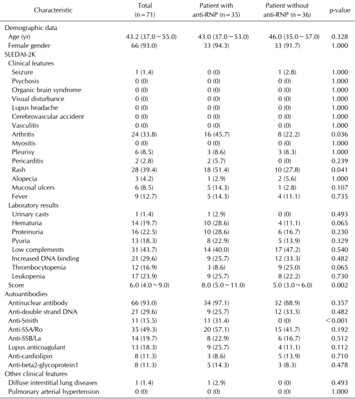
The baseline characteristics are described in Table 1. The median age of the total cohort of 71 patients (5 men, 66 women) was 43.2 years. The most common clinical fea- ture was rash (39.4%), followed by arthritis (33.8%) and fever (12.7%). Of the laboratory results, low complement levels were found in 31 of 71 patients (43.7%), whereas increased DNA binding (anti-ds DNA) and leukopenia were observed in 21 patients (29.6%) and 17 patients (23.9%), respectively. The overall median SLEDAI-2K score was 6.0 (4.0 to 9.0).
Anti-RNP was detected in 35 of 71 patients (49.3%).
When patients were divided into 2 groups according to the presence of anti-RNP, SLE patients with anti-RNP more frequently manifested rash (18 [51.4%] vs. 10 [27.8%], p=0.041) and arthritis (16 [45.7%] vs. 8 [22.2%], p=0.036) than did those without anti-RNP at SLE diagnosis. The two groups did not differ significantly in other clinical features and laboratory results relevant to SLEDAI-2K. However, SLE patients with anti-RNP had a higher median baseline SLEDAI-2K score than did those without anti-RNP (8.0 vs. 5.0, p=0.002). Anti-Smith an- tibodies were only detected in 11 SLE patients with an- ti-RNP (p<0.001).
Comparison of variables related to the follow-up SLEDAI-2K scores and SLE flare rate during the first year of follow-up, according to the presence of anti-RNP
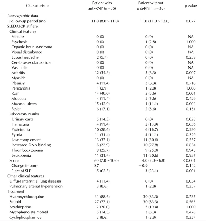
During the first follow-up year, SLE patients with RNP at diagnosis more frequently presented with mucosal ul- cers (15 [42.9%] vs. 4 [11.1%], p=0.003), rash (14 [40.0%] vs. 2 [5.6%], p=0.001), and arthritis (12 [34.3%] vs. 3 [8.3%], p=0.007) than did those without anti-RNP. However, the groups did not differ significantly with regard to diffuse interstitial lung disease or pulmo- nary arterial hypertension (Table 2). SLE patients with anti-RNP had a higher median follow-up SLEDAI-2K score, compared to those without anti-RNP (9.0 vs. 4.0, p<0.001). The median changes in SLEDAI-2K scores in patients with and without anti-RNP were 0.7 and −0.9, respectively, a non-significant difference. However, the SLE flare incidence was remarkably higher in patients with anti-RNP than in those without anti-RNP (62.5%
vs. 23.1%, p=0.001). Furthermore, patients with anti- RNP at the time of SLE diagnosis had a significantly high- er risk of experienced any SLE flare during the first year of follow-up, compared to those without anti-RNP (OR=
8.250, 95% confidence interval [CI] 2.121 to 32.090).
The 71 patients were further divided into 2 groups ac- cording to the presence or absence of anti-Smith anti- bodies at diagnosis (11 patients with anti-Smith vs. 60 without), and the groups were compared with respect to the SLE flare rate. Notably, this rate (change in SLEDAI-2K score ≥4) was significantly higher in pa- tients with anti-Smith antibodies than in those without anti-Smith antibodies (54.6% vs. 20.0%, p=0.025).
Furthermore, patients with anti-Smith antibodies at the time of SLE diagnosis had a significantly higher risk of ex- periencing SLE flare during the first year of follow-up (OR=4.800, 95% CI 1.251 to 18.421).
Anti-RNP titers were measured in 64 patients (31 pa- tients with RNP, 33 patients without RNP). Consequently, a positive correlation was observed between the RNP titer and the SLE flare incidence (correlation coefficient=
0.422, p=0.001). Finally, 57 of 71 patients (80.3%) re- ceived systemic steroid treatment, and additional im- munosuppressants (azathioprine, mycophenolate mofe- til, cyclophosphamide) were administered to some pa- tients (azathioprine 19.7%, mycophenolate mofetil 11.3%, cyclophosphamide 5.6%). However, no statisti- cally significant differences were observed between the two groups (Table 2).
DISCUSSION
In this study, we found that SLE patients with anti-RNP at the time of the initial SLE diagnosis exhibited larger in- creases in SLEDAI-2K scores during the first year of fol- low-up than did patients without anti-RNP. In addition, the presence of anti-RNP appeared to predict an approx- imately 8-fold increase in the SLE flare incidence during the same follow-up period. Moreover, our results sug- gested that the presence of anti-RNP at diagnosis is asso- ciated a greater incidence of arthritis and rash and may significantly promote the development of arthritis, rash, and mucosal ulcers, thus contributing to SLE flare accord- ing to the SLEDAI-2K definition, a finding that was con- sistent with the results of a previous study [9]. On the other hand, in contrast to previously reported results, the incidence of diffuse interstitial lung disease and pulmo- nary arterial hypertension did not differ according to the
Table 1. Baseline characteristics of patients with systemic lupus erythematosus and comparison of variables between patients with and without anti-RNP
Characteristic Total
(n=71)
Patient with anti-RNP (n=35)
Patient without
anti-RNP (n=36) p-value
Demographic data
Age (yr) 43.2 (37.0∼55.0) 43.0 (37.0∼53.0) 46.0 (35.0∼57.0) 0.328
Female gender 66 (93.0) 33 (94.3) 33 (91.7) 1.000
SLEDAI-2K Clinical features
Seizure 1 (1.4) 0 (0) 1 (2.8) 1.000
Psychosis 0 (0) 0 (0) 0 (0) 1.000
Organic brain syndrome 0 (0) 0 (0) 0 (0) 1.000
Visual disturbance 0 (0) 0 (0) 0 (0) 1.000
Lupus headache 0 (0) 0 (0) 0 (0) 1.000
Cerebrovascular accident 0 (0) 0 (0) 0 (0) 1.000
Vasculitis 0 (0) 0 (0) 0 (0) 1.000
Arthritis 24 (33.8) 16 (45.7) 8 (22.2) 0.036
Myositis 0 (0) 0 (0) 0 (0) 1.000
Pleurisy 6 (8.5) 3 (8.6) 3 (8.3) 1.000
Pericarditis 2 (2.8) 2 (5.7) 0 (0) 0.239
Rash 28 (39.4) 18 (51.4) 10 (27.8) 0.041
Alopecia 3 (4.2) 1 (2.9) 2 (5.6) 1.000
Mucosal ulcers 6 (8.5) 5 (14.3) 1 (2.8) 0.107
Fever 9 (12.7) 5 (14.3) 4 (11.1) 0.735
Laboratory results
Urinary casts 1 (1.4) 1 (2.9) 0 (0) 0.493
Hematuria 14 (19.7) 10 (28.6) 4 (11.1) 0.065
Proteinuria 16 (22.5) 10 (28.6) 6 (16.7) 0.230
Pyuria 13 (18.3) 8 (22.9) 5 (13.9) 0.329
Low complements 31 (43.7) 14 (40.0) 17 (47.2) 0.540
Increased DNA binding 21 (29.6) 9 (25.7) 12 (33.3) 0.482
Thrombocytopenia 12 (16.9) 3 (8.6) 9 (25.0) 0.065
Leukopenia 17 (23.9) 9 (25.7) 8 (22.2) 0.730
Score 6.0 (4.0∼9.0) 8.0 (5.0∼11.0) 5.0 (3.0∼6.0) 0.002
Autoantibodies
Antinuclear antibody 66 (93.0) 34 (97.1) 32 (88.9) 0.357
Anti-double strand DNA 21 (29.6) 9 (25.7) 12 (33.3) 0.482
Anti-Smith 11 (15.5) 11 (31.4) 0 (0) <0.001
Anti-SSA/Ro 35 (49.3) 20 (57.1) 15 (41.7) 0.192
Anti-SSB/La 14 (19.7) 8 (22.9) 6 (16.7) 0.512
Lupus anticoagulant 13 (18.3) 9 (25.7) 4 (11.1) 0.112
Anti-cardiolipin 8 (11.3) 3 (8.6) 5 (13.9) 0.710
Anti-beta2-glycoprotein1 8 (11.3) 5 (14.3) 3 (8.3) 0.478
Other clinical features
Diffuse interstitial lung diseases 1 (1.4) 1 (2.9) 0 (0) 0.493
Pulmonary arterial hypertension 0 (0) 0 (0) 0 (0) 1.000
Values are presented as median (interquartile range) or number (%). SLEDAI-2K: systemic lupus erythematosus Disease Activity Index 2000, RNP: ribonucleoprotein, SSA/Ro: Sjögren's syndrome-related antigen A, SSB/La: Sjögren's syndrome-related antigen B.
presence of anti-RNP [10]. However, a 1-year follow-up period might not be sufficient to identify histological al- terations that lead to clinical symptoms.
In general clinical situations, physicians tend to monitor follow-up anti-ds DNA titer and complement component levels to assess SLE disease activity; however, no current
Table 2. Comparison of variables of the follow-up SLEDAI-2K scores and SLE flare rate during the first follow-up year between SLE patients with and without anti-RNP
Characteristic Patient with
anti-RNP (n=35)
Patient without
anti-RNP (n=36) p-value
Demographic data
Follow-up period (mo) 11.0 (8.0∼11.0) 11.0 (11.0∼12.0) 0.077
SLEDAI-2K at flare Clinical features
Seizure 0 (0) 0 (0) NA
Psychosis 0 (0) 1 (2.8) 1.000
Organic brain syndrome 0 (0) 0 (0) NA
Visual disturbance 0 (0) 0 (0) NA
Lupus headache 2 (5.7) 0 (0) 0.239
Cerebrovascular accident 0 (0) 0 (0) NA
Vasculitis 0 (0) 0 (0) NA
Arthritis 12 (34.3) 3 (8.3) 0.007
Myositis 0 (0) 0 (0) NA
Pleurisy 4 (11.4) 3 (8.3) 0.710
Pericarditis 1 (2.9) 1 (2.8) 1.000
Rash 14 (40.0) 2 (5.6) 0.001
Alopecia 4 (11.4) 2 (5.6) 0.429
Mucosal ulcers 15 (42.9) 4 (11.1) 0.003
Fever 6 (17.1) 2 (5.6) 0.151
Laboratory results
Urinary casts 5 (14.3) 0 (0) 0.025
Hematuria 4 (11.4) 5 (13.9) 0.036
Proteinuria 10 (28.6) 6 (16.7) 0.230
Pyuria 11 (31.4) 4 (11.1) 0.329
Low complement 13 (37.1) 11 (30.6) 0.557
Increased DNA binding 8 (22.9) 10 (27.8) 0.634
Thrombocytopenia 9 (25.7) 9 (25.0) 0.945
Leukopenia 11 (31.4) 11 (30.6) 0.937
Score 9.0 (7.0∼10.0) 4.0 (2.0∼6.8) <0.001
Change in score 0.7 −0.9 0.142
Flare of SLE 15 (62.5) 3 (23.1) 0.001
Other clinical features
Diffuse interstitial lung diseases 4 (11.4) 0 (0) 0.054
Pulmonary arterial hypertension 3 (8.6) 1 (2.8) 0.357
Treatment
Hydroxychloroquine 31 (88.6) 30 (83.3) 0.735
Steroid 27 (77.1) 30 (83.3) 0.563
Azathioprine 7 (20.0) 7 (19.4) 1.000
Mycophenolate mofetil 5 (14.3) 3 (8.3) 0.478
Cyclophosphamide 3 (8.6) 1 (2.8) 0.357
Values are presented as median (interquartile range) or number (%). NA: not available, RNP: ribonucleoprotein, SLE: systemic lupus erythematosus, SLEDAI-2K: SLE Disease Activity Index 2000.
recommendations encourage the evaluation of other au- toantibodies, including anti-RNP. In contrast to anti-ds DNA, anti-RNP can be detected in the peripheral blood for more than 1 year. In addition, because anti-RNP is produced by long-lived plasma cells and supported by the enhanced differentiation of anti-RNP-related memory B
cells to plasma cells, the circulating anti-RNP concen- tration might also be maintained despite the lack of an ex- act established blood-survival time [14,17]. The long sta- bility of anti-RNP renders it unsuitable as a marker of rap- id alterations in SLE disease activity. Nevertheless, an- ti-RNP may be useful for anticipating SLE flare, improve-
ment, or remission during a lengthy follow-up period once its clinical relevance has been determined. Accor- dingly, our present study of the potentially predictive and reflective role of anti-RNP positivity at the time of SLE di- agnosis with regard to alterations in SLE disease activity within the first year of follow-up year after diagnosis was conducted against the backdrop of these concepts and characteristics of anti-RNP.
Because the anti-ds DNA titer and complement compo- nent levels are SLEDAI-2K items and are known to be re- flective of changes in activity within a relatively short time period, we did not analyze their predictive potential for SLE flare in this study. In contrast, we assumed that the presence of anti-Smith antibodies might be predictive of SLE flare in the early phase of disease, as all 11 patients with anti-Smith antibodies were also anti-RNP-positive.
Anti-Smith antibodies and anti-RNP might be simulta- neously detected at a high rate in SLE patients, as these autoantibodies were classified and analyzed in the same cluster in previous studies [9,17]. In the present study;
however, anti-Smith antibodies were only detected in pa- tients having anti-RNP, and therefore we could not dis- tinguish the direct effect of anti-Smith antibodies from the mutual effect of both types of autoantibodies.
Therefore, the predictive potential of anti-Smith anti- bodies with regard to SLE flare should be addressed in fu- ture studies involving a greater number of anti-Smith an- tibody-positive SLE patients.
One feature of our study that we consider to be a strength is that it is the first to propose the potential of anti-RNP at diagnosis to predict the development of SLE flare during the first year of follow-up using the well- documented SLEDAI-2K at each visit. However, our study also had sev- eral limitations. First, the study featured a small number of SLE patients and a relatively short follow-up period be- cause of the exclusion criteria for concomitant MCTD and the retrospective design. Second, we were unable to ex- plain the mechanism linking anti-RNP and the clinical items of SLEDAI-2K that contribute to SLE flare. Third, given the small number of patients, differences in baseline SLEDAI-2K scores between the two groups might have af- fected the SLE flare rate, despite speculation regarding the relationship between the presence of anti-RNP and base- line SLEDAI-2K scores. Future studies will be needed to compensate for these limitations and clarify the clinical role of anti-RNP for the prediction of the disease activity in newly diagnosed SLE patients.
CONCLUSION
In conclusion, our study determined that among Korean SLE patients, those with anti-RNP at the time of diag- nosis were 8.3-fold more likely to experience an SLE flare according to the SLEDAI-2K definition during the first year of follow-up relative to those without anti-RNP, thus indicating the potential predictive value of this auto- antibody.
CONFLICT OF INTEREST
No potential conflict of interest relevant to this article was reported.
REFERENCES
1. Mok CC, Lau CS. Pathogenesis of systemic lupus erythema- tosus. J Clin Pathol 2003;56:481-90.
2. Rahman A, Isenberg DA. Systemic lupus erythematosus. N Engl J Med 2008;358:929-39.
3. Benito-Garcia E, Schur PH, Lahita R; American College of Rheumatology Ad Hoc committee on immunologic testing guidelines. Guidelines for immunologic laboratory testing in the rheumatic diseases: anti-Sm and anti-RNP antibody tests. Arthritis Rheum 2004;51:1030-44.
4. Greidinger EL, Hoffman RW. The appearance of U1 RNP antibody specificities in sequential autoimmune human an- tisera follows a characteristic order that implicates the U1-70 kd and B'/B proteins as predominant U1 RNP immunogens. Arthritis Rheum 2001;44:368-75.
5. Cappelli S, Bellando Randone S, Martinović D, Tamas MM, Pasalić K, Allanore Y, et al. "To be or not to be," ten years af- ter: evidence for mixed connective tissue disease as a dis- tinct entity. Semin Arthritis Rheum 2012;41:589-98.
6. Lundberg I, Hedfors E. Clinical course of patients with an- ti-RNP antibodies. A prospective study of 32 patients. J Rheumatol 1991;18:1511-9.
7. Arbuckle MR, McClain MT, Rubertone MV, Scofield RH, Dennis GJ, James JA, et al. Development of autoantibodies before the clinical onset of systemic lupus erythematosus. N Engl J Med 2003;349:1526-33.
8. Cozzani E, Drosera M, Gasparini G, Parodi A. Serology of lu- pus erythematosus: correlation between immunopatho- logical features and clinical aspects. Autoimmune Dis 2014;2014:321359.
9. Li PH, Wong WH, Lee TL, Lau CS, Chan TM, Leung AM, et al. Relationship between autoantibody clustering and clin- ical subsets in SLE: cluster and association analyses in Hong Kong Chinese. Rheumatology (Oxford) 2013;52:337-45.
10. Allen D, Fischer A, Bshouty Z, Robinson DB, Peschken CA, Hitchon C, et al. Evaluating systemic lupus erythematosus patients for lung involvement. Lupus 2012;21:1316-25.
11. Carpintero MF, Martinez L, Fernandez I, Romero AC, Mejia C, Zang YJ, et al. Diagnosis and risk stratification in patients with anti-RNP autoimmunity. Lupus 2015;24:1057-66.
12. Hochberg MC. Updating the American College of Rheumatology revised criteria for the classification of sys- temic lupus erythematosus. Arthritis Rheum 1997;40:1725.
13. Gladman DD, Ibañez D, Urowitz MB. Systemic lupus eryth- ematosus disease activity index 2000. J Rheumatol 2002;29:288-91.
14. Amigues JM, Cantagrel A, Abbal M, Mazieres B.
Comparative study of 4 diagnosis criteria sets for mixed connective tissue disease in patients with anti-RNP antibodies. Autoimmunity Group of the Hospitals of Toulouse. J Rheumatol 1996;23:2055-62.
15. Touma Z, Urowitz MB, Ibañez D, Gladman DD. SLEDAI-2K
10 days versus SLEDAI-2K 30 days in a longitudinal evaluation. Lupus 2011;20:67-70.
16. Barst RJ, McGoon M, Torbicki A, Sitbon O, Krowka MJ, Olschewski H, et al. Diagnosis and differential assessment of pulmonary arterial hypertension. J Am Coll Cardiol 2004;43(12 Suppl S):40S-7S.
17. Weinstein JS, Delano MJ, Xu Y, Kelly-Scumpia KM, Nacionales DC, Li Y, et al. Maintenance of anti-Sm/RNP au- toantibody production by plasma cells residing in ectopic lymphoid tissue and bone marrow memory B cells. J Immunol 2013;190:3916-27.