Multiple calcifying hyperplastic dental follicle (MCHDF) is an extremely rare condition characterized by multiple impacted teeth and enlarged dental follicles that contain abundant calcifications and rests of odontogenic epitheli- um. A PubMed search with the keywords “calcification AND dental follicle” and “hyperplastic dental follicle”
made in May 2013 and examination of the references of the relevant articles revealed that, up to that date, 11 cases had been reported.1-6Two of these cases were originally published as odontogenic fibroma-like cases.2,4Later, when Gardner and Radden1analyzed these cases together with two additional cases, they suggested the term MCHDF and proposed that the condition is sufficiently distinctive to be considered a pathologic entity.
In all of the reported cases, the patients were male and younger than 40 years. The impacted teeth were mostly third or second molar, canine, or second premolar teeth, and they were associated with pericoronal radiolucencies delineated by sclerotic borders.1-6In three cases, focal dis-
crete radiopacities within the pericoronal radiolucencies were detected on the radiographs; however, calcification in the enlarged dental follicles was a significant histopa- thologic feature of this entity.1-3
The purpose of the present report was to describe the clinical, radiographic, and histopathologic features of a case of MCHDF.
Case Report
A 31-year-old female visited our clinic with the com- plaints of pain and gingival swelling on the left maxillary alveolar region and consequent difficulty of eating. Her dental history revealed that she had a dental visit due to her carious and crowded teeth when she was 17 years old.
Her carious molar teeth had been extracted and she had applied for orthodontic treatment. The palatally positioned left maxillary incisor teeth were treated for a four-year period. At that time, her impacted teeth were diagnosed on routine radiographs, and she had a surgical operation for the extraction of one of her impacted right mandibular teeth at the age of 18. Her orthodontist followed up on the eruption of the other impacted teeth yearly until she was 21 years old, but then she interrupted her dental treatment
Multiple calcifying hyperplastic dental follicles: A case report
Ulkem Aydin1, Timucin Baykul2, Benay Yildirim3, Derya Yildirim4, Esin Bozdemir4,*, Ayse Karaduman5
1Department of Dentomaxillofacial Radiology, Baskent University Faculty of Dentistry, Ankara, Turkey
2Department of Oral and Maxillofacial Surgery, Suleyman Demirel University Faculty of Dentistry, Isparta, Turkey
3Department of Oral Pathology, Gazi University Faculty of Dentistry, Ankara, Turkey
4Department of Dentomaxillofacial Radiology, Suleyman Demirel University Faculty of Dentistry, Isparta, Turkey
5Atlas Dent Dental Health Center, Aydin, Turkey
ABSTRACT
This report describes a 31-year-old female patient with six impacted teeth. The crowns of the impacted teeth were surrounded with cyst-like lesions with a mixed internal structure and well-defined cortical borders. Microscopic examination of the specimen obtained from the follicle of the left mandibular third molar tooth revealed loose to moderately dense collagenous connective tissue with abundant calcified material and sparse epithelial islands. A diagnosis of multiple calcifying hyperplastic dental follicles was made. (Imaging Sci Dent 2013; 43 : 303-8) KEY WORDS: Dental Follicle; Tooth, Impacted; Calcification
Received January 28, 2013; Revised March 24, 2013; Accepted June 4, 2013
*Correspondence to : Prof. Esin Bozdemir
Suleyman Demirel Üniversitesi Dishekimligi Fakultesi, Agiz Dis ve Cene Radyolojisi AD, Dogu Kampusu, 32260, Isparta, Turkey
Tel) 90-246-211-8752, Fax) 90-246-237-0607, E-mail) [email protected]
Copyright ⓒ 2013 by Korean Academy of Oral and Maxillofacial Radiology
This is an Open Access article distributed under the terms of the Creative Commons Attribution Non-Commercial License (http://creativecommons.org/licenses/by-nc/3.0) which permits unrestricted non-commercial use, distribution, and reproduction in any medium, provided the original work is properly cited.
Imaging Science in Dentistry∙pISSN 2233-7822 eISSN 2233-7830
and follow-up visits until our examination.
The patient had also been diagnosed with hypothyroidi- sm when she was five years old. She was undergoing lev- othyroxine sodium (Levotiron®, Abdi .
Ibrahim, Istanbul,
Turkey) therapy, and her T3, T4, and TSH hormone levels were normal. On extraoral examination, she was approxi- mately 1.50 meters tall and had sparse hair. Intraoral exam- ination revealed missing teeth, diastemata, macroglossia, and buccolingual expansion of the alveolar bone (Fig. 1).
A panoramic radiograph revealed six impacted perma- nent teeth: the third molar teeth in all quadrants and the maxillary canine teeth. The crowns of these impacted teeth were surrounded with cyst-like lesions containing radio- paque foci, and well-defined corticated borders. The im- pacted mandibular molar teeth were located in the apical direction near the inferior cortex of the mandible, and the impacted maxillary teeth were located close to the floor of the maxillary sinus (Fig. 2). For further information, com- puted tomography was performed. Axial sections of the mandible revealed rounded radiolucent lesions associated with the crowns of the right and left mandibular molar teeth. These lesions contained radiopaque foci and were interpreted as calcification (Fig. 3A). In axial sections of
Fig. 1.An intraoral photograph reveals missing teeth, diastemata, and buccolingual expansion of the alveolar bone.
Fig. 2.A cropped panoramic radio- graph demonstrates six impacted teeth, crowns surrounded with cyst- like lesions having mixed internal structure, and well-defined cortical borders.
Fig. 3.A. An axial computed tomo- graphy scan shows rounded hypo- dense lesions containing hyperdense foci with well-defined borders, asso- ciated with the crowns of mandibu- lar molar teeth. B. An axial comput- ed tomography scan reveals the mix- ed internal structure of the maxil- lary lesions.
A B
the maxilla, the mixed appearance of the lesions with well- defined borders was noteworthy (Fig. 3B).
After surgically exposing the impacted left mandibular third molar tooth, an incisional biopsy specimen was obta- ined from the center of the lesion, under direct vision. Sub- sequent to biopsy, histopathological examination of the specimen obtained from the follicle of the tooth was made.
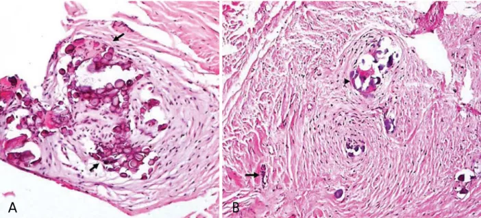
Microscopic examination revealed loose to moderately dense collagenous connective tissue with abundant calci- fied material and sparse epithelial islands. The calcifica- tions were mostly acellular type 1 calcification but scanty cellular type II calcifications were also detected. Inflam- matory cell infiltration was not observed (Fig. 4). A diag-
nosis of dental follicles with calcifications was made.
The patient refused a surgical operation for removal of the impacted teeth, and she was scheduled for a follow-up visit. One year later, there was not any alteration in the radiographic appearance of the lesions, and then the pati- ent was lost to follow-up. Five years later, the patient show- ed up for a follow-up appointment. A new panoramic ra- diograph revealed slight enlargement of the calcified fol- licles and an increase in the density of the calcifications.
A focal increase in the radiopacity associated with the crown of the right mandibular third molar tooth, a small radiopaque prominence associated with the inferomedial aspect of the follicle of the left mandibular third molar
Fig. 4.A. Microscopic examination shows type 1 calcifications arranged in whorled structures. Liesegang ring-like structures are promi- nent (arrows) (H&E stain, 200x). B. Type II calcifications (arrowhead) and an odontogenic epithelial rest (arrow) in a collagenized dental follicle are seen (H&E stain, 200x).
A B
Fig. 5.A cropped panoramic radio- graph taken five years later shows a focal increase in the radiopacity associated with the crown of the right mandibular third molar tooth and a small radiopaque prominence associated with the inferomedial aspect of the follicle of the left man- dibular third molar tooth. The hyper- plastic follicles reveal an irregular contour.
tooth, and irregularity of the contours of the hyperplastic follicles were detectable on the radiograph made five years after initial presentation (Fig. 5). The patient refused any surgical operation or prosthetic restoration again.
Discussion
MCHDFs are atypical follicles or hamartomatous ano- malies.2However, the lesions radiographically and histo- logically resemble many other benign neoplasms and may pose diagnostic problems for clinicians including maxillo- facial radiologists and pathologists who are not familiar with this condition.2,4,6,7
The crowns of unerupted teeth are normally surrounded by a dental follicle, and radiographically, the dental follicle appears as a radiolucent halo that has a thin radiopaque border. The follicular spaces of long-standing impacted teeth are frequently diminished, with the exception of max- illary canine teeth having enlarged follicles.8Histopatho- logic examination of an enlarged dental follicle sometimes reveals no pathosis, while at the same time, radiographi- cally normal dental follicles may exhibit histopathological changes.9,10Despite the controversy in the literature regard- ing the establishment of criteria for the differentiation of incipient pathoses and enlarged dental follicles, the width of the pericoronal space has been used as a criterion to dis- tinguish between a normal and an abnormal follicle. When the follicular radiolucency reaches 2.5 mm in width on the periapical and 3 mm on the panoramic radiograph, pathosis is suggested, and if the radiopaque border, which is the radiographic image of the surrounding cortical plate, is not well-defined, this is also a sign of pathologic change.8In the present case, all of the follicular spaces of the impacted teeth exceeded 3 mm in diameter, and the cortical plates around the right mandibular and maxillary third molar and the left maxillary canine teeth were well defined, whereas the right maxillary canine, left maxillary molar, and man- dibular third molar teeth were surrounded by roughly well- defined radiopaque borders, suggesting pathosis.
Radiographically, calcifying cystic odontogenic tumor, adenomatoid odontogenic tumor (AOT), ameloblastic fibro- odontoma, and calcifying epithelial odontogenic tumor (CEOT) may show radiopaque foci, and they may be asso- ciated with unerupted teeth.7,11-13Another rare entity that shows radiopaque foci and may be associated with unerupt- ed teeth is MCHDF.7
Calcifying cystic odontogenic tumor tends to occur an- terior to the first molar and is especially associated with cuspids and incisors, and most AOTs occur in the anterior
maxilla, especially the canine region.14 In our case, only two of the enlarged follicular spaces containing radiopaque foci were associated with canine teeth whereas there were four others associated with molar teeth, and slow-growing, painless jaw swelling was absent, which was reported to be a feature of these two pathologic entities. Ameloblastic fibro-odontoma and CEOT both have a predilection for the posterior mandible; however, ameloblastic fibro-odon- toma occurs predominantly in children and young adults, and jaw expansion in CEOT is a regular feature but was not observed in the present case.7,12,15
Calcifying cystic odontogenic tumor, AOT, ameloblastic fibro-odontoma, and CEOT are mostly encountered as solitary lesions, and multifocal cases are very rarely re- ported.16,17Therefore, when multiple impacted teeth show enlarged follicular spaces containing radiopaque foci, MCHDF should be considered.1-3
Including the case presented, the average age of all re- ported cases is 20.1 with a range of 11-40 years. A review of the previous cases revealed no other report of this phe- nomenon in females.1-6Clinically, diastemata and buccol- ingual expansion of the alveolar bone, which were also features of our case, were observed in one previously re- ported case, and in another report, firm, well localized asymptomatic expansions were noted in the mandibular and maxillary premolar regions.2,5The vast majority of the involved teeth were the canines, second and third molars, and premolars.1-6 In the present case, the impacted teeth were also the third molar and canine teeth. Congenitally missing or supernumerary teeth were previously reported but, at present, no other dental anomaly was known to be associated with MHCDF.4,5In the present case, there were missing teeth and a history of extraction of carious molar teeth, but it was not definitely known if there were any congenitally missing teeth or extracted supernumerary teeth.
Recently, Cho et al5reported five MCHDF cases; all of the patients had initially visited local clinics for the eval- uation of delayed eruption of permanent molar teeth.
According to the initial evaluation, all of the panoramic radiographs showed multiple odontogenic cysts, and the radiologists originally interpreted the enlarged follicles as dentigerous cysts.5Also, in other cases, the radiographic appearances of the lesions were described to be radiolu- cent areas around the crowns of the unerupted teeth.1-4,6 The authors did not mention any radiopacities associated with the enlarged dental follicles. On the other hand, in three of the reported cases and in the case presented here, focal discrete radiopacities were detected within the peri-
coronal radiolucencies.1-3Dare et al18stated that intraoral periapical radiographs enabled the detection of the radio- pacities in adenomatoid odontogenic tumor within radiol- ucency even with minimal calcified deposits, and panora- mic radiography was often unable to demonstrate radiopa- cities in adenomatoid odontogenic tumor when the calcifi- cation was minimal. Therefore, when radiolucent lesions are encountered on panoramic radiographs, periapical ra- diography should be useful to detect radiopaque foci, if present. Follow-up radiographs taken in one patient reveal- ed a slight increase in calcification within some of the peri- coronal lesions.2 The differences in exposure parameters and patient positioning make it difficult to compare the panoramic radiographs; however, radiographic examina- tion in the present case also revealed that the calcification process and follicular hyperplasia were continuing. The alterations were not detectable on the first follow-up radio- graph made one year after diagnosis but became prominent five years later. Therefore, long-term follow-up is suggest- ed for untreated cases.
In our case, microscopic examination revealed various spherical calcifications in the fibrous connective tissue and the histopathologic diagnosis was dental follicle containing calcifications. The histopathological features were abun- dant calcifications and rests of odontogenic epithelium in an enlarged dental follicle. It was not unusual to find inac- tive rests of odontogenic epithelium and calcified droplets of cementum-like material in follicles of normally devel- oping or unerupted teeth, which have been associated with the induction of the dental ectomesenchyme.19The distinct feature of our case was multiplicity of unerupted teeth. The radiographic features were suggestive of MCHDF, and the histopathological features, hyperplastic dense fibrous con- nective tissue with numerous deposits of type I and type II calcifications and rests of odontogenic epithelium, observ- ed in the present case were identical to the previous cases of MCHDF.1,2,5
The mesenchymal cells of the dental follicles play an im- portant role in the signalling pathway to induce eruption.20 Defective regulation of matrix metalloproteinases media- ting connective tissue remodelling in non-syndromic hyper- plastic dental follicles was suggested as a cause of abnor- mal tooth eruption.21Thus, retarded eruption or impaction of teeth may be associated with the calcifying phenomenon that originates from the mesenchymal cells. According to Gardner and Radden,1there was no apparent relationship of the present condition to either cleidocranial dysplasia22 or to Gardner’s syndrome,23both of which involve multiple impacted teeth. Hypothyroidism (juvenile), which was also
a feature of our case, is a condition related to delayed erup- tion.14,24In our case, it could be speculated that hyperplas- tic follicles hindered eruption of the involved teeth, which were probably delayed in eruption because of hypothy- roidism. On the other hand, hypothyroidism as an etiologic factor in MCHDF remains to be elucidated. In 2 out of 11 previously reported cases,2,5 the medical history included disorders that were not known to be associated with tooth impaction, and in the other cases, the medical history was either unremarkable or not reported. In one case, the pati- ent was known to have tuberous sclerosis complex.6Accor- ding to some authors, this multi-system disease was asso- ciated with the presence of impacted teeth.6,25Further stud- ies may be able to shed light on the etiology of MCHDF and mechanism of tooth eruption, and the association of MCHDF with medical conditions.
In conclusion, when multiple impacted teeth with enlarg- ed follicles are encountered, the lesions should be investi- gated using appropriate radiographic techniques, and mul- tiple calcifying hyperplastic dental follicles should be in- cluded in the differential diagnosis.
References
1. Gardner DG, Radden B. Multiple calcifying hyperplastic den- tal follicles. Oral Surg Oral Med Oral Pathol Oral Radiol En- dod 1995; 79: 603-6.
2. Sandler HJ, Nersasian RR, Cataldo E, Pochebit S, Dayal Y.
Multiple dental follicles with odontogenic fibroma-like changes (WHO type). Oral Surg Oral Med Oral Pathol 1988; 66: 78-84.
3. Gomez RS, Silva EC, Silva-Filho EC, Castro WH. Multiple calcifying hyperplastic dental follicles. J Oral Pathol Med 1998;
27: 333-4.
4. Lukinmaa PL, Hietanen J, Anttinen J, Ahonen P. Contiguous enlarged dental follicles with histologic features resembling the WHO type of odontogenic fibroma. Oral Surg Oral Med Oral Pathol 1990; 70: 313-7.
5. Cho YA, Yoon HJ, Hong SP, Lee JI, Hong SD. Multiple cal- cifying hyperplastic dental follicles: comparison with hyper- plastic dental follicles. J Oral Pathol Med 2011; 40: 243-9.
6. Magliocca KR, Bhattacharyya I, Wolfrom RB, Cohen DM.
Multiple impacted teeth and associated pericoronal tissue abnor- mality in tuberous sclerosis complex. J Oral Maxillofac Surg 2012; 70: 2581-4.
7. Wood NK, Goaz PW, Lehnert JF. Mixed radiolucent-radiopa- que lesions associated with teeth. In: Wood NK, Goaz PW.
Differential diagnosis of oral and maxillofacial lesions. 5th ed.
St Louis, MO: Mosby; 1997. p. 415-32.
8. Wood NK, Kuc IM. Pericoronal radiolucencies. In: Wood NK, Goaz PW. Differential diagnosis of oral and maxillofacial les- ions. 5th ed. St Louis, MO: Mosby; 1997. p. 279-95.
9. Sun CX, Ririe C, Henkin JM. Hyperplastic dental follicle-re- view of literature and report of two cases in one family. Chin J Dent Res 2010; 13: 71-5.
10. Baykul T, Saglam AA, Aydin U, Basak K. Incidence of cystic changes in radiographically normal impacted lower third molar follicles. Oral Surg Oral Med Oral Pathol Oral Radiol Endod 2005; 99: 542-5.
11. Buchner A. The central (intraosseous) calcifying odontogenic cyst: an analysis of 215 cases. J Oral Maxillofac Surg 1991;
49: 330-9.
12. Handschel JG, Depprich RA, Zimmermann AC, Braunstein S, Kübler NR. Adenomatoid odontogenic tumor of the mandible:
review of the literature and report of a rare case. Head Face Med 2005; 1: 3.
13. Philipsen HP, Reichart PA. Calcifying epithelial odontogenic tumour: biological profile based on 181 cases from the litera- ture. Oral Oncol 2000; 36: 17-26.
14. White SC, Phaorah M. Oral radiology: principles and interpre- tation. 3rd ed. St. Louis, MO: Mosby; 2008.
15. Li TJ, Yu SF. Clinicopathologic spectrum of the so-called cal- cifying odontogenic cysts: a study of 21 intraosseous cases with reconsideration of the terminology and classification. Am J Surg Pathol 2003; 27: 372-84.
16. Sedghizadeh PP, Wong D, Shuler CF, Linz V, Kalmar JR, Allen CM. Multifocal calcifying epithelial odontogenic tumor.
Oral Surg Oral Med Oral Pathol Oral Radiol Endod 2007; 104:
e30-4.
17. Bartake AR, Punnya VA, Sudeendra P, Rekha K. Two adeno-
matoid odontogenic tumours of the maxilla: a case report. Br J Oral Maxillofac Surg 2009; 47: 638-40.
18. Dare A, Yamaguchi A, Yoshiki S, Okano T. Limitation of panoramic radiography in diagnosing adenomatoid odontogenic tumors. Oral Surg Oral Med Oral Pathol 1994; 77: 662-8.
19. Odell EW, Morgan PR. Biopsy pathology of the oral tissues.
London: Chapman & Hall Medical; 1998.
20. Wise GE. Cellular and molecular basis of tooth eruption. Orth- od Craniofac Res 2009; 12: 67-73.
21. Kim SG, Kim MH, Chae CH, Jung YK, Choi JY. Downregula- tion of matrix metalloproteinases in hyperplastic dental folli- cles results in abnormal tooth eruption. BMB Rep 2008; 41:
322-7.
22. Mohan RP, Suma GN, Vashishth S, Goel S. Cleidocranial dys- plasia: clinico-radiological illustration of a rare case. J Oral Sci 2010; 52: 161-6.
23. Bradley JF, Orlowski WA. Multiple osteomas, impacted teeth and odontomas - a case report of Gardner’s Syndrome. J N J Dent Assoc 1977; 48: 32-3.
24. Suri L, Gagari E, Vastardis H. Delayed tooth eruption: patho- genesis, diagnosis, and treatment. A literature review. Am J Orthod Dentofacial Orthop 2004; 126: 432-45.
25. Cutando A, Gil JA, López J. Oral health management implica- tions in patients with tuberous sclerosis. Oral Surg Oral Med Oral Pathol Oral Radiol Endod 2000; 90: 430-5.