Copyright ⓒ 2016 by Korean Academy of Oral and Maxillofacial Radiology
This is an Open Access article distributed under the terms of the Creative Commons Attribution Non-Commercial License(http://creativecommons.org/licenses/by-nc/3.0) which permits unrestricted non-commercial use, distribution, and reproduction in any medium, provided the original work is properly cited.
Imaging Science in Dentistry·pISSN 2233-7822 eISSN 2233-7830
http://dx.doi.org/10.5624/isd.2016.46.1.17
Introduction
The interaction between orthodontic forces and the peri- odontal ligament leads to an inflammatory phenomenon that induces apical resorption by clastic activity1 without clinical symptoms. This phenomenon is known as external apical root resorption(EARR), and it is an undesirable2 and irreversible side effect of orthodontic treatment.
The reported occurrence of EARR is between 48% and
66% according to radiographic studies and more than 90%
based on histologic analyses.3,4 Fortunately, most cases exhibit resorption of no greater than 1mm, which does not impair tooth function. However, higher degrees of root shortening are observed in approximately 8% of patients one year after orthodontic treatment.5
Several factors are associated with EARR, including in- creased treatment duration, direction of tooth movement, the loading regimen,2 tooth extraction,1 the type of the tooth and the malocclusion, and patient-related factors in- cluding certain systemic conditions, age, and gender.4
The early detection of initial resorptive lesions during orthodontic treatment is essential for identifying teeth at risk of severe resorption.6-8 The subsequent interruption
A posteriori registration and subtraction of periapical radiographs for the evaluation of external apical root resorption after orthodontic treatment
Eliane Maria Kreich1,*, Ana Cláudia Chibinski2, Ulisses Coelho3, Letícia Stadler Wambier2, Rosário de Arruda Moura Zedebski1, Mari Eli Leonelli de Moraes4, Luiz Cesar de Moraes4
1Department of Dental Radiology, School of Dentistry, Ponta Grossa State University, Ponta Grossa, Paraná, Brazil
2Department of Pediatric Dentistry, School of Dentistry, Ponta Grossa State University, Ponta Grossa, Paraná, Brazil
3Department of Orthodontics, School of Dentistry, Ponta Grossa State University, Ponta Grossa, Paraná, Brazil
4Department of Dental Radiology, School of Dentistry, State University of São Paulo, São José dos Campos, São Paulo, Brazil
AbstrAct
Purposes: This study employed a posteriori registration and subtraction of radiographic images to quantify the apical root resorption in maxillary permanent central incisors after orthodontic treatment, and assessed whether the external apical root resorption(EARR) was related to a range of parameters involved in the treatment.
Materials and Methods: A sample of 79 patients(mean age, 13.5±2.2 years) with no history of trauma or endo- dontic treatment of the maxillary permanent central incisors was selected. Periapical radiographs taken before and after orthodontic treatment were digitized and imported to the Regeemy software. Based on an analysis of the post- treatment radiographs, the length of the incisors was measured using Image J software. The mean EARR was described in pixels and relative root resorption(%). The patient’s age and gender, tooth extraction, use of elastics, and treatment duration were evaluated to identify possible correlations with EARR.
results: The mean EARR observed was 15.44±12.1 pixels(5.1% resorption). No differences in the mean EARR were observed according to patient characteristics(gender, age) or treatment parameters(use of elastics, treatment duration). The only parameter that influenced the mean EARR of a patient was the need for tooth extraction.
conclusion: A posteriori registration and subtraction of periapical radiographs was a suitable method to quantify EARR after orthodontic treatment, and the need for tooth extraction increased the extent of root resorption after orthodontic treatment.(Imaging Sci Dent 2016; 46: 17-24)
Key words: Root Resorption; Orthodontics; Subtraction Technique; Image Processing, Computer-Assisted; Tooth Extraction
Received October 1, 2015; Revised November 4, 2015; Accepted November 22, 2015
*Correspondence to : Prof. Eliane Maria Kreich
Department of Dental Radiology, School of Dentistry, Ponta Grossa State University, General Carlos Cavalcanti Avenue, #4748, CEP 84030-900, Ponta Grossa, Paraná, Brazil
Tel) 55-42-99171983, Fax) 55-42-32203102, E-mail) [email protected]
of active treatment can help to reduce adverse outcomes during later stages of treatment,7 as well as avoiding or limiting the potential damage to the patient.9
The tools most commonly used to detect EARR are periapical1,8 and panoramic radiographs.10 This is probably due to the frequent use of these exams during the stages of orthodontic treatment11,12 and the good cost-benefit out- comes for the patient. Nevertheless, the visual comparison of radiographs before and after orthodontic treatment to measure the EARR is subject to discrepancies in clinical practice, and this technique is not able to detect minor changes in sequential images.13,14
Moreover, both radiographic techniques have limita- tions. Panoramic radiographs show a substantial amount of magnification and do not allow the clear visualization of the premaxilla.11 Although they exhibit much less dis- tortion,11 periapical radiographs require a certain degree of root shortening to have taken place before it is detect- able visually on the radiograph.12,14
In order to overcome these problems, cone-beam com- puted tomography has been suggested as an alternative to periapical and panoramic radiographs. The scientific literature indicates that this technique is suitable for the detection of early EARR.1,12 However, this exam makes orthodontic treatment more expensive and subjects the patient to an additional dose of radiation.
As an alternative, this paper proposes a posteriori reg- istration and subtraction of radiographic images and the use of computerized techniques to quantify the EARR us- ing periapical radiographs. The procedure of a posteriori registration and subtraction corrects the discrepancies of the geometric projection and equalizes the density and the contrast of the radiographic images before and after treat- ment, enabling the comparison of the two radiographs and improving the sensitivity and the accuracy of the evalua- tion, which is performed using specific software.
Although this method has been extensively tested in vitro in extracted teeth with simulated resorption,5,7,15-19 the use of periapical radiographs from actual orthodontic treatments,6,20 as proposed in this paper, is not a common feature of the dental literature on digital subtraction(DSR) radiography and EARR.
The null hypotheses tested were that a posteriori regis- tration and subtraction of periapical radiographic images cannot be used to quantify the EARR following orthodon- tic treatment and that patient-related factors(gender and age) and treatment-related factors(tooth extraction, use of intermaxillary elastics, and duration of orthodontic treat- ment) do not affect the amount of root resorption.
Therefore, the objective of this retrospective study was to evaluate whether a posteriori registration and subtrac- tion of radiographic images could be used to quantify api- cal root resorption in maxillary permanent central incisors after orthodontic treatment, as well as to determine whe- ther the EARR was related to the parameters involved in the treatment.
Materials and Methods
This study was approved by the Ethical Committee of São Paulo State University(Campus of São José dos Campos) under protocol #041/2009.
Sample selection
A careful analysis of 300 orthodontic clinical records from patients treated by dentists who attended a postgrad- uate dental education program in orthodontics was per- formed. The inclusion criteria were: orthodontic treatment with the standard edgewise technique; no history of trau- ma, wear, or endodontic treatment in the maxillary central incisors; complete radiographic exams, including baseline and final periapical radiographs of the maxillary central incisors and cephalometric analysis; the absence of synd- romic or skeletal disorders; and complete root formation in the maxillary central incisors. After this screening, 79 patients were selected. The sample number was above the calculated required sample size(n=64; α=0.5; 1-β=
0.9), but all the selected patients were kept in the sample to compensate for possible losses during the study.
Radiographic analysis: sequence of procedures Periapical radiographs were taken using the modified parallel technique and standardized exposure parameters:
Heliodent 70(Siemens, Erlangen, Germany) X-ray unit, 70kVp, 7mA, 0.4 seconds. The radiographs were digital- ized using a scanner with transparency adapter(HP Scan- jet G4050, Hewlett-Packard, Palo Alto, CA, USA) with a resolution of 300dpi and 8-bit gray scale. The images were stored as maximum-quality TIFF format files.
All the images were imported into the Regeemy Image Registration and Mosaicking 0.2.43-RCB software(DPI- INPE, São José dos Campos, São Paulo, Brazil). This software provides image registration and subtraction al- gorithms(i.e., it corrects geometric discrepancies, equal- izes the contrast of two sequential radiographs to make them comparable, and subtracts the analog pixels values from two sequential images).
The radiographic image obtained at baseline(before or-
thodontic treatment) was termed Image 1 and used as the reference image. The radiographic image obtained after orthodontic treatment was referred to as Image 2.
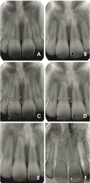
Reference points(fixed points in both radiographs) were selected on both images manually(Fig. 1). These refer- ence points served as coordinates for the software to align and correct the geometry of the second image according to the reference image(Image 1). Clearly distinguishable
structures were selected as reference points, such as the cement-enamel junction and the incisal margin of the max- illary central incisors.
This process generated Image 3, which was the retros- pectively corrected form of Image 2. The quality of the image correction was visually determined using the image subtraction routine(Image 1-Image 3). The image regis- tration was considered adequate when the structural noise on the teeth of interest was reduced to the lowest possible level, as indicated by a minimal or nonexistent discrepan- cy in the geometrical position between images, which ap- pears as brighter or darker shades of gray in the subtract- ed radiograph.
Therefore, the corrected image(Image 3) became the new follow-up radiographic image, since small differences in projection angles during exposure or contrast/density during processing were corrected(Fig. 1).
The evaluation of EARR was performed using Image 1 (baseline) and Image 3(the corrected image)(Fig. 2). The measurement of the long axis of the central maxillary incisors was done using the UTHSCSA Image Tool(Uni- versity of Texas Health Science Center at San Antonio;
http://compdent.uthscsa.edu/dig/itdesc.html). The length of the tooth was considered to be the distance between the root apex and the incisal edge at a specific point cor- responding to the mean distance between the mesial and distal angles. The above-described protocol was carried out separately for the right and left maxillary central inci- sors.
The complete procedures of a posteriori registration and subtraction and DSR were completed by one experienced investigator trained in this methodology. The measurement of the length of the tooth was repeated three times and the
Fig. 1. Illustrative images of the post-processing procedures of periapical radiographs for the right maxillary central incisor. Peri- apical radiographs at baseline(A, Image 1) and after orthodontic treatment(B, Image 2) were digitalized. The same reference points are tagged in the pre-treatment (C) and post-treatment (D) radiographs to align the images and to generate the posteriori reg- istration of Image 3(E). The quality of the registered image (E) is confirmed by the fact that the subtracted image (F) exhibited the least possible structural noise.
A B
C D
E F
Fig. 2. External apical root resorption is evaluated by the measure- ment of the long axis of the maxillary central incisors at baseline(A, Image 1) and in the registered image(B, Image 3).
A B
final result consisted of the mean of the three evaluations.
Intraexaminer agreement was calculated using Cohen’s kappa(k=0.87).
The EARR in the central maxillary incisors was deter- mined by the difference between baseline(Image 1) and post-treatment(Image 3) tooth lengths. These values were obtained in pixels. The difference between the values be- fore and after treatment(mean EARR) was obtained and described in terms of pixels and relative root resorption(%).
Evaluation parameters
In order to identify possible correlations with factors that are commonly associated with EARR, data regarding patient characteristics such as gender and age were col- lected from the clinical records. The features involved in the orthodontic treatment that were considered for analy- sis were the need for maxillary first bicuspid extraction, the use of elastics, and the treatment duration. The mea- sures 1.NA(angle formed by the maxillary incisor long axis and the nasion line) and 1-NA(linear distance be- tween the most anterior point of the maxillary central incisor and the NA line) were also obtained, since these are part of the cephalometric analysis that determines the dental pattern of the maxillary incisors.
Statistical analysis
The obtained data showed a normal distribution(Sha- piro-Wilk). Therefore, the comparisons between mean tooth length, 1.NA, and 1-NA before and after orthodontic treatment were made using the paired t-test. The Student’s t-test was used to compare the parameters of gender, age, tooth extraction, elastics use, and treatment duration. The statistical analysis was conducted using SigmaPlot 12.0 (Systat Software Inc, San Jose, CA, USA), with the sig- nificance level set at p=5%.
results
The final sample was composed of 79 patients(mean
age, 13.5±2.2 years; range, 10-19 years; 22 males and 57 females). The mean duration of the treatment was 25.8±
6.2 months(range, 10-38 months).
The tooth length before and after orthodontic treatment was 302.9±30.3 pixels and 287.2±32.3 pixels for the right maxillary central incisors; and 302.4±30.2 pixels and 287.2±31.1 pixels for the left maxillary central inci- sors. No significant differences were observed when the right and left central incisors were compared(p>0.05), and all subsequent analyses were therefore made using the mean of the measurements of the left and right incisors.
The mean EARR observed was 15.4±12.1 pixels, repre- senting a resorption of 5.1%(Table 1).
The mean length of the maxillary central incisors, as well as the mean values of the cephalometric measures 1.NA and 1-NA before and after orthodontic treatment are shown on Table 1. These parameters exhibited significant decreases after treatment.
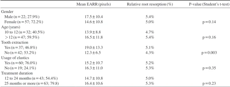
No differences in the mean EARR were observed ac- cording to gender, age, the use of elastics, or treatment duration. The only parameter that influenced the patient’s mean EARR was the need for tooth extraction as part of orthodontic treatment(Table 2).
discussion
The data obtained in this study allowed to reject the first hypothesis tested, since a posteriori registration and sub- traction of periapical radiographic images performed us- ing specific software was able to quantify the EARR after orthodontic treatment. The second hypothesis was par- tially rejected, because the parameter of tooth extraction influenced the extent of root resorption.
In our sample, all the patients exhibited EARR to some degree. This finding was expected and has been widely documented in the literature.1,2,4,6,9,12,17,21-24 The most com- monly affected teeth are the maxillary incisors,3,25-27 which is why this paper evaluated resorption in this specific type of tooth.
Table 1. Mean values of the length of the upper central incisors, angle formed by the maxillary incisor long axis and the nasion line(1.NA), and linear distance between the most anterior point of the maxillary central incisor and the NA line(1-NA) before and after orthodontic treatment
Length(pixels) 1.NA(°) 1-NA(mm)
Pre-orthodontic treatment 302.7±30.2 23.6±7.0 5.8±2.4
Post-orthodontic treatment 287.2±31.6 22.2±6.3 5.0±2.2
Difference 15.4±12.1
(5.1%) 1.4±8.7
(6.0%) 0.9±4.0
(15.0%)
P-value by paired t-test p<0.001 p=0.152 p=0.0207
Most previous studies investigating EARR used the Malmgren scale to evaluate EARR.9,12,28,29 The scale con- sists of four scores, varying from no resorption to resorp- tion beyond the apical third of the tooth. It is considered a subjective method, and the analysis of two radiographs taken at a temporal interval may introduce some bias, reflecting the instruction, training, and experience of the examiner.5,13 Additionally, the diagnosis of root resorption by the comparison of periapical radiographs is only possi- ble after five to six months.5
The use of a posteriori registration and subtraction over- came these drawbacks. The accuracy of the DSR method has been confirmed by in vitro studies using extracted tooth.5,7,8,16 A posteriori registration and subtraction of periapical radiographic images, the accuracy of which was checked with DSR, has been proven to be a method that can quantify small changes associated with EARR in vivo;
in our study, it was possible to diagnose early root loss (as reflected by changes as small as 0.6 pixels), and our technique corrected for possible changes in tooth position resulting from orthodontic treatment.26 In daily practice, this process can be performed easily. However, the stan- dardization of the follow-up periapical radiographs should be further developed, since the accuracy of a posteriori registration may have been higher if the vertical and hor- izontal angulations did not show variations greater than 20° and 10°, respectively.5
Our results showed that the length of the maxillary incisors diminished by a mean of 15.4±12.1 pixels or by approximately 5.1%. These values were measured in pixels and as relative root resorption(%) because these
measurement units overcome the inherent variation of the direct measurement of root length,30 and this is probably the best way to standardize the results, allowing the fair- est comparisons between different papers that evaluate EARR in a quantitative manner.
Higher mean values have been reported when panoram- ic radiographs were evaluated(19.5±12.6 pixels),26 as well as periapical radiographs(mean EARR percentage of 9.77% after 12 months of orthodontic treatment).31 This may be expected, since the evaluation of EARR using panoramic radiographs tends to overestimate the amount of tooth loss by 20% or more when compared to periapi- cal radiography,11,12 and the evaluation using periapical radiographs without geometric correction may induce shortening or lengthening of the image, thereby interfer- ing with the diagnosis.8
Orthodontic treatment produces an apical displacement of the maxillary incisors. It is highly correlated with api- cal root resorption32 and with consequent changes in the values of 1.NA and 1-NA. In our study, both cephalomet- ric measures decreased after orthodontic treatment, which means that the maxillary central incisors experienced in- clination and overall movement in the lingual direction.
Significant changes were observed in the 1-NA measures, and a reduction of 15% was noted in the protrusion of the maxillary central incisors after orthodontic treatment;
therefore, the final 1-NA mean value was closer to the reference value of 4mm.
The other treatment-related factor evaluated was the need for tooth extraction, which was the only factor that showed a significant association with the prevalence of
Table 2. Mean values of external apical root resorption(EARR) and relative root resorption after orthodontic treatment according to dif- ferent parameters.
Mean EARR(pixels) Relative root resorption(%) P-value(Student’s t-test) Gender
Male(n=22; 27.9%) 17.5±10.4 5.4%
Female(n=57; 72.2%) 14.6±10.8 5.0% p=0.14
Age(years)
10 to 12(n=32; 40.5%) 13.9±8.8 4.7%
>12(n=47; 59.5%) 16.5±11.8 5.4% p=0.16
Tooth extraction
Yes(n=37; 46.8%) 19.0±13.3 5.1%
No(n=42; 53.2%) 12.3±6.5 4.3% p=0.003
Usage of elastics
Yes(n=60; 76.0%) 15.2±10.7 5.2%
No(n=19; 24.1%) 16.3±11.0 5.3% p=0.35
Treatment duration
12 to 24 months(n=43; 54.4%) 14.7±10.8 5.0%
25 months or more(n=63; 79.8) 16.4±10.6 5.3% p=0.23
EARR. Patients who underwent extractions showed a level of relative root resorption that was 1% higher than observed in the patients who were treated without extrac- tions. This pattern has also been observed by several oth- er studies.1,9,23,25,27,30,31 When tooth extraction is required, the maxillary incisors move greater distances than any other tooth,27,32 with substantial apical displacement.28,31 Therefore, the amount of movement is a risk factor for apical resorption of the maxillary incisors.9,20
The need for tooth extraction and the necessity of great- er tooth movement have frequently been associated with longer treatment duration.9,25 Some authors have associat- ed root resorption with tooth extraction and the duration of the treatment,9,25,29-31 while others have found no such correlation.33-35 A systematic review reported that “it is unclear in the literature whether treatment duration is re- lated to root resorption.”3
It must be considered that confounding variables, such as appointment intervals or the lack of patient coopera- tion, may increase the treatment duration9 without involv- ing long periods of active forces to the teeth.32 This con- sideration is the most likely explanation of the conflicting results in the literature.
In our study, the mean duration of the treatment was 25.8±6.2 months and it showed no relation with EARR.
Severe EARR has been reported after longer treatment durations(seven years)9 and in a sample that included adult patients.29 These differences must be taken into ac- count when comparing our results with those previously reported in the literature.
The use of elastics has been associated with severe root resorption when used for more than six months.36 Other- wise, reports have indicated that the use of elastics had no significant effect on root resorption.35,37 The same results were obtained in our study. The use of elastics for treat- ment finishing is common. This practice is patient-depen- dent and can influence the treatment duration, which may partially explain the conflicting results in the literature.
The severity of root resorption cannot be fully explained only by treatment-related factors.29 Therefore, the possi- ble association of gender and age with resorption was evaluated. These factors were selected because they are considered potential co-factors of EARR, and clinical tri- als match samples by these parameters in order to mini- mize bias.28-30,38 Nonetheless, in our study, EARR was not influenced by gender or age.
Although a recent paper showed a trend for female pa- tients to exhibit 3% less resorption than male patients,30 other reports have found that male patients have a higher
rate of EARR,35 but most studies have reported that gen- der had no influence at all on EARR.6,9,25,26,38-40
Regarding to the age criterion, it is often stated that adults experience more root resorption than teenagers un- dergoing orthodontic treatment.3,39 This may be related to the creation of more hyalinized areas, longer hyalinization duration, and slower healing patterns in adults.31,35 Con- trastingly, a systematic review has affirmed that chrono- logical age is not a primary indicator of root resorption,3 but the degree of root formation may be.3,9,41
In our study, the patients’ age varied between 10 and 19 years. Since complete root formation was an inclusion criterion for our sample, all patients had completed root formation. Our upper limit confined the sample to teen- age patients. Therefore, the absence of a statistically sig- nificant age difference in our sample was expected and agrees with the observations of maxillary incisors made by other researchers.25,26,39,42 Nonetheless, it is important to point out that the criteria of gender and age may not be reliable predictors of root shortening after orthodontic treatment.
The multifactorial etiology of EARR complicates the establishment of a definitive relation with the several fac- tors that cause resorption. This is also related to the het- erogeneity of the study designs. The methodologies used to measure EARR are not standardized, and variations are present in the radiographic images used for analysis. In this regard, our study made the contribution of proposing a methodology that reduces subjectivity when evaluating root resorption and facilitates early diagnosis. Regardless, further research on this topic is needed, especially con- trolled clinical trials with longer follow-up periods.
It may be concluded that a posteriori registration and subtraction of periapical radiographs is a suitable method for quantifying EARR after orthodontic treatment, and that the need for tooth extraction increased the extent of root resorption after orthodontic treatment.
references
1. de Freitas JC, Lyra OC, de Alencar AH, Estrela C. Long-term evaluation of apical root resorption after orthodontic treat- ment using periapical radiography and cone beam computed tomography. Dental Press J Orthod 2013; 18: 104-12.
2. Roscoe MG, Meira JB, Cattaneo PM. Association of ortho- dontic force system and root resorption: a systematic review.
Am J Orthod Dentofacial Orthop 2015; 147: 610-26.
3. Tieu LD, Saltaji H, Normando D, Flores-Mir C. Radiologi- cally determined orthodontically induced external apical root resorption in incisors after non-surgical orthodontic treatment
of class II division 1 malocclusion: a systematic review. Prog Orthod 2014; 15: 48.
4. Weltman B, Vig KW, Fields HW, Shanker S, Kaizar EE. Root resorption associated with orthodontic tooth movement: a sys- tematic review. Am J Orthod Dentofacial Orthop 2010; 137:
462-76.
5. Ono E, Medici Filho E, Faig Leite H, Tanaka JL, De Moraes ME, De Melo Castilho JC. Evaluation of simulated external root resorptions with digital radiography and digital subtrac- tion radiography. Am J Orthod Dentofacial Orthop 2011; 139:
324-33.
6. Sunku R, Roopesh R, Kancherla P, Perumalla KK, Yudhistar PV, Reddy VS. Quantitative digital subtraction radiography in the assessment of external apical root resorption induced by orthodontic therapy: a retrospective study. J Contemp Dent Pract 2011; 12: 422-8.
7. Eraso FE, Parks ET, Roberts WE, Hohlt WF, Ofner S. Density value means in the evaluation of external apical root resorp- tion: an in vitro study for early detection in orthodontic case simulations. Dentomaxillofac Radiol 2007; 36: 130-7.
8. Gegler A, Fontanella V. In vitro evaluation of a method for obtaining periapical radiographs for diagnosis of external api- cal root resorption. Eur J Orthod 2008; 30: 315-9.
9. Maués CP, do Nascimento RR, Vilella Ode V. Severe root re- sorption resulting from orthodontic treatment: prevalence and risk factors. Dental Press J Orthod 2015; 20: 52-8.
10. Mahida K, Agrawal C, Baswaraj H, Tandur AP, Patel B, Chokshi H. Root resorption: an abnormal consequence of the orthodontic treatment. Int J Contemp Dent 2015; 6: 7-9.
11. Sameshima GT, Asgarifar KO. Assessment of root resorption and root shape: periapical vs panoramic films. Angle Orthod 2001; 71: 185-9.
12. Dudic A, Giannopoulou C, Leuzinger M, Kiliaridis S. Detec- tion of apical root resorption after orthodontic treatment by using panoramic radiography and cone-beam computed to- mography of super-high resolution. Am J Orthod Dentofacial Orthop 2009; 135: 434-7.
13. Chibinski AC, Reis A, Kreich EM, Tanaka JL, Wambier DS.
Evaluation of primary carious dentin after cavity sealing in deep lesions: a 10- to 13-month follow-up. Pediatr Dent 2013;
35: E107-12.
14. Patil SR, Prabhu A, Ranjan R. Quantitative digital subtraction radiography(DSR) as an approach for evaluating crestal alve- olar bone density changes around teeth following orthodontic tooth movement. Int J Clin Dent Sci 2011; 2: 94-100.
15. Goorabjavari NM, Talaeipour A, Ezoddini-Ardakani F, Safi Y, Shamloo N. Evaluation of diagnostic efficacy of digital subtraction radiography in the diagnosis of simulated external root resorption: an in vitro study. Health 2015; 7: 439-48.
16. Eraso FE, Parks ET, Roberts WE, Hohlt WF, Van Dis ML.
Digital subtraction evaluation for external apical root resorp- tion in orthodontic case simulations. Oral Surg Oral Med Oral Pathol Oral Radiol Endod 2006; 101: E6-7.
17. Heo MS, Lee SS, Lee KH, Choi HM, Choi SC, Park TW.
Quantitative analysis of apical root resorption by means of digital subtraction radiography. Oral Surg Oral Med Oral Pathol Oral Radiol Endod 2001; 91: 369-73.
18. Hintze H, Wenzel A, Andreasen FM, Swerin I. Digital subtrac-
tion radiography for assessment of simulated root resorption cavities. Performance of conventional and reverse contrast modes. Endod Dent Traumatol 1992; 8: 149-54.
19. Kravitz LH, Tyndall DA, Bagnell CP, Dove SB. Assessment of external root resorption using digital subtraction radiogra- phy. J Endod 1992; 18: 275-84.
20. Artun J, Van ‘t Hullenaar R, Doppel D, Kuijpers-Jagtman AM.
Identification of orthodontic patients at risk of severe apical root resorption. Am J Orthod Dentofacial Orthop 2009; 135:
448-55.
21. Brezniak N, Wasserstein A. Orthodontically induced inflam- matory root resorption. Part II: the clinical aspects. Angle Orthod 2002; 72: 180-4.
22. Jacobs C, Gebhardt PF, Jacobs V, Hechtner M, Meila D, Weh- rbein H. Root resorption, treatment time and extraction rate during orthodontic treatment with self-ligating and conven- tional brackets. Head Face Med 2014; 10: 2.
23. McNab S, Battistutta D, Taverne A, Symons AL. External apical root resorption following orthodontic treatment. Angle Orthod 2000; 70: 227-32.
24. Walker SL, Tieu LD, Flores-Mir C. Radiographic comparison of the extent of orthodontically induced external apical root resorption in vital and root-filled teeth: a systematic review.
Eur J Orthod 2013; 35: 796-802.
25. Jung YH, Cho BH. External root resorption after orthodontic treatment: a study of contributing factors. Imaging Sci Dent 2011; 41: 17-21.
26. Ioannidou-Marathiotou I, Papadopoulos MA, Kondylidou- Sidira A, Kokkas A, Karagiannis V. Digital subtraction radi- ography of panoramic radiographs to evaluate maxillary cen- tral incisor root resorption after orthodontic treatment. World J Orthod 2010; 11: 142-52.
27. Ramanathan C, Hofman Z. Root resorption during orthodontic tooth movements. Eur J Orthod 2009; 31: 578-83.
28. de Freitas MR, Beltrão RT, Janson G, Henriques JF, Chiqueto K. Evaluation of root resorption after open bite treatment with and without extractions. Am J Orthod Dentofacial Orthop 2007; 132: 143.e15-22.
29. Sharab LY, Morford LA, Dempsey J, Falcão-Alencar G, Ma- son A, Jacobson E, et al. Genetic and treatment-related risk factors associated with external apical root resorption(EARR) concurrent with orthodontia. Orthod Craniofac Res 2015; 18 Suppl 1: 71-82.
30. Pereira SA, Lopez M, Lavado N, Abreu JM, Silva H. A clinic- al risk prediction model of orthodontic-induced external apical root resorption. Rev Port Estomatol Med Dent Cir Maxilofac 2014; 55: 66-72.
31. Mohandesan H, Ravanmehr H, Valaei N. A radiographic anal- ysis of external apical root resorption of maxillary incisors during active orthodontic treatment. Eur J Orthod 2007; 29:
134-9.
32. Segal G, Schiffman P, Tuncay O. Meta analysis of the treat- ment-related factors of external apical root resorption. Orthod Craniofac Res 2004; 7: 71-8.
33. Mirabella AD, Årtun J. Risk factors for apical root resorption of maxillary anterior teeth in adult orthodontic patients. Am J Orthod Dentofacial Orthop 1995; 108: 48-55.
34. Linge BO, Linge L. Apical root resorption in upper anterior
teeth. Eur J Orthod 1983; 5: 173-83.
35. Zahed Zahedani S, Oshagh M, Momeni Danaei Sh, Roein- peikar S. A comparison of pical root resorption in incisors after fixed orthodontic treatment with standard edgewise and straight wire(MBT) method. J Dent(Shiraz) 2013; 14: 103-10.
36. Mavragani M, Vergari A, Selliseth NJ, Bøe OE, Wisth PL. A radiographic comparison of apical root resorption after ortho- dontic treatment with a standard edgewise and a straight-wire edgewise technique. Eur J Orthod 2000; 22: 665-74.
37. Motokawa M, Terao A, Kaku M, Kawata T, Gonzales C, Dar- endeliler MA, et al. Open bite as a risk factor for orthodontic root resorption. Eur J Orthod 2013; 35: 790-5.
38. Lempesi E, Pandis N, Fleming P, Mavragani M. A compari- son of apical root resorption after orthodontic treatment with surgical exposure and traction of maxillary impacted canines
versus that without impactions. Eur J Orthod 2014; 36: 690-7.
39. Sameshima GT, Sinclair PM. Predicting and preventing root resorption: Part I. Diagnostic factors. Am J Orthod Dentofa- cial Orthop 2001; 119: 505-10.
40. Nanekrungsan K, Patanaporn V, Janhom A, Korwanich N. Ex- ternal apical root resorption in maxillary incisors in orthodon- tic patients: associated factors and radiographic evaluation.
Imaging Sci Dent 2012; 42: 147-54.
41. Mavragani M, Bøe OE, Wisth PJ, Selvig KA. Changes in root length during orthodontic treatment: advantages for immature teeth. Eur J Orthod 2002; 24: 91-7.
42. Jacobson A. A prospective study of apical root resorption during orthodontic treatment and into retention. Am J Orthod Dentofacial Orthop 2001; 119: 457.