bloodresearch.or.kr Blood Res 2016;51:204-14.
Letters to the Editor 207
Fig. 1. (A) Peripheral blood shows neutrophils with toxic granules and two intra-cytoplasmic yeast forms of Histoplasma capsulatum in the cell on the left (May-Grun- wald Giemsa, ×1,000). Inset: The fungus was brightly positive for periodic acid–Schiff stain (hema- toxylin counterstain, ×1,000). (B) Hemodilute bone marrow smears showed dysgranulopoiesis with 5% blasts (May-Grunwald Giemsa,
×1,000). Inset: Ring sideroblasts comprised 32% of all cells (Perls’
Prussian Blue reaction with neutral red counterstain, ×1,000). (C) Bone marrow biopsy showed in- tracellular yeast forms (hemato- xylin and eosin, ×400). Inset:
These were positive for Grocott’s silver methenamine stain (methyl green counterstain, ×1,000). (D) Lactophenol cotton blue wet- mount preparation of the isolated mold shows thick-walled and tu- berculate macroconidia, with a close-up in the inset (lactophenol cotton blue stain, ×1,000).
Acknowledgments
The authors thank Dr. Nandini Sethuraman for help in microbiological testing.
AuthorsÊ Disclosures of Potential Conflicts of Interest No potential conflicts of interest relevant to this article were reported.
REFERENCES
1. Vardiman JW, Bennett JM, Bain BJ, Baumann I, Thiele J, Orazi A. Myelodysplastic/myeloproliferative neoplasms, unclassifi- able. In: Swerdlow SH, Campo E, Harris NL, et al, eds. WHO clas- sification of tumours of haematopoietic and lymphoid tissues.
Lyon, France: IARC Press, 2008:85-6.
2. Sharma P, Tyagi S. An unusual cause of eosinophilia in aml-m4 without the Inv(16) abnormality. J Blood Disord Transfus 2010;1:104.
3. Mazzone A, Porta C, Fossati G, Gritti D, Mazzucchelli I, Ricevuti G. Granulocyte dysplasia and dysfunction, and CD11/CD18 de- fects in myelodysplastic syndromes. Leuk Lymphoma 1996;23:
267-75.
A case of thrombotic
thrombocytopenic purpura in late pregnancy
TO THE EDITOR: Thrombotic thrombocytopenic purpura (TTP) was first described by Eli Moschowitz about 90 years ago [1]. TTP is caused by a severe deficiency in the protein A disintegrin and metalloprotease with thrombospondin 1 motifs 13 (ADAMTS13), a metalloprotease that cleaves ul- tra-large von Willebrand factor (vWF) multimers [2].
Deficiency in this enzyme causes the accumulation of large vWF multimers, which increase platelet adhesiveness and impair fibrinolytic activity with subsequent thrombotic oc- clusion of the microvasculature. TTP is a rare and potentially life-threatening disorder. The incidence rate of suspected TTP-hemolytic uremic syndrome (TTP-HUS) in the general population is 11 cases per 1,000,000 people [3], compared to the estimated incidence of one in 25,000 deliveries [4].
The clinical features of TTP may resemble those of the more common pregnancy complications, such as pre- eclampsia or of hemolytic anemia, elevated liver enzymes, and low platelet count (HELLP) syndrome. However, the management of TTP is completely different from that of preeclampsia/HELLP. Here, we present the case of a young woman who developed TTP in late gestation.
Blood Res2016;51:204-14. bloodresearch.or.kr
208 Letters to the Editor
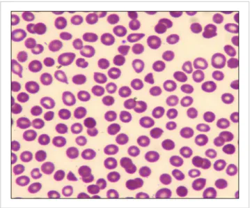
Fig. 1. Peripheral blood smear showing schistocytes and decreased
platelets, characteristics of microangiopathic hemolytic anemia (×500). Fig. 2. Platelet count during the patient’s hospital stay.
A 22-year-old, apparently healthy, Caribbean woman at 39 weeks of gestation was admitted from the outpatient obstetrics clinic for proteinuria and decreased fetal heart rate. She did not have any complications during previous pregnancies. She denied having fever, cough, abdominal pain, headache, blurry vision, recent infection, contacts with sick persons, or experiencing bleeding. Upon physical ex- amination, the patient’s blood pressure was 130/70 mmHg, and her heart rate was 103 beats per minute. She was alert, awake, and oriented. Abdominal examination revealed a non-tender gravid uterus of about 36-week size, and a vagi- nal examination showed a closed cervical os. A few petechiae were noted on the lower extremities. The rest of the physical examination was not significant.
Laboratory tests performed on admission revealed a hemo- globin concentration of 10 g/dL, white blood cell count of 12.3×10³/L, platelet count of 12×103/L, and creatinine concentration of 0.6 mg/dL. Her coagulation profile, fibri- nogen level, and liver function tests were normal, thus ruling out HELLP syndrome and disseminated intravascular coagu- lation (DIC). She had an elevated lactate dehydrogenase level of 560 mg/dL, reticulocyte count of 3.2%, and a very low haptoglobin level (<8 mg/dL). Autoimmune hemolysis was excluded by a normal direct antiglobulin test. A HIV test and her viral hepatitis profiles (hepatitis A, B, and C) were negative. A peripheral blood smear examination re- vealed the presence of occasional schistocytes (2–3 schisto- cytes per high power field) and markedly decreased platelet count (Fig. 1). A presumed TTP diagnosis was made based on these clinical and laboratory findings. Tests for ADAMTS13 activity and ADAMTS13 inhibitor level were sent, and arrangements were made to initiate plasma ex- change (PEX).
The patient underwent a cesarean section and was trans- fused with 2 units of single-donor platelets and 2 units
of fresh frozen plasma during both the pre- and intra-oper- ative periods. She delivered a healthy full-term baby boy without any complications. Daily one-plasma-volume PEX was initiated immediately after delivery. Because of the concern for delayed wound healing, prednisone was not initially started. She was also given folic acid. Her platelet count began to increase on day 3 of PEX. Her hospital course was complicated by healthcare-associated pneumo- nia, and she was also treated with broad-spectrum anti- biotics. The results of the ADAMTS13 activity and ADAMTS13 inhibitor tests were returned on the fifth day of PEX, and were reported as <3% and 1.7 Bethesda units/mL, respectively, confirming the diagnosis of acquired TTP (normal <0.4 Bethesda units/mL). Prednisone (1 mg/kg) was initiated on day 7 of PEX. Platelet count was normalized after two weeks (Fig. 2); the delayed platelet recovery could be due to concurrent infection and medi- cations. The patient was discharged on day 18 of hospital- ization and followed-up with as an outpatient. Due to a slight drop in platelet count, she continued to receive five more PEX sessions in the two weeks following discharge.
Five weeks after her delivery, she was maintaining a stable platelet count and the prednisone was tapered off.
TTP occurring in association with pregnancy was first reported in 1955 by Miner et al. [5]. In a review of 45 cases of pregnancy-associated thrombotic microangiopathies between 1966 and 1988, a maternal mortality of 44% and an associated fetal loss rate of 80% were reported [6].
Historically, TTP had a mortality rate as high as 90% when left untreated [7]. Prompt recognition and initiation of early therapy have drastically reduced the mortality rate to 10–
20% [8].
Pregnancy is a commonly recognized risk factor for trig- gering an acute episode of TTP. The plausible contributing factors include an increase in concentration of procoagulant factors, decrease in fibrinolytic activity, loss of endothelial cell thrombomodulin, and progressive ADAMTS13 defi- ciency over the course of pregnancy [9].
Causes of TTP-HUS during pregnancy include familial
bloodresearch.or.kr Blood Res 2016;51:204-14.
Letters to the Editor 209
TTP (Upshaw-Schulman syndrome, caused by congenital ADAMTS13 deficiency), congenital complement-mediated HUS, and acquired TTP-HUS. In a prospective series of women from the United Kingdom TTP registry, 23 (66%) of 35 pregnant women who presented with TTP were classi- fied as having congenital TTP, and 12 cases (34%) were acquired [10]. In contrast, in an earlier series of 42 patients with a first episode of TTP during pregnancy, only 10 cases (24%) were attributable to congenital TTP [11]. In a larger series of 166 pregnancies, the median time of TTP onset was 23–24 weeks with 12%, 55%, and 33% of TTP occurring in the first, second, and third trimesters, respectively [12].
The classic “pentad” of fever, hemolytic anemia, thrombo- cytopenia, renal impairment, and neurologic manifestations is not always present. It is of utmost importance to have a high clinical vigilance in TTP recognition and treatment.
It is also important to differentiate TTP from other serious pregnancy complications such as DIC and preeclampsia/
eclampsia/HELLP. A complete blood count and review of the peripheral blood smear for microangiopathic hemolytic anemia and thrombocytopenia play a crucial role in the diagnosis of TTP. Coagulation testing should also be done to exclude the possibility of DIC. Additional testing includes a chemistry profile to determine renal function, serum lac- tate dehydrogenase and bilirubin to assess hemolysis, and measuring ADAMTS13 activity, with testing for an inhibitor level (anti-ADAMTS13 autoantibody) if activity is <10%.
PEX is the mainstay for TTP treatment. Once TTP is suspected, PEX should be initiated immediately. PEX re- moves the ADAMTS13 autoantibody and replenishes ADAMTS13 levels. Fresh frozen plasma could be infused to increase ADAMTS13 levels if PEX is not available immediately. A meta-analysis of six randomized controlled trials found that PEX is more effective than plasma infusion in improving overall survival rates [13]. The use of cortico- steroids in TTP is controversial. Further prospective studies are required to define the benefit of the addition of steroids to PEX compared to PEX alone.
Patients with severe ADAMTS13 deficiency exhibit varia- ble clinical courses. Twice daily PEX may be performed in some of the patients who fail to respond to the initial daily PEX or have exacerbation of symptoms. If response to PEX is insufficient, administration of weekly rituximab should be considered in addition to daily PEX. In an analysis of 100 patients with acute refractory or chronic relapsing idiopathic TTP treated with rituximab, complete remission was seen in 98% of patients, while 2% were non-responsive to treatment, and 9% relapsed after complete remission.
Testing positive for ADAMTS13 inhibitor and severe ADAMTS13 deficiency were both found to be highly pre- dictive of response to rituximab [14]. Once a normal platelet count is achieved, PEX may be discontinued abruptly or tapered gradually, and corticosteroids should be quickly tapered.
Many women may subsequently have successful preg- nancy outcomes after recovery from TTP. Women with
congenital TTP have a far higher chance of developing recurrent TTP episodes in subsequent pregnancies unless appropriate prophylaxis with plasma infusion is provided.
On the other hand, the risk of recurrence in women with acquired TTP in subsequent pregnancies is low [8]. In an analysis of 16 pregnancies in 10 women who had recovered from acquired TTP associated with severe acquired ADAMTS13 deficiency (activity <10%), successful full-term deliveries were reported in 13 pregnancies (81%) [15].
Studies suggest that severe ADAMTS13 deficiency or the presence of an inhibitor may be useful in relapse prediction.
The risk of recurrence may be monitored by performing the serial measurements of ADAMTS13 activity during preg- nancy and elective PEX should be initiated and im- munosuppressive treatments could be considered in women with ADAMTS13 deficiency.
In conclusion, although the occurrence of TTP is rare in pregnancy, clinicians should be highly vigilant of its potential presentation in pregnant women with thrombocy- topenia. If TTP is clinically suspected, PEX should be promptly instituted while waiting for the results of ADAMTS13 activity level tests. Because of the potential risk of recurrent TTP, pregnant women with a history of TTP should be monitored carefully by a multidisciplinary team that includes an obstetrician, a hematologist, and a neonatologist.
Aye Min Soe, Nay Min Tun, Elizabeth Guevara, Maxim Shulimovich
Division of Hematology and Oncology, The Brooklyn Hospital Center, New York, NY, USA
Correspondence to: Aye Min Soe Division of Hematology and Oncology, The Brooklyn
Hospital Center, 121 DeKalb Avenue, Brooklyn, New York 11201, USA E-mail: [email protected]
Received on Nov. 11, 2015; Revised on Dec. 12, 2015; Accepted on Jan. 15, 2016 http://dx.doi.org/10.5045/br.2016.51.3.207
AuthorsÊ Disclosures of Potential Conflicts of Interest No potential conflicts of interest relevant to this article were reported.
REFERENCES
1. Moschcowitz E. Hyaline thrombosis of the terminal arterioles and capillaries: a hitherto undescribed disease. Proc N Y Pathol Soc 1924;24:21-4.
2. Levy GG, Nichols WC, Lian EC, et al. Mutations in a member of the ADAMTS gene family cause thrombotic thrombocytopenic purpura. Nature 2001;413:488-94.
3. Terrell DR, Williams LA, Vesely SK, Lämmle B, Hovinga JA, George JN. The incidence of thrombotic thrombocytopenic pur- pura-hemolytic uremic syndrome: all patients, idiopathic pa- tients, and patients with severe ADAMTS-13 deficiency. J
Blood Res2016;51:204-14. bloodresearch.or.kr
210 Letters to the Editor
Thromb Haemost 2005;3:1432-6.
4. Dashe JS, Ramin SM, Cunningham FG. The long-term con- sequences of thrombotic microangiopathy (thrombotic throm- bocytopenic purpura and hemolytic uremic syndrome) in pregnancy. Obstet Gynecol 1998;91:662-8.
5. Miner PE, Nutt RL, Thomas ME. Thrombotic thrombocytopenic purpura occurring in pregnancy. Am J Obstet Gynecol 1955;70:611-7.
6. Weiner CP. Thrombotic microangiopathy in pregnancy and the postpartum period. Semin Hematol 1987;24:119-29.
7. Shepard KV, Bukowski RM. The treatment of thrombotic throm- bocytopenic purpura with exchange transfusions, plasma in- fusions, and plasma exchange. Semin Hematol 1987;24:178-93.
8. Scully M, Hunt BJ, Benjamin S, et al. Guidelines on the diagnosis and management of thrombotic thrombocytopenic purpura and other thrombotic microangiopathies. Br J Haematol 2012;158:
323-35.
9. George JN. The association of pregnancy with thrombotic thrombocytopenic purpura-hemolytic uremic syndrome. Curr Opin Hematol 2003;10:339-44.
10. Scully M, Thomas M, Underwood M, et al. Thrombotic thrombo- cytopenic purpura and pregnancy: presentation, management, and subsequent pregnancy outcomes. Blood 2014;124:211-9.
11. Moatti-Cohen M, Garrec C, Wolf M, et al. Unexpected frequency of Upshaw-Schulman syndrome in pregnancy-onset thrombotic thrombocytopenic purpura. Blood 2012;119:5888-97.
12. Martin JN Jr, Bailey AP, Rehberg JF, Owens MT, Keiser SD, May WL. Thrombotic thrombocytopenic purpura in 166 pregnancies:
1955-2006. Am J Obstet Gynecol 2008;199:98-104.
13. Altuntas F, Aydogdu I, Kabukcu S, et al. Therapeutic plasma ex- change for the treatment of thrombotic thrombocytopenic pur- pura: a retrospective multicenter study. Transfus Apher Sci 2007;36:57-67.
14. Tun NM, Villani GM. Efficacy of rituximab in acute refractory or chronic relapsing non-familial idiopathic thrombotic throm- bocytopenic purpura: a systematic review with pooled data analysis. J Thromb Thrombolysis 2012;34:347-59.
15. Jiang Y, McIntosh JJ, Reese JA, et al. Pregnancy outcomes follow- ing recovery from acquired thrombotic thrombocytopenic purpura. Blood 2014;123:1674-80.
A case of atypical hemolytic uremic syndrome associated with the
c.1273C > T mutation in the complement C3 gene
TO THE EDITOR: Hemolytic uremic syndrome (HUS) is characterized by non-immune hemolytic anemia, thrombo- cytopenia, and renal impairment [1]. The disease typically develops in children under the age of 5 years and is preceded by bloody diarrhea caused by infection associated with Shiga
toxin-producing Escherichia coli or Shiga-like toxin-pro- ducing bacteria (STEC-HUS) [2].
Atypical HUS (aHUS), which comprises 5–10% of HUS cases, is not associated with a prodrome of diarrhea and has a worse prognosis than that caused by STEC-HUS [3].
Uncontrolled complement activation, whether sporadic or familial, plays a major role in the pathogenesis of aHUS.
Genetic abnormalities in the complement system that lead to uncontrolled complement activation have been demon- strated in 60% of aHUS cases [4]. The most common muta- tion in aHUS occurs in complement factor H (CFH), followed by membrane cofactor protein (MCP), complement factor I (CFI), thrombomodulin, and complement component 3 (C3) [3]. So far, only 11 patients with aHUS associated with mutations in CFH, CFI, MCP, and diacylglycerol kinase epsilon have been reported in Korea, owing to the rarity of this syndrome and the lack of a suitable laboratory for its genetic diagnosis [5–7].
C3 mutation occurs in about 4–10% of all aHUS cases [3]. This mutation results in a resistance to C3b inactivation caused either by decreased regulatory binding of MCP, CFH, and CFI to C3, or by increased binding of C3 to complement factor B (CFB) to produce a high amount of C3 convertase [8]. The prognosis of aHUS with a C3 mutation is known to be poor, where about 50% of the cases recur after the initial treatment, and the rate of death or development of end-stage renal disease is about 60% [3].
Although C3 mutations are a well-known etiology of aHUS, the c.1273C>T mutation has never been reported in Caucasians and Koreans. Herein, we report a case of aHUS with C3 mutation in Korea and review of the literature.
CASE
A 66-year-old woman was admitted to the hospital owing to altered mental status. She had previously been diagnosed with systemic lupus erythematosus (SLE) and diabetes melli- tus and was on medications. She was treated with a 1,000 mg intravenous steroid pulse regimen for cerebral infarction and SLE. Five days after steroid treatment, she was dis- charged home with clinical improvement.
A month after the discharge, her mental status became aggravated again and was re-admitted to the department of nephrology under the impression of thrombotic thrombo- cytopenic purpura. Her complete blood count showed a hemoglobin level of 11.2 g/dL, reticulocyte count of 0.2%, platelet count of 60,000 /mm3, and white blood cell count of 13,450 /mm3. The prothrombin time was 12 s, and the activated partial thromboplastin time was 42 s. The periph- eral blood smear revealed many schistocytes. In the blood chemistry, her blood urea nitrogen and creatinine levels were increased to 46.3 mg/dL and 1.25 mg/dL, respectively.
Her total and direct bilirubin levels were 0.58 mm/dL and 0.19 mm/dL, respectively. Her lactated dehydrogenase (LDH) level was elevated to 1,192 U/L. The complement levels (reference ranges in parentheses) were as follows: