ISSN: 2233-601X (Print) ISSN: 2093-6516 (Online)
− 61 −
Received: April 12, 2017, Revised: June 21, 2017, Accepted: June 26, 2017, Published online: February 5, 2018
Corresponding author: Hyung Gon Je, Department of Thoracic and Cardiovascular Surgery, Pusan National University Yangsan Hospital, 20 Geumo-ro, Mulgeum-eup, Yangsan 50612, Korea
(Tel) 82-55-360-2127 (Fax) 82-55-360-2157 (E-mail) [email protected]
© The Korean Society for Thoracic and Cardiovascular Surgery. 2018. All right reserved.
This is an open access article distributed under the terms of the Creative Commons Attribution Non-Commercial License (http://creativecommons.org/
licenses/by-nc/4.0) which permits unrestricted non-commercial use, distribution, and reproduction in any medium, provided the original work is properly cited.
Minimally Invasive Mitral Valve Repair in a Woman with Marfan Syndrome and Type B Dissection
Mi Hee Lim, M.D., Hyung Gon Je, M.D., Sang Kwon Lee, M.D.
Department of Thoracic and Cardiovascular Surgery, Pusan National University Yangsan Hospital, Medical Research Institute of Pusan National University
We report the case of a patient with mitral regurgitation complicated by type B dissection and Marfan syn- drome (MFS) who was managed successfully with minimally invasive mitral valve repair. Without type A aortic dissection or aortic root dilation, MFS patients may develop mitral valve regurgitation, as in this case, and need valve surgery to improve their symptoms and long-term survival. However, it is not clear that a full sternotomy and prophylactic aortic surgery are necessary. Although retrograde perfusion to the dissected aorta is controversial, our approach minimizes the risk of future anticipated aortic surgery in MFS patients.
Key words: 1. Minimally invasive surgical procedures 2. Mitral valve annuloplasty
3. Aortic dissection 4. Marfan syndrome
Case report
Cardiovascular complications, such as aortic rup- ture and mitral valve dysfunction, affect the life ex- pectancy of patients with Marfan syndrome (MFS) [1]. While aortic disease is responsible for most cas- es of sudden premature mortality in adults with MFS, mitral valve dysfunction is the most common cause of morbidity and mortality in MFS.
A 46-year-old woman presented to the emergency department with acute chest pain and shortness of breath (New York Heart Association class II) . A phys- ical examination showed that she was 173 cm tall, weighed 46.9 kg, and had a regular heartbeat with a diastolic murmur. Her mother and 2 brothers had died suddenly of unknown causes and her sister had been diagnosed with valvular heart disease.
Computed tomography angiography (CTA) revealed
acute type B dissection from the left subclavian ar- tery to the iliac bifurcation (Fig. 1). Based on her family history, physical examination, and imaging studies, we suspected MFS and confirmed the diag- nosis of MFS through a genetic study. Transthoracic echocardiography (TTE) and transesophageal echo- cardiography (TEE) revealed severe mitral valve re- gurgitation and no aortic valve regurgitation, with a sinus of Valsalva diameter of 33.5 mm (Fig. 2).
On reconstructed CTA, her right femoral artery was not dissected and branched from the true lumen of the abdominal aorta (Fig. 1). Therefore, we de- cided on a minimally invasive approach with cannu- lation of the right femoral artery and vein. A right thoracotomy was performed with a 6-cm sub- mammary incision and the chest was opened through the fourth intercostal space. When cardiopulmonary bypass (CPB) was initiated, real-time TEE confirmed
Korean J Thorac Cardiovasc Surg 2018;51:61-63 □ CASE REPORT □
https://doi.org/10.5090/kjtcs.2018.51.1.61
Mi Hee Lim, et al
− 62 −
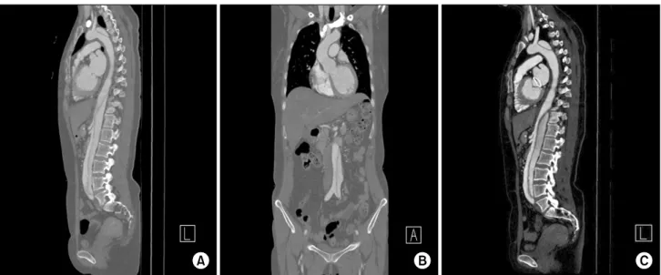
Fig. 1. (A) Initial CT shows type B dissection. (B) The right common iliac artery branched from the true lumen of the dissected abdominal aorta. (C) No significant change was noted in the descending aorta on a CT scan taken 1 year postoperatively. CT, computed tomography.
Fig. 2. (A) Transthoracic echocardiography shows a normal-sized aortic root, sinotubular junction, and ascending aorta. (B) A pre- operative transesophageal echocardiography image shows mitral regurgitation with anterior mitral leaflet prolapse (B). Ao, ascending aorta; LV, left ventricle; LA, left atrium; RV, right ventricle; RVOT, right ventricular outflow tract.
no changes in the descending thoracic aorta and sta- ble perfusion through the true lumen. After trans- thoracic aortic cross-clamping with a Chitwood clamp (Scanlan International Inc., St. Paul, MN, USA) through the third intercostal space, cardioplegic arrest was induced with Custodiol cardioplegia via root cannula- tion. After a left atriotomy through the interatrial groove, the mitral valve was repaired with limited triangular resection of the A3 leaflet and new chor- dae were made with 5-0 Gore-Tex sutures (W. L.
Gore & Associates Inc., Flagstaff, AZ, USA), as de- scribed previously [2]. Mitral annuloplasty was per- formed with a 28-mm Physio ring (Edward Life Science, Irvine, CA, USA).
Weaning from CPB was uneventful. The aortic cross-clamp time was 63 minutes and the CPB time was 94 minutes. The patient was extubated in the operating room and recovered uneventfully. A TTE on the second postoperative day showed trivial re- sidual mitral regurgitation, and the patient was dis-
Minimal MVP for MFS
− 63 − charged without complications the next day. No sig- nificant changes were observed in the descending aorta on a computed tomography scan taken 1 year after the operation (Fig. 1).
Discussion
Our patient presented with acute type B aortic dis- section and had a family history of sudden death. In addition, the patient’s relatively young age increased the possibility of aortic growth and aortic events during follow-up [3]. Prophylactic aortic surgery via a median sternotomy was inappropriate because the ascending aorta in our patient was only 30 mm in diameter.
With this clinical information, we planned mini- mally invasive mitral valve repair to avoid a full ster- notomy, considering the possibility of future aortic surgery, based on the site of peripheral cannulation, and repair over replacement of the mitral valve.
Minimally invasive mitral valve surgery in patients with MFS has been reported to be feasible [4].
Regarding the site of arterial cannulation for type B dissection patient, we typically use antegrade per- fusion via the axillary artery. In this case, however, we chose right femoral cannulation to preserve the axillary artery for future aortic surgery. Although femoral arterial cannulation is usually contraindicated for aortic dissection, we obtained retrograde perfu- sion with careful surveillance using intraoperative TEE. Intraoperative TEE is helpful for the early rec- ognition and proper treatment of iatrogenic aortic
dissection, particularly in patients with collagen vas- cular diseases such as MFS [5]. We delayed the oper- ation for 4 weeks to stabilize the dissected aorta, and 3 serial CTAs showed no interval changes in the dissected flap.
In conclusion, minimally invasive mitral valve re- pair is an option for patients with MFS with type B aortic dissection. Intraoperative TEE surveillance is indispensable for this approach.
Conflict of interest
No potential conflict of interest relevant to this ar- ticle was reported.
References
1. Silverman DI, Burton KJ, Gray J, et al. Life expectancy in the Marfan syndrome. Am J Cardiol 1995;75:157-60.
2. Park JM, Je HG, Lee SK. Single-suture neochorda-folding plasty for mitral regurgitation. Korean J Thorac Cardiovasc Surg 2016;49:70-2.
3. Van Bogerijen GH, Tolenaar JL, Rampoldi V, et al.
Predictors of aortic growth in uncomplicated type B aortic dissection. J Vasc Surg 2014;59:1134-43.
4. Kato H, Ohtake H, Yamaguchi S, et al. Minimally invasive cardiac surgery for a young woman with Marfan syndrome and mitral regurgitation. Gen Thorac Cardiovasc Surg 2010;58:531-3.
5. Vollroth M, Seeburger J, Garbade J, Borger MA, Misfeld M, Mohr FW. Conversion rate and contraindications for mini- mally invasive mitral valve surgery. Ann Cardiothorac Surg 2013;2:853-4.