ISSN: 2233-601X (Print) ISSN: 2093-6516 (Online)
Department of Thoracic and Cardiovascular Surgery, Konkuk University Medical Center, Konkuk University School of Medicine Received: September 16, 2014, Revised: October 28, 2014, Accepted: November 7, 2014, Published online: October 5, 2015 Corresponding author: Je Kyoun Shin, Konkuk University Medical Center, 120-1 Neungdong-ro, Gwangjin-gu, Seoul 05030, Korea
(Tel) 82-2-2030-7595 (Fax) 82-2-2030-5009 (E-mail) [email protected]
C
The Korean Society for Thoracic and Cardiovascular Surgery. 2015. All right reserved.
CC
This is an open access article distributed under the terms of the Creative Commons Attribution Non-Commercial License (http://creative- commons.org/licenses/by-nc/4.0) which permits unrestricted non-commercial use, distribution, and reproduction in any medium, provided the original work is properly cited.
Postoperative Outcomes of Mitral Valve Repair for Mitral Restenosis after Percutaneous Balloon Mitral Valvotomy
Seong Lee, M.D., Hyun Keun Chee, M.D., Jun Seok Kim, M.D., Myong Gun Song, M.D., Jae Bum Park, M.D., Je Kyoun Shin, M.D.
Background: There have been a number of studies on mitral valve replacement and repeated percutaneous mitral balloon valvotomy for mitral valve restenosis after percutaneous mitral balloon valvotomy. However, studies on mitral valve repair for these patients are rare. In this study, we analyzed postoperative outcomes of mitral valve repair for mitral valve restenosis after percutaneous mitral balloon valvotomy. Methods: In this study, we assessed 15 pa- tients (mean age, 47.7±9.7 years; 11 female and 4 male) who underwent mitral valve repair between August 2008 and March 2013 for symptomatic mitral valve restenosis after percutaneous mitral balloon valvotomy. The mean in- terval between the initial percutaneous mitral balloon valvotomy and the mitral valve repair was 13.5±7 years. The mean preoperative Wilkins score was 9.4±2.6. Results: The mean mitral valve area obtained using planimetry in- creased from 1.16±0.16 cm
2to 1.62±0.34 cm
2(p=0.0001). The mean pressure half time obtained using Doppler ul- trasound decreased from 202.4±58.6 ms to 152±50.2 ms (p=0.0001). The mean pressure gradient obtained using Doppler ultrasound decreased from 9.4±4.0 mmHg to 5.8±1.5 mmHg (p=0.0021). There were no early or late deaths. Thromboembolic events or infective endocarditis did not occur. Reoperations such as mitral valve repair or mitral valve replacement were not performed during the follow-up period (39±16 months). The 5-year event-free survival was 56.16% (95% confidence interval, 47.467–64.866). Conclusion: On the basis of these results, we could not conclude that mitral valve repair could be an alternative for patients with mitral valve restenosis after percutaneous balloon mitral valvotomy. However, some patients presented with results similar to those of mitral valve replacement. Further studies including more patients with long-term follow-up are necessary to determine the possibility of this application of mitral valve repair.
Key words: 1. Restenosis 2. Mitral valve, repair
3. Percutaneous mitral balloon valvotomy 4. Complication
INTRODUCTION
Percutaneous mitral balloon valvotomy (PMV) has been used widely for mitral stenosis since it was introduced in 1984 by Inoue et al. [1]. Further, symptomatic mitral valve restenosis after PMV occurs at the rate of 7% to 23% [2,3].
Treatment modalities in these cases include mitral valve re-
placement and repeated PMV. With respect to repeated PMV,
recent studies have presented good intermediate and long-term
outcomes [4,5]. Nevertheless, mitral valve replacement is usu-
ally preferred [6,7]. A number of studies have demonstrated
the possibility of mitral valve repair for rheumatic mitral
http://dx.doi.org/10.5090/kjtcs.2015.48.5.328
Table 1. Preoperative baseline characteristics of patients
Variable Value
Age (yr) 47±10 (31–64)
Sex (male/female) 4/11
Interval time (yr) between PMV and mitral valvuloplasty
13±7 (3–25)
Functional class (New York Heart Association)
I 0
II 6 (40)
III 8 (53.3)
IV 1 (6.7)
Rhythm
Atrial fibrillation 9 (60)
Previous thromboembolism 2 (13.3)
Left atrial thrombosis 2 (13.3)
Second PMV 4 (26.7)
Echocardiographic score (Wilkins score) 9.4±2.6 Left atrial dimension (mm) 56.9±12.3 Mitral regurgitation
Grade 1 3 (20)
Grade 2 2 (13.3)
Grade 3 0
Grade 4 0
Tricuspid regurgitation
Grade 1 5 (33.3)
Grade 2 3 (20)
Grade 3 0
Grade 4 0
Values are presented as mean±standard deviation (range), num- ber (%), or mean±standard deviation.
PMV, percutaneous balloon mitral valvotomy.
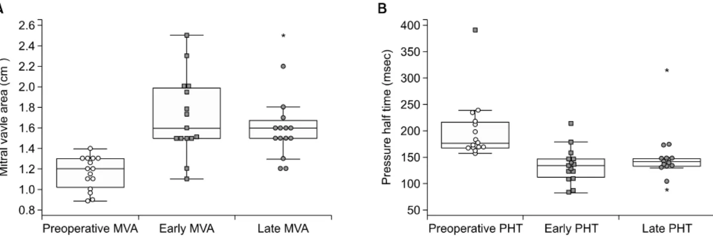
Fig. 1. (A) Comparison of preoperative and postoperative mitral valve area. (B) Comparison of preoperative and postoperative pressure half time. MVA, mitral valve area; PHT, pressure half time.
valve diseases, which were mostly mixed with stenosis and regurgitation [8-14]. However, mitral valve repair for rheu- matic mitral valve stenosis is still uncommon. Accordingly, few reports on mitral valve repair for mitral valve restenosis after PMV have been published. In this study, we assessed postoperative outcomes of mitral valve repair for symptomatic mitral valve restenosis after PMV.
METHODS
Between August 2008 and March 2013, 22 patients under- went mitral valve surgery for mitral valve disease at our cen- ter after they had undergone PMV for rheumatic mitral valve stenosis at different hospitals. Patients were excluded if they had significant (>grade 2) mitral regurgitation because this factor could affect the analyses by confounding variables and we were concerned about mitral valve restenosis after PMV.
Three patients did not undergo follow-up echocardiography.
In the end, 15 patients formed the subject population of this study. Preoperative baseline characteristics are given in Table 1.
Mitral valve replacement was recommended to eight patients (53.3%) at different centers before visits to our center. The mean preoperative left ventricular ejection fraction was 59.5%
±7.9%. The mean mitral valve area was 1.15±0.16 cm
2(Fig.
1A), and the mean pressure half time was 202.4±58.6 ms (Fig. 1B).
Right anterolateral thoracotomy was performed in 13 pa-
tients as a standard approach of surgery for mitral valve dis-
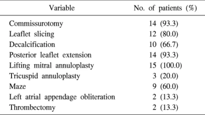
Table 2. Surgical technique for mitral valve repair and concomitant procedures
Variable No. of patients (%)
Commissurotomy 14 (93.3)
Leaflet slicing 12 (80.0)
Decalcification 10 (66.7)
Posterior leaflet extension 14 (93.3) Lifting mitral annuloplasty 15 (100.0)
Tricuspid annuloplasty 3 (20.0)
Maze 9 (60.0)
Left atrial appendage obliteration 2 (13.3)
Thrombectomy 2 (13.3)
Fig. 2. Comparison of preoperative and postoperative New York Heart Association (NYHA) functional class.
ease or tricuspid valve disease at our center. Median sternot- omy was performed in two patients who had very moderate forced expiratory volume to achieve one-lung ventilation.
Each valve was assessed in direct vision and via intraope- rative transesophageal echocardiography. All operations were performed by the same surgeon. Surgical techniques utilized for mitral valve repair and concomitant procedures are listed in Table 2. Posterior leaflet extension was performed with bovine pericardium (Supple Peri-Guard Repair Patch, 6×8 cm Synovis; Jisang International Inc., Seoul, Korea). Lifting mi- tral annuloplasty was performed with a mitral strip (Mitracon, Mitral Annuloplasty Ring, Mitracon Strip; Sciencity Co. Ltd., Seoul, Korea). Tricuspid annuloplasty was performed with a Carpentier- Edward ring.
Demographic clinical variables were collected retrospec- tively with a review of medical records. The optimal echo- cardiographic outcome was defined as a mitral valve area of more than 1.5 cm
2and a pressure half time of less than 150 ms. Event-free survival means that in addition to these hemo- dynamic parameters, the New York Heart Association func- tional class (NYHA Fc) was less than III without reoperation events such as mitral valve repair and mitral valve replace- ment [4,5].
Statistical analyses were performed with PASW SPSS ver.
18.0 (SPSS Inc., Chicago, IL, USA). Categorical variables are expressed as percentages or numbers, and continuous varia- bles are expressed as means with standard deviations. After testing for the normality of distribution, continuous variables were compared using the paired samples t-test or the Wilcoxon signed-rank test. The cumulative event-free survival curve
was determined according to the Kaplan–Meier method. All p-values of less than 0.05 were considered statistically significant.
RESULTS
All patients underwent follow-up for a mean duration of 39±16 months. There were no early or late deaths. One pa- tient suffered from postoperative bleeding due to intercostal artery injury. There were no morbidities such as thromboem- bolic events, infective endocarditis, reoperations, or complica- tions with respect to the use of warfarin during the follow-up period. Atrial fibrillation persisted in two of the nine patients who underwent the maze procedure. Thirteen patients pre- sented with a sinus rhythm. The functional capacity of 12 pa- tients (80%) improved from NYHA Fc II to NYHA Fc I.
Three patients (20%) improved from NYHA Fc III or IV to NYHA Fc II (Fig. 2). Postoperatively, 13 patients were treat- ed with warfarin for 2 months. Two patients with persistent atrial fibrillation were treated with aspirin alone after cessa- tion of warfarin treatment. Two patients who had atrial thro- mbosis preoperatively were treated with warfarin for 6 months.
When echocardiography revealed no atrial thrombosis 6 months later, these two patients were treated with aspirin alone. The range of the target international normalized ratio was 2.0 to 3.0.
1) Early postoperative echocardiographic results
In a comparison of early postoperative echocardiographic
results with preoperative echocardiographic results, we found
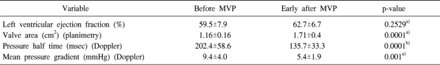
Table 3. Comparison of preoperative with early echocardiographic results after MVP
Variable Before MVP Early after MVP p-value
Left ventricular ejection fraction (%) 59.5±7.9 62.7±6.7 0.2529
a)Valve area (cm
2) (planimetry) 1.16±0.16 1.71±0.4 0.0001
a)Pressure half time (msec) (Doppler) 202.4±58.6 135.7±33.3 0.0001
b)Mean pressure gradient (mmHg) (Doppler) 9.4±4.0 5.4±1.9 0.001
a)Values are presented as mean±standard deviation.
MVP, mitral valve repair.
a)
By paired samples t-test.
b)By Wilcoxon signed-rank test.
Table 4. Comparison of preoperative with late echocardiographic results after MVP
Variable Before MVP Late after MVP p-value
Left ventricular ejection fraction (%) 59.5±7.9 64.5±4.9 0.0276
a)Valve area (cm²) (planimetry) 1.16±0.16 1.62±0.34 0.0001
b)Pressure half time (msec) (Doppler) 202.4±58.6 152±50.2 0.0001
b)Mean pressure gradient (mmHg) (Doppler) 9.4±4.0 5.8±1.5 0.0021
a)Values are presented as mean±standard deviation.
MVP, mitral valve repair.
a)