ISSN: 2233-601X (Print) ISSN: 2093-6516 (Online)
Department of Thoracic and Cardiovascular Surgery, University of Ulsan College of Medicine
Received: May 13, 2015, Revised: June 23, 2015, Accepted: June 23, 2015, Published online: June 5, 2016
Corresponding author: Seung-Il Park, Department of Thoracic and Cardiovascular Surgery, Asan Medical Center, University of Ulsan College of Medicine, 88 Olympic-ro 43-gil, Songpa-gu, Seoul 05505, Korea
(Tel) 82-2-3010-3904 (Fax) 82-2-3010-6966 (E-mail) [email protected]
C
The Korean Society for Thoracic and Cardiovascular Surgery. 2016. All right reserved.
CC
This is an open access article distributed under the terms of the Creative Commons Attribution Non-Commercial License (http://creative- commons.org/licenses/by-nc/4.0) which permits unrestricted non-commercial use, distribution, and reproduction in any medium, provided the original work is properly cited.
Clinical Outcomes of Heart-Lung Transplantation: Review of 10 Single-Center Consecutive Patients
Jae Kwang Yun, M.D., Se Hoon Choi, M.D., Seung-Il Park, M.D., Asan Medical Center Heart-Lung Transplantation Team
Background: Heart-lung transplantation (HLT) has provided hope to patients with end-stage lung disease and irre- versible heart dysfunction. We reviewed the clinical outcomes of 10 patients who underwent heart-lung trans- plantation at Asan Medical Center. Methods: Between July 2010 and August 2014, a total of 11 patients under- went HLT at Asan Medical Center. After excluding one patient who underwent concomitant liver transplantation, 10 patients were enrolled in our study. We reviewed the demographics of the donors and the recipients’ baseline in- formation, survival rate, cause of death, and postoperative complications. All patients underwent follow-up, with a mean duration of 26.1±16.7 months. Results: Early death occurred in two patients (20%) due to septic shock.
Late death occurred in three patients (38%) due to bronchiolitis obliterans (n=2) and septic shock (n=1), although these patients survived for 22, 28, and 42 months, respectively. The actuarial survival rates at one year, two years, and three years after HLT were 80%, 67%, and 53%, respectively. Conclusion: HLT is a procedure that is rarely performed in Korea, even in medical centers with large heart and lung transplant programs. In order to ach- ieve acceptable clinical outcomes, it is critical to carefully choose the donor and the recipient and to be certain that all aspects of the transplant procedure are planned in advance with the greatest care.
Key words: 1. Heart-lung transplantation 2. Complication
3. Mortality
INTRODUCTION
Since the first case of heart-lung transplantation (HLT) was conducted at Stanford University in 1981 [1], 3,755 adult HLTs (including re-transplantation) have been performed worldwide as of June 2013 [2]. Initially, the majority of HLTs were performed for pulmonary vascular disease and cystic fibrosis, which had previously been treated with lung transplantation alone. Recently, the major indications for adult HLT have been expanded to include congenital heart disease
and idiopathic pulmonary artery hypertension [2]. HLT recipi- ents have survival rates of 63% at one year, 52% at three years, and 45% at five years [2].
In Korea, a total of 17 HLTs have been performed be- tween July 2010 and August 2014, 11 of which were per- formed at Asan Medical Center, according to the official re- port of the Korean Network for Organ Sharing (http://www.
konos.go.kr). However, the survival rate of the Korean HLT recipients has not yet been investigated due to the low num- ber of cases of HLT. Therefore, the purpose of this study
http://dx.doi.org/10.5090/kjtcs.2016.49.3.157
was to evaluate the clinical outcomes of HLT at a single medical center over five years.
METHODS 1) Patients and surgical indications
Our study population consisted of all patients who under- went HLT between July 2010 and August 2014 at Asan Medical Center in Seoul, Korea. Both pediatric and adult pa- tients were included. The patients were sorted through our prospectively maintained HLT registry database containing clinical information regarding the recipients and donors.
Clinical outcomes including deaths and complications follow- ing HLT were meticulously reviewed using the patients’ elec- tronic medical records of Asan Medical Center. The in- dications for HLT have changed over time. Initially, pulmo- nary hypertension with congenital or acquired heart disease were the main indications for HLT. As time passed, the in- dications expanded to include end-stage lung disease with pulmonary hypertension (HTN) caused by right heart failure.
For all patients, the decision to perform concomitant HLT was confirmed by a team of both cardiologists and pulmonol- ogists, who have performed 260 cases of heart transplantation and 40 cases of lung transplantation over 15 years. This study was approved by the ethics committee of institutional review board of Asan Medical Center. And the requirement for informed consent was waived.
2) Donor selection, organ procurement, and transplant technique
We have paid special attention to manage the donor organs properly from the donor selection stage. Techniques used to assess organ quality included chest radiography, arterial blood gas determination, bronchoscopy, echocardiography, and visu- al inspection of the heart and lungs [3,4]. We tried to per- form flexible bronchoscopy in every case to evaluate the air- ways and to remove retained secretion.
The donor and recipient operations were performed using previously described techniques [5]. Following sternotomy, we opened mediastinal pleura of both side and carefully ex- amined the condition of donor’s lung. If there was atelectasis, we requested to adjust the ventilation setting to expand the
lungs fully. After dissection of the trachea, the superior vena cava (SVC), inferior vena cava (IVC), ascending aorta, and main pulmonary artery, cannulae were placed in order to pro- vide preservative solutions to heart and lung. Before aorta cross-clamping, 25,000 units of heparin were administered in- travenously and prostaglandin E1 (500 μg) was administered into the main pulmonary artery. When the systolic blood pressure dropped to under 90 mmHg following the admin- istration of prostaglandin E1, aortic cross-clamping was car- ried out and cardioplegic infusion was started. The left atrial appendage was amputated in order to ensure complete empty- ing of the heart. The heart was preserved using 2 L of histi- dine-tryptophan-ketoglutarate solution (Custodiol HTK;
Essential Pharmaceuticals, Newtown, PA, USA) infused through the aortic root cannula. The lungs were preserved us- ing antegrade pulmoplegia with 5 L of low-potassium dextran solution (Perfadex; Vitrolife AB, Kungsbacka, Sweden) through the main pulmonary artery, with venting through the IVC and LA auricle. If the cold ischemic time was predicted to be more than 120 minutes, 1 L of Perfadex solution was infused again into the lung allograft at recipient’s operation room before anastomosis. At the time of tracheal stapling, the lungs were inflated enough to prevent atelectasis. Cold HTK solution was used for graft storage during returning to recipi- ent’s operation room.
Standard median sternotomy was performed in eight recipi- ents, while two patients underwent the clamshell approach due to severe adhesion. We used standard cardiopulmonary bypass (CPB) strategy including aortic and bicaval cannula- tions, and tried to cannulate the aorta, the SVC, and the IVC as distally as possible. After the initiation of CPB and cool- ing using ice slush, recipient cardiectomy and bilateral pneu- monectomies were completed immediately after the arrival of the donor heart-lung bloc. The tracheal anastomosis was done first using a running polydioxanone 4-0 suture (PDS II;
Johnson & Johnson K.K., Seoul, Korea) on the posterior side,
and intermittent suturing was later performed on the anterior
side using the same suture material. Next, the aortic anasto-
mosis was performed. After the cross-clamp was removed us-
ing standard de-airing maneuvers, 500 mg of methyl-
prednisolone was administered intravenously. Anastomosis of
the SVC was performed, followed by anastomosis of the
IVC. After weaned off from CPB, routinely 1 pericardial soft drain (Hemovac Evacuators; Zimmer, Warsaw, IN, USA), 1 mediastinal tube, and 2 pleural chest tubes in each side were placed. In every case, a pair of ventricular pacing wires was placed also.
3) Antimicrobial prophylaxis, immunosuppression, and postoperative management
As a prophylactic antimicrobiotics, cefepime was intra- venously administered in patients who were reported as spu- tum-culture negative, or the appropriate antibiotics was chos- en according to the result of the donor and recipient sputum cultures. For antiviral prophylaxis, Intravenous ganciclovir was given at a dose of 5 mg/kg every 24 hours from the first week to fourth week after the HLT, followed by 900 mg of oral valganciclovir once daily for six months. As antifungal prophylaxis, Voriconazole was intravenously administered 4 mg/kg every 12 hours with the target trough level of 1.5–5.5 mg/dL. After the recipient resumed normal diet, oral vor- iconazole was given. The total duration of voriconazole pro- phylaxis was six months. Cytomegalovirus and Aspergillus antigenemia assays were performed regularly for up to six months after HLT. Trimethoprim-sulfamethoxazole was then administered to prevent Pneumocystis jirovecii pneumonia throughout the patient’s life.
In all patients, induction of immune suppression was made using anti-interleukin-2 receptor monoclonal antibody (basiliximab) and mycophenolate mofetil (CellCept; Roche Laboratories, Nutley, NJ, USA). Basiliximab was intra- venously administered one hour before the surgery and on the fourth post-transplant day at a dose of 20 mg in patients weighing over 35 kg. Mycophenolate mofetil was started pre- operatively and continued following surgery at a dose of 1.0 g every 12 hours, with a target trough level of 2 μg/mL (range, 1 to 3 μg/mL).
Postoperative immunosuppression consisted of a tri- ple-maintenance therapy based on steroids, mycophenolate mofetil, and a calcineurin inhibitor. Among the calcineurin in- hibitors, tacrolimus (FK506) was used first and cyclosporine (CsA) was used as a second choice when complications re- lated to tacrolimus occurred. Tacrolimus was begun on the second postoperative day at a dose of 0.05–0.1 mg/kg for 24
hours, and the target continuing level was 10–15 ng/mL for six months and 8–12 ng/mL after three days. If CsA was used rather than tacrolimus, it was usually administered on the second postoperative day at an initial dose of 2–3 mg/kg or half the dose as a four-hour infusion twice a day, and was continued until the patient was able to take oral medication.
A 500-mg dose of methylprednisolone was given intra- operatively after weaning of CPB. Two additional post- operative doses were given (1 mg/kg every 12 hours for 24 hours) until the day after the operation, and a half dose was then given for two weeks, based on the assumption that this would decrease the incidence of bronchial anastomotic complications. After two weeks, steroids were changed to oral prednisone with a daily dosage of 0.6 mg/kg, tapering to 0.2 mg/kg/day at six weeks and 0.1 mg/kg/day at six months.
All patients were required to undergo follow-up at our transplant outpatient clinic, which is associated with the de- partment of pulmonology and critical care medicine. Peak ex- piratory flow was calculated daily in all patients using a port- able peak flow meter to detect acute rejection or early lung injury. Routine spirometry was performed during the first six months after transplantation. Thereafter, bronchoscopies were only performed for special indications such as infection or rejection. Acute rejection was diagnosed by chest computed tomography and bronchoscopy if pulmonary function decreased. Chronic rejection, including restrictive allograft syndrome or bronchiolitis obliterans syndrome (BOS), was al- so made using concurrent forced vital capacity and forced ex- piratory volume in one second [6].
4) Surveillance and treatment of rejection
The patients did not undergo surveillance endomyocardial biopsies, but did have a routine transthoracic echocardiogram once a week until discharge and two, four, six, and 12 months after discharge.
The surveillance bronchoscopy was routinely performed at
two, four, six, and 12 months and annually thereafter. Either
endomyocardial or lung biopsies were obtained only when re-
jection was suggested clinically. Patients were followed up at
the out-patient clinic of the department of pulmonology and
critical care medicine. In cases of the patients showed signs
of moderate or severe rejection, intravenous methylpredni-
Table 1. Demographic and clinical characteristics of the heart-lung transplant recipients
Characteristic Value
Age (yr) 44 (5–62)
Gender (male) 6 (60)
Disease leading to transplantation
Interstitial lung disease 3 (30)
Toxic ARDS 2 (20)
Ischemic cardiomyopathy with ARDS 1 (10) Dilated cardiomyopathy with pulmonary HTN 1 (10) Atrial septal defect with pulmonary HTN 1 (10) Bronchiectasis with corpulmonale 1 (10)
Angiosarcoma 1 (10)
Preoperative extracorporeal membrane oxygenation support
Patients 6 (60)
Duration (day) 14.5 (2 –100)
Preoperative mechanical ventilation support
Patients 8 (80)
Duration (day) 20.5 (2–104)
Wait for transplantation
Duration (day) 31.5 (3 –180)
Values are presented as median (interquartile range) or num- bers of patients (%), unless otherwise indicated.
ARDS, acute respiratory distress syndrome; HTN, hypertension.
Table 2. Donor demographics
Variable Value
Donor age (yr) 29.1±13.7 (range, 11–52)
10–19 2 (20)
20 –29 5 (50)
30 –39 1 (10)
50–59 2 (20)
Gender (male/female) 5/5
Cause of death
Cerebrovascular accident 4 (40)
Head trauma 3 (30)
Hanging 2 (20)
Traffic accident 1 (10)
Location within Korea
Seoul (≤3 hr of transportation time) 5 (50) Gyeonggi-do, Jeolla-do, Daejeon
(3 –4 hr of transportation time)
4 (40) Ulsan (5–6 hr of transportation time) 1 (10)
Values are presented as mean±standard deviation or numbers of patients (%), unless otherwise indicated.
solone (1 g/day for three days) was injected, followed by a two-week tapering course of prednisone. Persistent rejection was treated with an additional course of steroids followed by everolimus and low-dose cyclosporine.
5) Statistical analysis
Statistical analysis was performed using IBM SPSS soft- ware ver. 22.0 (IBM Co., Armonk, NY, USA). Data are shown as the mean plus or minus the standard error of the mean. The overall survival included all deaths from any cause during the follow-up period, whereas live patients were right-censored at the last available follow-up.
RESULTS 1) Patient demographics
Between July 2010 and August 2014, a total of 11 patients underwent HLT in our medical institution, one of whom was excluded due to undergoing simultaneous heart, lung, and liv-
er transplantation. Table 1 shows the demographic and clin- ical characteristics of these patients. The median patient age was 44 years, ranging from five to 62 years. The reasons for HLT were interstitial lung disease with rapidly deteriorating pulmonary function (n=3), toxic acute respiratory distress syn- drome (n=2), atrial septal defect with pulmonary HTN (n=1), dilated cardiomyopathy with pulmonary HTN (n=1) with a previous heart transplantation, ischemic cardiomyopathy with acute respiratory distress syndrome (n=1), angiosarcoma (n=1), and bronchiectasis (n=1). The mean patient height was 165 cm (range, 109 to 170 cm), and the mean weight was 62 kg (range, 18 to 74 kg). Eight patients (80%) were on pre- operative ventilatory support, the median duration of which was 20.5 days (range, 2 to 104 days). Six patients (60%) re- quired extracorporeal membrane oxygenation (ECMO) therapy preoperatively, the median duration of which was 14.5 days, (range, 2 to 100 days). The median waiting time from regis- tration to transplantation was 31.5 days (range, 3 to 180 days). The mean follow-up time was 26.1±16.7 months.
2) Organ donor and transplant operations
Five patients were female and five were male. The mean
donor age was 29.1±13.7 years (range, 11 to 52 years) (Table 2).
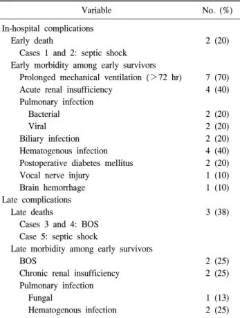
Table 3. Complications and causes of death
Variable No. (%)
In-hospital complications
Early death 2 (20)
Cases 1 and 2: septic shock Early morbidity among early survivors
Prolonged mechanical ventilation (>72 hr) 7 (70)
Acute renal insufficiency 4 (40)
Pulmonary infection
Bacterial 2 (20)
Viral 2 (20)
Biliary infection 2 (20)
Hematogenous infection 4 (40)
Postoperative diabetes mellitus 2 (20)
Vocal nerve injury 1 (10)
Brain hemorrhage 1 (10)
Late complications
Late deaths 3 (38)
Cases 3 and 4: BOS Case 5: septic shock
Late morbidity among early survivors
BOS 2 (25)
Chronic renal insufficiency 2 (25)
Pulmonary infection
Fungal 1 (13)
Hematogenous infection 2 (25)
BOS, bronchiolitis obliterans syndrome.
The causes of brain death were cerebrovascular accident (40%), head trauma (30%), hanging (20%), and traffic acci- dent (10%). The median total ischemic time of the donor har- vest was 192 minutes (range, 100 to 313 minutes). The me- dian warm ischemic time was 89 minutes (range, 69 to 127 minutes), and the cold ischemic time was 104 minutes (range, 28 to 245 minutes). Five donors were in Seoul (mean ische- mic time of less than three hours) and four donors were in Gyeonggi-do, Jeolla-do, and Daejeon (mean ischemic time of less than four hours). One donor was located in Ulsan, which led to an ischemic time of 331 minutes. During the HLT, the median surgery and CPB times were 587 minutes (range, 384 to 663 minutes) and 262 minutes (range, 181 to 439 mi- nutes), respectively, and the median duration of aortic cross- clamping was 140 minutes (range, 128 to 173 minutes).
3) Morbidity and mortality
The morbidity and mortality data are presented in Table 3.
Early mortality (less than 30 days following surgery or in-hospital mortality) occurred in 20% (n=2) of the patients and was caused by septic shock. One of the patients who was diagnosed with dilated cardiomyopathy underwent heart transplantation 15 days before HLT. On postoperative day 1, the patient required ECMO support for right heart failure, and progressive pulmonary HTN made it impossible to wean the patient from ECMO. After two episodes of bleeding control surgery, the patient expired nine days later due to septic shock. The other patient underwent ECMO–assisted emer- gency coronary artery bypass grafting surgery due to an acute myocardial infarction three months before HLT. After post- operative bleeding control, acute renal failure and acute respi- ratory distress syndrome also took place, followed by con- tinuous renal replacement therapy and tracheostomy. The pa- tient underwent HLT, although the total ischemic time of the donor heart-lung bloc was 313 minutes. Eventually, the pa- tient suffered from low cardiac output syndrome and expired 31 days following the surgery due to septic shock.
The immediate postoperative course was complicated in one patient (10%) by excessive bleeding that required re-ster- notomy within the first 24 hours. The bleeding focus of the patient was proven to be from the collateral vessels in the posterior mediastinum or in the chest wall. The most com- mon postoperative complication was respiratory failure requir- ing prolonged mechanical ventilation (defined as assisted ven- tilation for >72 hours or reintubation), which occurred in seven early survivors (70%), all of whom required tracheo- stomy. The median ventilation support time was 12 days (range, 3 to 87 days). Acute renal failure requiring dialysis occurred in four patients (40%), three of whom already re- quired continuous postoperative renal replacement therapy.
The median length of the intensive care unit (ICU) stay among the survivors was 31 days (range, 5 to 124 days), and the median length of their hospital stay was 88 days (range, 42 to 379 days). Eight early survivors among the transplant recipients were discharged from the hospital. No prolonged air leak or acute graft failure was observed among the early survivors.
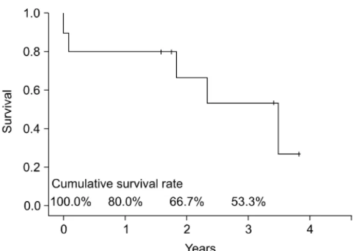
Late mortality (after postoperative day 30) occurred in 38%
(3/8) of the remaining patients. The causes of late mortality
included chronic allograft failure as a result of BOS (n=2)
Fig. 1. Overall survival curves after heart-lung transplantation, us- ing the Kaplan-Meier method.
and septic shock (n=1). The survival duration of these pa- tients was 22, 28, and 42 months, respectively. The actuarial survival rates at one, two, and three years after HLT were 80%, 67%, and 53%, respectively (Fig. 1). The common late complications were BOS in two patients (25%), chronic renal failure in two patients (25%), and hematogenous infection in two patients (25%). All survivors were in New York Heart Association class I or II after discharge.
DISCUSSION
In this study we presents the clinical outcomes of HLTs performed at the Asan Medical Center over a five-year peri- od, with acceptable short- and mid-term survival rates. The survival rates at one year and three years in this study were 80% and 53%, whereas they were 63% and 52% in the (ISHLT) study [2]. According to the ISHLT study, the most common causes of death during the first 30 days after heart-lung transplantation were graft failure, technical compli- cations, and non-cytomegalovirus infections. In late period, BOS/late graft failure (lung or heart) and non-cytomegalovi- rus infections were the common causes of death [2]. Due to the low number of cases, we were unable to analyze the cause of early and late deaths in order to determine statisti- cally significant trends in the cause of death. However, the leading causes of early and late mortality were septic shock and BOS.
During the last 34 years, HLT has provided hope to a
large number of patients and a great benefit to many.
According to the International Society for Heart and Lung Transplantation study [2], the major indications for HLT are congenital heart disease (35.5%), idiopathic primary pulmo- nary hypertension (27.4%), cystic fibrosis (13.9%), and ac- quired heart disease (5.4%). The decision to list patients with idiopathic primary pulmonary hypertension for HLT rather than for bilateral lung transplantation was made depending on the right heart function and its predicted recovery after iso- lated lung transplantation [7]. However, until recently it has been difficult to assess the reversibility of right ventricular function in patients with right heart failure due to the lack of definite criteria. Consequently, the decision to perform HLT was made by experienced cardiologists who had specialized in performing heart transplantation for 20 years in con- sultation with pulmonologists based on the echocardiogram, cardiac computed tomography, and/or information derived from cardiac catheterization. According to the Asan Medical Center protocol, six patients with end-stage lung disease un- derwent HLT rather than isolated lung transplantation due to moderate-to-severe right heart failure with pulmonary HTN, although the ejection fraction was sometimes within the nor- mal range. Three patients had heart failure (congenital or ac- quired) and their pulmonary function was irreversibly aggravated.
Chronic allograft rejection after lung transplantation is manifested by ‘constrictive’ BOS and is physiologically char- acterized by progressive expiratory airflow obstruction seen on pulmonary function testing [7]. With regard to HLT, the lung grafts seem to protect the heart such that pulmonary in- filtration usually precedes or prevents myocardial infiltration [8].
Meanwhile, as the heart does not provide immunoprotective effects for the lungs, HLT recipients exhibit BOS at a rate similar to lung transplantation recipients [9]. Therefore, BOS is a major factor limiting long-term survival after HLT as well as lung transplantation, affecting approximately one third of all patients within three years and half of all patients with- in five years [10]. In the past, BOS was considered an irre- versible and incurable condition, whereas azithromycin can now improve airflow limitation in a significant proportion of patients with even long-standing BOS [11]. Tacrolimus con- version from CsA [12] or extracorporeal photopheresis [13]
has also shown to be an effective adjunctive treatment.
According to our transplant protocol, oral azithromycin is routinely administered at a dosage of 250 mg three times per week in order to prevent BOS. If BOS is suspected or diag- nosed, CsA is switched to tacrolimus and high-dose steroids are used. In our study, BOS was diagnosed in two patients by transbronchial biopsy and both of them died, 22 months and 28 months following their surgery, respectively.
ECMO is a well-established bridging therapy in patients with cardiac or pulmonary failure and is used in order to maintain organ function. The use of ECMO as a bridge to lung transplantation is already an accepted therapy for pa- tients with end-stage lung disease, although their survival rate is markedly worse when ECMO is necessary preoperatively [14]. Patients requiring mechanical ventilation (MV) at the time of lung transplantation are also known to have worse clinical outcomes [15]. In HLT patients, recipients bridged by MV or ECMO have been shown to have increased short- term. Long-term mortality, and longer hospital stays [16].
However, in our study, we could not confirm significant dif- ference of survival outcomes of the patients who required ECMO support (n=6) and MV (n=8) before HLT when com- pared to unsupported group. This finding might be due to the small number of patients. Early mortality occurred pre- operatively in 17% (1/6) of the ECMO-supported patients and in 25% (1/4) of the unsupported patients. The survival rate of the ECMO-supported patients was 83% over the course of one to three years, whereas the survival rate of the un- supported patients was 75% at one year and 25% at three year, although this was not a statistically significant differ- ence (p=0.495). The MV-supported patients had longer pre- operative ICU stays than the unsupported patients (45 days vs. eight days), although this difference did not reach stat- istical significance (p=0.248). It is likely that these patients are susceptible to the same factors that cause worse outcomes in mechanically ventilated HLT patients, such as airway colo- nization and the risk of post-transplantation pneumonia [17].
Patients requiring MV are also often deconditioned [18,19], which can also delay their recovery from transplant surgery.
The main limitation of this study is that the number of the patients is small. However our study does provide valuable, recent, and detailed morbidity and mortality information re- garding post-HLT patients in a Korean cohort. A cumulative
analysis of HLT cases and a multicenter study will be re- quired in order to further evaluate the actual survival out- comes and prognosis after HLT in Korean patients.
In conclusion, HLT is a procedure that is infrequently per- formed even in medical centers with large heart and lung transplant programs. It is critical that recipients be carefully chosen and that all aspects of the transplant procedure be me- ticulously planned in advance. These challenging procedures require experienced heart and lung surgeons with expertise in HLT in order to ensure the optimal utilization of these pre- cious organs. Major advances in infection prophylaxis and treatment as well as continuously improved immunosuppre- ssive regimens, together with our increasing clinical experi- ence, allow us to expect a higher rate of mid- and long-term survivors in the future.
CONFLICT OF INTEREST
No potential conflict of interest relevant to this article was reported.
ACKNOWLEDGMENTS
The following members of Asan Medical Center Heart-Lung Transplantation Team contributed data and par- ticipated in the discussions: Hyeong Ruyl Kim
1, Yong-Hee Kim
1, Dong Kwan Kim
1, Sung-Ho Jung
1, Tae-Jin Yun
1, Jae Won Lee
1, Jae Suk Yoo
2, Kyung-Wook Jo
3, Tae Sun Shim
3, Sang-Bum Hong
3, Jae Joong Kim
4, Sang-Oh Lee
51
Department of Thoracic and Cardiovascular Surgery, University of Ulsan College of Medicine,
2Department of Thoracic and Cardiovascular Surgery, Sejong General Hospital, Departments of
3Pulmonology and Critical Care Medicine,
4