Pure 3D laparoscopy versus open right hemihepatectomy in a donor with type II and III portal vein variations
Kyungho Park1,*, Ahmed Shehta1,2,*, Jeong-Moo Lee1, Suk Kyun Hong1, Kyung Chul Yoon3, Jae-Hyung Cho1, Nam-Joon Yi1, Kwang-Woong Lee1, and Kyung-Suk Suh1
1Department of Surgery, Seoul National University College of Medicine, Seoul, Korea,
2Liver Transplantation Unit, Gastrointestinal Surgery Center, College of Medicine, Mansoura University, Mansoura, Egypt, 3Department of Surgery, Korea University College of Medicine, Seoul, Korea
Backgrounds/Aims: Pure laparoscopic living donor right hemihepatectomy (PLDRH) has been performed in many expe- rienced centers. However, portal vein variations still remain challenging thus disturbing the widespread of PLDRH in many centers. PLDRH when integrated with 3-dimensional laparoscopy and indocyanine green (ICG) near-infrared fluo- rescence cholangiography is safe and feasible. Methods: We reviewed 19 donors with separated right anterior and right posterior portal veins who underwent living donor right hemihepatectomy between January 2014 and December 2016. We compared the clinical outcomes of PLDRH and conventional open right hemihepatectomy (CDRH). Results:
6 donors (31.6%) underwent PLDRH while 13 donors (68.4%) underwent CDRH. There was no intraoperative complica- tions, transfusions and open conversions in the PLDRH donors. The total operative time was longer in PLDRH (356.5 vs.
244.5 minutes, p=0.003). However, the length of hospital stay (8.5 vs. 9.0 days, p=0.703), blood loss (450.0 vs. 393.6 ml, p=0.557) and complication rate (16.6% vs.27.3%; p=0.327) did not differ between the two groups. Conclusions: PLDRH is safe and feasible in donors with type II and III portal vein variations. Further prospective comparative studies are needed to prove the safety and efficacy of PLDRH. (Ann Hepatobiliary Pancreat Surg 2019;23:313-318)
Key Words: Portal vein variations; Pure laparoscopic donor hepatectomy; Open donor hepatectomy
Received: August 13, 2019; Revised: October 8, 2019; Accepted: October 23, 2019 Corresponding author: Jeong-Moo Lee
Department of Surgery, Seoul National University College of Medicine, 101 Daehak-ro, Jongno-gu, Seoul 03080, Korea Tel: +82-2-2072-2817, Fax: +82-2-766-3975, E-mail: [email protected]
*Kyungho Park and Ahmed Shehta contributed equally to this work as co-first authors.
Copyright Ⓒ 2019 by The Korean Association of Hepato-Biliary-Pancreatic Surgery
This is an Open Access article distributed under the terms of the Creative Commons Attribution Non-Commercial License (http://creativecommons.org/
licenses/by-nc/4.0) which permits unrestricted non-commercial use, distribution, and reproduction in any medium, provided the original work is properly cited.
Annals of Hepato-Biliary-Pancreatic Surgery ∙ pISSN: 2508-5778ㆍeISSN: 2508-5859
INTRODUCTION
Laparoscopic surgery is widely and diversely utilized in various surgical areas providing several advantages compared to open surgery in terms of faster postoperative recovery, shorter hospital stays, and better cosmetic out- comes.1,2 Laparoscopic hepatectomy has developed rela- tively slowly compared to other abdominal surgeries due to the long learning curve, technical difficulties, risk of bleeding and questionable long-term outcomes. However, recent studies have reported excellent results of laparo- scopic liver resection. Since then, utilization of laparo- scopy in liver resection has gradually expanded.3,4
Great concerns have been raised regarding the applica- tion of pure laparoscopic living donor hepatectomy in liv- ing donor liver transplantation. This is attributed to the
technical challenges of the procedure and concerns regard- ing donor safety.5 Several studies from highly specialized centers reported satisfactory outcomes of pure laparo- scopic living donor hepatectomy for adults. However, most of these studies are based on donors selected under strict selection criteria.6-9
Anatomical variations of the portal vein represent a ma- jor obstacle to pure laparoscopic donor hepatectomy. Type II and III variations of the right portal vein represent a major obstacles in donor surgery and can be a reason for donor exclusion since additional manipulations of the liver graft is required such as venoplasty or grafting. In addi- tion, bile duct variation can also be accompanied.
Due to advances in laparoscopic equipment such as 3-dimensional (3D) flexible scope and indocyanine green (ICG) near-infrared fluorescence cholangiography, laparo-
scopic donor surgery has become a safe alternative to rou- tine open donor hepatectomy.
The aim of this study is to evaluate the safety and fea- sibility of pure laparoscopic donor right hemi-hepatec- tomy (PLDRH) in donors with type II and III portal vein anatomic variations compared to conventional open donor hemi-hepatectomy (CDRH).
MATERIALS AND METHODS
Study design
Donors with separate anterior and posterior portal veins who underwent living-donor right hemihepatectomy at Seoul National University Hospital (SNUH), South Korea from January 2014 to December 2016 were retrospectively re- viewed and analyzed. The clinical outcomes of donors who underwent PLDRH and CDRH groups were com- pared. This study was approved by the institutional review board at SNUH. Informed consent was waived by the IRB due to the retrospective study design.
Donor evaluation
The donor evaluation process practiced at Seoul Nation- al University Hospital is described in details elsewhere.10,11 Dynamic triphasic computed tomography (CT) and mag- netic resonance cholangiography (MRCP) using specific contrast media (PRIMOVIST) were taken to confirm the preoperative anatomic variation.
Surgical procedure
The surgical technique of PLDRH has been described elsewhere.11 The donor was placed supine, with legs apart, in the reversed Trendelenburg position. The surgeon stands between the donor’s legs, and the assistant and camera op- erator stand on the donor’s left side. Pneumoperitoneum was established and maintained at 12 mmHg. Four 12-mm trocars and 15-mm trocar were inserted.
While viewing with the Endoeye Flex 3D laparoscope (Olympus, Tokyo, Japan), the right liver was mobilized by dividing the round, falciform, right coronary and trian- gular ligaments with the Thunderbeat (Olympus Tokyo, Japan). The middle hepatic vein (MHV) and right hepatic vein (RHV) were exposed from above. The right portion of segment I was mobilized to enable dissection of the anterior aspect of the inferior vena cava (IVC) by dividing
small venous branches with intracorporeal ties or endo- clips. The right inferior hepatic vein was divided using Hem-O-Lok clips (Weck Closure System, Research Triangle Park, NC) or transected later with the RHV using an en- dostapler (Echelon Flex Powered Vascular Stapler, Ethicon, Somerville, NJ) if the size was considered large enough for anastomosis. The area between the liver and IVC was carefully dissected as high as possible, followed by in- sertion of the Goldfinger (Ethicon Endosurgery, Cincinnati, OH) to create a tunnel. A nelaton tube was inserted through the tunnel to lift the cutting area of the liver in the posterior-to-anterior direction. After dividing the cyst- ic artery and duct, the right side of the hilum was exposed and dissected. The right hepatic artery (RHA) and right portal vein (RPV) branches were identified. The exact transection plane of the liver is determined by ICG near- infrared fluorescence camera (PINPOINT Endoscopic Fluo- rescence Imaging System, NOVADAQ, Mississauga, ON, Canada).
The superficial layer of the liver parenchyme is divided using an energy device (Thunderbeat, Olympus, Tokyo, Japan). The deep portion of the parenchyme is transected by cavitron ultrasonic suction aspirator (CUSA®, Valleylab, Inc., Boulder, CO, USA) along the MHV. Larger veins draining segments V and VIII are preserved, clipped and divided to be reconstructed on the back table.
Real-time ICG near-infrared fluorescence cholangiog- raphy is used to identify the ideal transection point of the right hepatic ducts (RHDs). The liver graft is placed in a laparoscopic endo-bag (LapBag®, Sejong Medical Co., Ltd., Gyeonggi-do, Korea). Pfannenstiel incision is pre- pared at the suprapubic area allowing for future extraction of the graft. First, the RHA is clipped and divided. Then, the RPV branches are divided with laparoscopic stapler.
Finally, the main RHV and the IVC ligament are divided with laparoscopic vascular staplers. The liver graft is ex- tracted through the pre-made Pfannenstiel incision.
The Pfannenstiel incision is closed and re-insufflation is done. The resection surface and vascular and bile duct stumps are checked for bleeding or bile staining. Fibrin glue sealant (Greenplast, Green Cross Corp., Seoul, Korea) is applied to the cut surface of the liver and bile duct stumps. The remnant left liver is then fixed by suturing the divided falciform ligament.
On the back table, Y-graft from recipient is used in
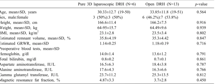
Table 1. Comparison of preoperative characteristics between pure 3D laparoscopic and conventional open donor right hemi-hepatectomy Pure 3D laparoscopic DRH (N=6) Open DRH (N=13) p-value
Age, mean±SD, years 30.33±12.7 (19-50) 33.85±11.8 (19-51) 0.564
Sex, male:female 3 (50%):3 (50%) 6 (46.2%):7 (53.8%)
Height, mean±SD, cm 166.6±11.4 166.2±7.5 0.916
Weight, mean±SD, kg 64-95±15.7 64.49±9.6 0.939
BMI, mean±SD, kg/m2 23.1±2.8 23.5±3.4 0.802
Estimated remnant volume, mean±SD, % 35.8±4.19 35.3±4.42 0.847
Estimated GRWR, mean±SD 1.14±0.25 1.18±0.19 0.716
Preoperative blood tests, mean±SD
Hemoglobin, g/dl 14.0±1.4 13.6±1.2 0.791
Total bilirubin, mg/dl 0.8±0.2 0.7±0.1 0.861
Aspartate aminotransferase, IU/L 16.5±6.3 18.4±3.8 0.787
Alanine aminotransferase, IU/L 17.6±4.3 16.3±6.6 0.766
Gamma glutamyl transferase, IU/L 23.7±11.2 25.3±13.5 0.812
Magnetic resonance fat fraction, % 4.87±3.3 3.7±2.8 0.450
SD, standard deviation; BMI, body mass index; GRWR, graft-to-recipient weight ratio; DRH, donor right hemi-hepatectomy case there is a distance between the right anterior portal
vein and right posterior portal vein as in type III and di- rect venoplasty was performed in trifurcations like Type II or relatively close distance in Type III.
Statistical analysis
Continuous variables were compared using Student’s t test and categorical variables using by the chi-square test or Fisher’s exact test, as appropriate. Continuous variables are expressed as median value with range or mean value with standard deviation while categorical variables are express- ed as numbers and percentages. A two-tailed p value <0.05 was considered statistically significant. Statistical analysis was performed using SPSS software version 22.0 (SPSS Inc., Chicago, IL).
RESULTS
During the study period, 19 donors with type II or type III portal vein variations underwent living donor right hemihepatectomy and were included in this study. Six do- nors (31.6%) underwent PLDRH, while 13 donors (68.4%) underwent CDRH.
Preoperative characteristics of the donors Table 1 summarizes the baseline characteristics of both groups. There was no significant difference between lapa- roscopic and open living donors.
Post-operative outcomes of the donors
Table 2 summarizes the operative outcomes of both groups.
There was no significant difference between the open and laparoscopic groups, except the operative time. The total operative time was longer in the laparoscopic group (356.5 vs. 262.8 minutes, p=0.02). None of the donors had intraoperative complications and none required perioper- ative transfusions. Most donors with type II or type III portal vein variations also had multiple bile duct opening regardless of the surgical approach (83.3% vs. 76.9%, p=0.627). There was no significant difference in the over- all complication rate between the two groups (16.7% vs.
27.3%, p=0.627) and the biliary complication rate was al- so insignificant (16.7% vs. 15.4%, p=0.316). All donors fully recovered without further complications and there was no reoperation or readmission.
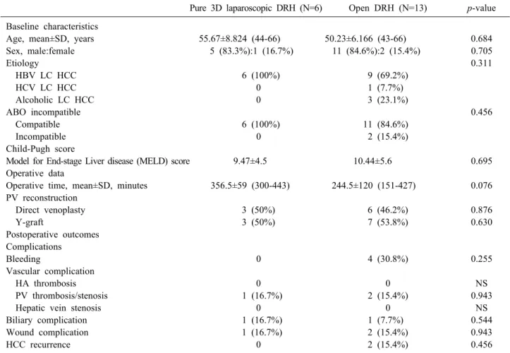
Recipient outcomes
The baseline characteristics and perioperative outcomes of the recipients are described in Table 3. There was no significant difference between the two groups regarding baseline clinical characteristics including age, sex, etiol- ogy, Child-Pugh and MELD score. In the PLDRH group, Y-grafts were used in thee (50%) recipients and three (50%) recipients underwent direct venoplasty. Y-grafts were used in seven (53.8%) recipients in the CDRH group while direct venoplasty was performed six (46.2%) recipi- ents.
There was no significant difference in the operative time
Table 2. Comparison of postoperative outcomes between pure 3D laparoscopic and conventional open donor right hemi-hep- atectomy
Pure 3D laparoscopic DRH (N=6) Open DRH (N=13) p-value Operative time, mean±SD, minutes 356.5±57 (300-443) 262.8±79 (151-427) 0.020
Graft weight, mean±SD, g 914.8±218 796±103 0.254
GRWR, mean±SD 1.43±0.41 1.18±0.19 0.094
Estimated blood loss, mean±SD, ml 450±89 (350-600) 406±201 (50-700) 0.620
Intraoperative transfusion, n (%) 0 0 NS
Multiple bile duct opening, n (%) 5 (83.3%) 10 (76.9%) 0.627
Peak total bilirubin, IU/L 2.75±1.5 (1.5-5.3) 3.26±0.88 (2.0-4.8) 0.345
Peak aspartate aminotransferase, IU/L 211±76 (141-453) 176±91 (32-378) 0.439 Peak alanine aminotransferase, IU/L 208±122 (95-449) 173±130 (48-577) 0.587
Overall complications 1 (16.7%) 3 (27.3%) 0.627
Bleeding 0 0 NS
Vascular complication 0 0 NS
Biliary complication 1 (16.7%) 2 (15.4%) 0.316
Wound complication 0 1 (7.7%) 0.684
Hospital stay, days 8.5±3.2 (6-15) 8.92±2.0 (7-15) 0.703
Rehospitalization, n (%) 0 0 NS
Reoperation 0 0 NS
SD, standard deviation; GRWR, graft-to-recipient weight ratio; DRH, donor right hemi-hepatectomy; NS, not significant
Table 3. Comparison of recipients perioperative characteristics of the between pure 3D laparoscopic and conventional open donor right hemi-hepatectomy
Pure 3D laparoscopic DRH (N=6) Open DRH (N=13) p-value Baseline characteristics
Age, mean±SD, years 55.67±8.824 (44-66) 50.23±6.166 (43-66) 0.684
Sex, male:female 5 (83.3%):1 (16.7%) 11 (84.6%):2 (15.4%) 0.705
Etiology 0.311
HBV LC HCC 6 (100%) 9 (69.2%)
HCV LC HCC 0 1 (7.7%)
Alcoholic LC HCC 0 3 (23.1%)
ABO incompatible 0.456
Compatible 6 (100%) 11 (84.6%)
Incompatible 0 2 (15.4%)
Child-Pugh score
Model for End-stage Liver disease (MELD) score 9.47±4.5 10.44±5.6 0.695
Operative data
Operative time, mean±SD, minutes 356.5±59 (300-443) 244.5±120 (151-427) 0.076 PV reconstruction
Direct venoplasty 3 (50%) 6 (46.2%) 0.876
Y-graft 3 (50%) 7 (53.8%) 0.630
Postoperative outcomes Complications
Bleeding 0 4 (30.8%) 0.255
Vascular complication
HA thrombosis 0 0 NS
PV thrombosis/stenosis 1 (16.7%) 2 (15.4%) 0.943
Hepatic vein stenosis 0 0 NS
Biliary complication 1 (16.7%) 1 (7.7%) 0.544
Wound complication 1 (16.7%) 2 (15.4%) 0.943
HCC recurrence 0 2 (15.4%) 0.456
SD, standard deviation; DRH, donor right hemi-hepatectomy; NS, not significant; HBV LC, hepatitis B virus related liver cirrhosis;
HCV LC, hepatitis C related liver cirrhosis; HCC, hepatocellular carcinoma; PV, portal vein; HA, hepatic artery
between the two groups (356.5 vs 244.5, p=0.076). One (16.7%) patient in the PLDRH group and two (15.4%) pa- tients in the CDRH group underwent vascular intervention due to portal vein stenosis and/or thrombosis.
DISCUSSION
LDLT is considered a safe alternative to deceased do- nor liver transplantation. Although a lifesaving procedure, any harm to the donor is unacceptable. With improve- ments in surgical techniques and perioperative patient care, adult donor hepatectomy has become a safe proce- dure with acceptable outcomes. However, living donor hepatectomy still remains a major surgical procedure en- tailing unpredictable morbidities.1,12,13 Most living donors have great concern that related with the large abdominal scar for CDRH. This sometimes caused a lot of psycho- logical stresses affecting their decision to donate. In addi- tion, the excess postoperative pain prolongs the hospital stay and delays their postoperative recovery.1,2
PLDRH was first reported by Soubrane et al. in 2013.14 Yet, unlike other abdominal surgeries, the expansion of PLDRH has been very slow. Various concerns have been raised regarding the safety of PLDRH. The second inter- national consensus on left lateral sectionectomy held in Morioka addressed that PLDRH is in the developmental phase with unclear benefit-to-risk ratio and that the long-term outcomes of both the donor and recipient is unknown.5
Several studies from highly specialized centers reported satisfactory outcomes of PLDRH. However, these studies emphasized the importance of careful selection of appro- priate donors for this approach. Furthermore, most of these studies report that donors with portal vein or hepatic duct anatomical variations or marginal liver grafts are considered unsuitable for PLDRH.6-9 PLDRH was first performed in 2015 at the SNUH.15 The center is highly experienced in adult LDLT with different anatomical var- iations providing excellent outcomes. At the same time, laparoscopic hepatectomy for hepatic neoplasm is fre- quently performed at this center with great experience.
This allowed for the easy and rapid adoption of PLDRH.
With accumulating experience, more than 90% of PLDRH are currently performed via pure laparoscopic approach.8,16
In this study, the safety and feasibility of PLDRH in
donors with type II and III portal vein variations has been evaluated. This is the first report that compares laparo- scopic and open donor hepatectomy in donors with type II or III variations of portal vein.
PLDRH requires a cautious technique to achieve the complex balance between donor safety and sufficient graft quality. The key to success in PLDRH is the technical standardization of the procedure. The key features are the use of flexible 3D scope and ICG near-infrared fluo- rescence cholangiography.8,16 In this study, there was no significant difference between open and laparoscopic ap- proaches regarding various operative variables except for the operative time. The total operative time was longer in laparoscopic approach compared to the standard open approach (356.5 vs. 262.8 minutes, p=0.02). This is re- lated to the learning curve of the laparoscopic approach during the transition from open right hepatectomy to PLDRH. With accumulating experience, shorter operation time was accomplished.
Donor safety in LDLT is the main goal in any donor surgery. A living donor is a healthy person who is inten- tionally exposed to major a surgical procedure, in which a dominant proportion of their liver is resected. Therefore, any harm to these living donors is unacceptable. In this study, none of the donors experienced intraoperative com- plications and none required perioperative transfusions.
While there was no harm observed or documented in living donors, there was no significant difference between open and laparoscopic approaches in respect to post- operative hospital stay and complication rate. All donors fully recovered without any postoperative complications.
Donors with dual portal vein variations were commonly associated with bile duct variations. The biliary complica- tion rate in the study donors was relatively higher in both groups when compared with those without variation.
However, the biliary complications rates were similar in both groups (16.6% vs. 27.3%, p=0.327).
The graft quality in laparoscopic donor hepatectomy may be inferior compared to its open counterpart in re- gards to the graft vasculature length. Therefore, the pres- ence of a highly skilled recipient team is essential to allow for safe graft implantation. In this study, in the PLDRH group, Y-grafts were used in three recipients and direct venoplasty was performed in three other recipients. In the CDRH group, Y-grafts were used in seven recipients in
the CDRH group and direct venoplasty was performed in six recipients. One patient in the PLDRH group and two patients in the CDRH group underwent vascular inter- vention due to portal vein stenosis and/or thrombosis.
These recipients fully recovered after the radiological interventions.
This study has some limitations. First, the number of donors included is small. However, this issue is related to the lack of PLDRH in donor with anatomic variations.
Moreover, the retrospective nature of the study may have some selection bias. This is related to that the cases with anatomical variation tends to be easily approached by the conventional open approach. In addition, this study is a single center experience with considerable experience in open liver surgery and LDLT.
In conclusion, PLDRH is a technically challenging pro- cedure requiring several complex laparoscopic techniques.
The presence of portal vein variations adds on further challenges. The current series support the safety and feasi- bility of PLDRH for donors with type II and III portal vein variations by surgeons with great experience in lapa- roscopic hepatobiliary surgery and LDLT.
REFERENCES
1. Samstein B, Griesemer A, Halazun K, Kato T, Guarrera JV, Cherqui D, et al. Pure laparoscopic donor hepatectomies: ready for widespread adoption? Ann Surg 2018;268:602-609.
2. Au KP, Chok KSH. Minimally invasive donor hepatectomy, are we ready for prime time? World J Gastroenterol 2018;24:2698- 2709.
3. Takahara T, Wakabayashi G, Beppu T, Aihara A, Hasegawa K, Gotohda N, et al. Long-term and perioperative outcomes of lapa- roscopic versus open liver resection for hepatocellular carcinoma with propensity score matching: a multi-institutional Japanese study. J Hepatobiliary Pancreat Sci 2015;22:721-727.
4. Han HS, Shehta A, Ahn S, Yoon YS, Cho JY, Choi Y. Laparo- scopic versus open liver resection for hepatocellular carcinoma:
case-matched study with propensity score matching. J Hepatol 2015;63:643-650.
5. Wakabayashi G, Cherqui D, Geller DA, Buell JF, Kaneko H, Han HS, et al. Recommendations for laparoscopic liver re- section: a report from the second international consensus confer- ence held in Morioka. Ann Surg 2015;261:619-629.
6. Han HS, Cho JY, Yoon YS, Hwang DW, Kim YK, Shin HK, et al. Total laparoscopic living donor right hepatectomy. Surg Endosc 2015;29:184.
7. Kim KH, Kang SH, Jung DH, Yoon YI, Kim WJ, Shin MH, et al. Initial outcomes of pure laparoscopic living donor right hepatectomy in an experienced adult living donor liver transplant center. Transplantation 2017;101:1106-1110.
8. Lee KW, Hong SK, Suh KS, Kim HS, Ahn SW, Yoon KC, et al. One hundred fifteen cases of pure laparoscopic living donor right hepatectomy at a single center. Transplantation 2018;102:
1878-1884.
9. Soubrane O, Kwon CH. Tips for pure laparoscopic right hep- atectomy in the live donor. J Hepatobiliary Pancreat Sci 2017;24:
E1-E5.
10. Yi NJ, Suh KS, Cho JY, Lee HW, Cho EH, Yang SH, et al.
Three-quarters of right liver donors experienced postoperative complications. Liver Transpl 2007;13:797-806.
11. Lee KW, Kim SH, Han SS, Kim YK, Cho SY, You T, et al.
Use of an upper midline incision for living donor partial hep- atectomy: a series of 143 consecutive cases. Liver Transpl 2011;
17:969-975.
12. Abecassis MM, Fisher RA, Olthoff KM, Freise CE, Rodrigo DR, Samstein B, et al. Complications of living donor hepatic lobec- tomy--a comprehensive report. Am J Transplant 2012;12:1208- 1217.
13. Brige P, Hery G, Chopinet S, Palen A, Azoulay D, Gregoire E.
Morbidity and mortality of hepatic right lobe living donors: sys- tematic review and perspectives. J Gastrointestin Liver Dis 2018;
27:169-178.
14. Soubrane O, Perdigao Cotta F, Scatton O. Pure laparoscopic right hepatectomy in a living donor. Am J Transplant 2013;13:2467- 2471.
15. Suh KS, Hong SK, Yi NJ, Lee KW, Kim HS, Yoon KC, et al.
Pure 3-dimensional laparoscopic extended right hepatectomy in a living donor. Liver Transpl 2016;22:1431-1436.
16. Suh KS, Hong SK, Lee KW, Yi NJ, Kim HS, Ahn SW, et al.
Pure laparoscopic living donor hepatectomy: focus on 55 donors undergoing right hepatectomy. Am J Transplant 2018;18:434-443.