Application of negative pressure wound therapy in patients with wound dehiscence after abdominal open surgery: a single center experience
Ji Young Jang, Hongjin Shim
1, Yun Jin Lee
2, Seung Hwan Lee, Jae Gil Lee
Department of Surgery, Yonsei University College of Medicine, Seoul,
1Department of Surgery, Yonsei University Wonju College of Medicine, Wonju,
2Department of Wound Ostomy Continence Nursing, Severance Hospital, Seoul, Korea
Journal of the Korean Surgical Society
JKSS
Purpose: Since the 1990’s, negative pressure wound therapy (NPWT) has been used to treat soft tissue defects, burn wounds, and to achieve skin graft fixation. In the field of abdominal surgery, the application of NPWT is increasing in cases with an open abdominal wound requiring temporary wound closure and a second look operation. In the present study, the authors analyzed patients that underwent NPWT for postoperative wound dehiscence.
Methods: The computerized records of patients that had undergone an abdominal operation from November 2009 to May 2012 were retrospectively analyzed.
Results: The number of total enrolled patients was 50, and 30 patients (60%) underwent an emergency operation. Diagnoses were as follows: panperitonitis or intra-abdominal abscess (24 cases, 48%), intestinal obstruction (10 cases, 20%), cancer (7 cases, 14%), mesentery ischemia (3 cases, 6%), and hemoperitoneum (1 case, 2%). NPWT was applied at a mean of 12.9 ± 8.2 days after surgery and mean NPWT duration was 17.9 days (2 to 96 days). The 11 patients (22%) with unsuccessful wound closure had a deeper and more complex wound than the other 39 patients (78%) (90.9% vs. 38.5%, P = 0.005). There were two complication cases (4%) due to delayed wound healing.
Conclusion: Most patients recovered well due to granulation formation and suturing.
NPWT was found to be convenient and safe, but a prospective comparative study is needed to confirm the usefulness of NPWT in patients whose wounds are dehisced.
INTRODUCTION
Negative pressure wound therapy (NPWT) was introduced as a vacuum-assisted closure (VAC) by Morykwas et al. [1,2] in the late 1990’s, and is currently used for wound management in various fields, such as, to manage soft tissue defects, fixate grafted skin, and to treat burn wounds. Whereas the application of NPWT to surgical abdominal wounds was initiated as a form of damage control surgery in trauma patients or for temporary wound closure prior to a second look operation in the 2000s [3,4]. Recently, NPWT has applied in patients who were diagnosed with abdominal compartment syndrome as an essential procedure of decompressive laparotomy [5,6]. The majority of previous studies on the topic have focused on effective closure of opened abdominal wounds using NPWT, and relatively few reports have been issued on the use of NPWT to treat postoperative wound complications. Here, we
Corresponding Author Jae Gil Lee
Department of Surgery, Severance Hospital, Yonsei University College of Medicine, 50 Yonsei-ro, Seodaemun-gu, Seoul 120-752, Korea
Tel: +82-2-2228-2127 Fax: +82-2-313-8289 E-mail: jakii@yuhs.ac
Key Words
Negative pressure wound therapy, Vacuum-assis- ted closure, Surgical wound dehiscence, Abdomen, Surgery
Received May 31, 2013 Revised July 20, 2013 Accepted July 21, 2013 J Korean Surg Soc 2013;85:180-184 http://dx.doi.org/10.4174/jkss.2013.85.4.180
Copyright © 2013, the Korean Surgical Society
cc Journal of the Korean Surgical Society is an Open Access Journal. All articles are distributed under the terms of the Creative Commons Attribution Non-Commercial License (http://
creativecommons.org/licenses/by-nc/3.0/) which permits unrestricted non-commercial use, distribution, and reproduction in any medium, provided the original work is properly cited.
Ji Young Jang, et al: NPWT in dehisced abdominal wound
describe our early experiences of the use of NPWT to treat for postoperative dehisced wounds.
METHODS
The electronic records of 55 patients whose wounds were managed by NPWT from November 2009 to May 2012 were analyzed retrospectively. NPWT was used; 1) to treat postoperative wound complications, 2) to prevent intra- abdominal wound infection during operation, and 3) for temporary wound closure after laparotomy. Among these cases, 50 patients with postoperative wound complication were enrolled in this study. When wound dressing was needed more than three times a day, because of a large amount of discharge from the dehisced wound or wound infection was identified, NPWT was applied. Wound complications were classified as superficial, deep or complex as per wound depth. Superficial wound complications were defined as invasion of the skin or subcutaneous fat layer, deep wounds were defined as those resulting in bowel or omentum exposure due to a fascia defect, and a complex wound complication was defined as an wound associated with an intra-abdominal infection. Times and durations of postoperative NPWT and whether wounds were repaired fully were noted. The negative pressure applied was a continuous 125 mmHg. Amounts and characteristics of drained fluids were also noted. When the fluid extracted
changed to serous and its volume decreased, the wound bed was confirmed and NPWT was stopped. In patients with successful wound healing, we evaluated whether the wound had been repaired by sutures or healed due to granulation tissue formation or due to a prosthesis. Complications associated with NPWT were also studied.
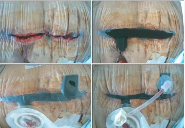
NPWT was obtained by applying a CuraVAC system (CuraVAC, Daewoong Bio, Seoul, Korea) (Fig. 1). The vacuum system was exchanged two or three times weekly. Additional exchange was performed depending on wound status. When the bowel was exposed by a deep or complex wound due to a fascia defect, an aseptic isolation plastic bag or vinyl film was applied under the vacuum system to protect the small bowel.
All analyses were performed using IBM SPSS ver. 20.0 (IBM Co., Armonk, NY, USA). Continuous variables were analyzed using the Student t-test, and results are expressed as means ± standard deviations and median (range). Categorical variables were analyzed using Fisher exact test.
RESULTS
The mean age of the 50 patients was 61.5 ± 16.2 years and 27 (54%) were men. Eight patients had diabetes mellitus (DM) and 4 (8%) were managed for inflammatory bowel disease.
Emergency surgery was performed in 30 patients (60%). NPWT was performed, on average, 12.9 ± 8.2 days after surgery and
Fig. 1. Application of negative pressure wound therapy for postoperative abdominal dehisced wound.
applied for a median of 17.9 days (2 to 96). Median hospital stay was 42 days (11 to 210 days) (Table 1). Regarding diagnoses, 24 patients (48%) had an intra-abdominal infection containing panperitonitis and 10 had anastomotic leakage or intestinal obstruction or strangulation. Cancer and hemoperitoneum were diagnosed in 7 (14%) and 3 (6%) respectively (Table 2).
Wounds were superficial in 25 patients (50%), deep in 11 (22%) and complex in 14 (28%). Wounds were repaired in 39 patients (78%); 29 patients by skin suturing, 9 by granulation tissue formation, and in one by prosthesis application. In 11 patients (22%), wound closure failed, because 10 patients succumbed to general deterioration and one patient with radiation enteritis was managed in an outpatient clinic.
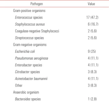
Peritoneal bacterial culture was positive in 36 patients (72%).
Enterococcus species was identified in 17 patients (47.2%), which was most commonly a gram positive bacterium, and Staphylococcus aureus and coagulase-negative Staphylococcus were found in 6 (16.7%) and 2 patients (5.6%), respectively.
Regarding gram-negative bacteria, Escherichia coli was found in 9 patients (25%), Pseudomonas aeruginosa in 4 (11.1%), Enterococcus species in 4 (11.1%), Acinetobacter baumannii in 4 (11.1%) and Citrobacter species in 3 (8.3%). An anaerobic bacterium was found in one patient (2.8%) and identified as Bacteroides species (Table 3).
When we compared patients that achieved wound closure and patients that did not, no significant difference was found
with respect to age, DM, emergency operation, diagnosis or infection. Deep and complex wound accounted for 90.9% of cases in the successful closure group, and 38.5%
in the unsuccessful group (P = 0.005) (Table 4). No severe complication, such as bowel perforation or an enterocutaneous fistula was encountered, but in 2 patients NPWT was administered for a protracted time (>4 weeks).
DISCUSSION
Infection is the most common cause of abdominal wound dehiscence. When wound dehiscence is identified, surgeons select mainly gauze dressing and change dressing many times a day in cases of intact fascia. If there is a large fascia defect and evisceration of the bowel, operation is usually performed to repair the fascia. In cases where abdominal distension is too severe to close the wound, temporary abdominal closure options include the Wittmann Patch, Bogota bag and NPWT [7,8]. Definitive reconstruction has been presented with mesh, biologic mesh, components separation, and rectus abdominis sheath turnover flap method, and so on [9,10].
The application of negative pressure to a wound increases dermal perfusion and stimulates the formation of granulation tissue, and thus, accelerates wound healing and decreases bacterial colonization because it reduces tissue edema and interstitial tissue fluid [2,11]. In addition, the reverse tissue expansion effect of negative pressure helps to approximate skin and fascia. The efficacy of NPWT has already been proven, and currently, it is used to treat trauma-induced soft tissue Table 3. Distribution of pathogens isolated from surgical wounds
Pathogen Value
Gram-positive organisms
Enterococcus species 17 (47.2)
Staphylococcus aureus 6 (16.7)
Coagulase-negative Staphylococci 2 (5.6)
Streptococcus species 2 (5.6)
Gram-negative organisms
Escherichia coli 9 (25)
Pseudomonas aeruginosa 4 (11.1)
Enterobacter species 4 (11.1)
Citrobacter species 3 (8.3)
Acinetobacter baumannii 4 (11.1)
Other 3 (8.3)
Anaerobic organism
Bacteroides species 1 (2.8)
Values are presented as number (%).
Table 1. Demographics of patients treated by NPWT
Variable Value
Male sex, n (%) 27 (54)
Age (yr), mean ± SD 61.5 ± 16.2
Diabetes mellitus, n (%) 8 (16)
IBD & auto immune disease, n (%) 4 (8) Hospital stay (day), median (range) 42 (11–210) Infection; positive peritoneal culture, n (%) 36 (72) NPWT, negative pressure wound therapy; SD, standard deviation; IBD, inflam- matory bowel disease.
Table 2. Patients diagnoses
Diagnosis Value Panperitonitis & intra-abdominal infection 24 (48)
Intestinal obstruction 10 (20)
Cancer (elective operation) 7 (14)
Mesenteric ischemia 3 (6)
Hemoperitoneum 1 (2)
Values are presented as number (%).
Ji Young Jang, et al: NPWT in dehisced abdominal wound
defects, necrotizing fasciitis, suppurative and extravasation injuries and burn wounds, and to promote skin graft fixation [12,13]. Recently, NPWT has been applied in the abdominal surgery field for temporary closure in cases of trauma and bowel strangulation, and to manage abdominal compartment syndrome when the abdomen is open [14,15]. Batacchi et al. [6], in a prospective study, found that the time to wound closure and intensive care unit stay in a NPWT group were shorter than in a Bogota bag group, and in another study, fascia- to-fascia closure was successfully achieved by applying an abdominal topical negative pressure in a patient with an intra- abdominal infection [16]. The majority of our 50 patients had profuse fluid drainage through the main wound caused by postoperative seroma or a partial fascial defect. In 25 of our patients, the wound was superficial, and wounds were closed in 24 of these patients (96%) (Table 4). Most patients with a superficial wound had no gross fascia defect but a large amount of discharge. This suggests that NPWT is effective in cases of intra-abdominal infection, if the strength of the fascia is maintained. Generally, a wet gauze dressing is applied after removing staples or sutures in cases of postoperative seroma or wound dehiscence, and these are applied many times a day until the wound bed is washed-up. On the other hand, NPWT is more convenient for dressing and preventing infections because the wound dressing only needs replacement 2 to 3 times per week. On the other hand, in 11 patients whose wounds failed to close, 10 patients died due to deterioration in general condition or sepsis, which implies that infection and the recovery of general condition are important. In the present study, the results of peritoneal bacterial cultures were
positive in 36 patients (72%), and as previously reported, when intra-abdominal infection is identified, source control and the appropriate use of antibiotics are essential [17]. Recently, various reports have described the effective management of abdominal wounds with intra-abdominal infection by NPWT, and have attributed this success to infection control and wound closure [4,16]. In the present study, the unsuccessful closure group had a significantly greater proportion of deep and complex wounds than the successful closure group (P = 0.005). Profuse fluid drainage through fascia defects probably makes wound closure difficult and this is undoubtedly exacerbated by abdominal sepsis caused by intra-abdominal infection caused by anastomotic leakage or bowel perforation.
This suggests that inappropriate control of the infection source has a negative impact on not only wound healing but also prognosis. Furthermore, the timing of NPWT application in the successful closure group was 11.3 ± 6.9 days, whereas in the unsuccessful closure group it was significantly greater at 18.9 ± 9.7 days. Thus, although the unsuccessful closure group had a greater proportion of deep and complex wounds (10, 90.9%), the later NPWT start in the unsuccessful closure group shows that intra-abdominal infections were difficult to treat because of inadequate source control. Baharestani and Gabriel [18] reported that NPWT resulted in successful wound closure in 86% of patients in whom mesh had failed to repair the abdominal wall, and that one of the important factors of successful healing was early application. In fact, if the identification of seroma or dehiscence is delayed in patients with an intact fascia or focal defect, infected fluid collection in dead space above the fascia and pocket formation caused by Table 4. Comparison between patients that did and did not achieve successful closure
Variable Successful wound closure (n=39) Unsuccessful wound closure (n=11) P-value
Age (yr) 61.2 ± 16.9 62.6 ± 14.2 0.803a)
Sex 0.014b)
Male 25 (64.1) 2 (18.2)
Female 14 (35.9) 9 (81.8)
Diabetes mellitus 6 (15.4) 2 (18.2) 1.000b)
Emergency operation 18 (46.2) 2 (18.2) 0.163b)
Diagnosis (panperitonitis & intra-abdominal infection) 19 (48.7) 5 (45.5) 0.848b)
Infection 27 (69.2) 9 (81.8) 0.705b)
Wound type 0.005b)
Superficial wound 24 (61.5) 1 (9.1)
Deep & complex wound 15 (38.5) 10 (90.9)
Point of application (day) 11.3 ± 6.9 18.8 ± 9.7 0.005a)
Values are presented as mean ± standard deviation or number (%).
a)Student t-test. b)Fisher exact test.
the abrading effect of suture material worsens infection [19].
This study has some limitations that should be considered.
First, it was difficult to determine the significance of differences because of the small cohort. Second, it is not a comparative study with patients in whom NPWT was not used. Third, it was difficult to evaluate the effects of NPWT in patients with wound dehiscence due to the heterogeneity shown by patients concerned and the retrospective nature of this study.
In conclusion, abdominal wound complications, such as large seroma or infections can be managed with NPWT. There was no harm or risk of bowel perforation or enterocutaneous fistula. However, a prospective comparative study is required to confirm the usefulness of NPWT in patients whose wounds are dehisced.
CONFLICTS OF INTEREST
No potential conflict of interest relevant to this article was reported.
REFERENCES
1. Argenta LC, Morykwas MJ. Vacuum-assisted closure: a new method for wound control and treatment: clinical experience.
Ann Plast Surg 1997;38:563-76.
2. Morykwas MJ, Argenta LC, Shelton-Brown EI, McGuirt W.
Vacuum-assisted closure: a new method for wound control and treatment: animal studies and basic foundation. Ann Plast Surg 1997;38:553-62.
3. Caro A, Olona C, Jimenez A, Vadillo J, Feliu F, Vicente V.
Treatment of the open abdomen with topical negative pressure therapy: a retrospective study of 46 cases. Int Wound J 2011;8:274-9.
4. D’Hondt M, D’Haeninck A, Dedrye L, Penninckx F, Aerts R.
Can vacuum-assisted closure and instillation therapy (VAC- Instill therapy) play a role in the treatment of the infected open abdomen? Tech Coloproctol 2011;15:75-7.
5. Plaudis H, Rudzats A, Melberga L, Kazaka I, Suba O, Pupelis G. Abdominal negative-pressure therapy: a new method in countering abdominal compartment and peritonitis - prospective study and critical review of literature. Ann Intensive Care 2012;2 Suppl 1:S23.
6. Batacchi S, Matano S, Nella A, Zagli G, Bonizzoli M, Pasquini A, et al. Vacuum-assisted closure device enhances recovery of critically ill patients following emergency surgical procedures.
Crit Care 2009;13:R194.
7. Wittmann DH, Aprahamian C, Bergstein JM, Edmiston CE,
Frantzides CT, Quebbeman EJ, et al. A burr-like device to facilitate temporary abdominal closure in planned multiple laparotomies. Eur J Surg 1993;159:75-9.
8. Kirshtein B, Roy-Shapira A, Lantsberg L, Mizrahi S. Use of the
“Bogota bag” for temporary abdominal closure in patients with secondary peritonitis. Am Surg 2007;73:249-52.
9. Arhi C, El-Gaddal A. Use of a silver dressing for management of an open abdominal wound complicated by an enterocutaneous fistula-from hospital to community. J Wound Ostomy Continence Nurs 2013;40:101-3.
10. Ramirez OM, Ruas E, Dellon AL. “Components separation”
method for closure of abdominal-wall defects: an anatomic and clinical study. Plast Reconstr Surg 1990;86:519-26.
11. Bovill E, Banwell PE, Teot L, Eriksson E, Song C, Mahoney J, et al. Topical negative pressure wound therapy: a review of its role and guidelines for its use in the management of acute wounds.
Int Wound J 2008;5:511-29.
12. Armstrong DG, Lavery LA; Diabetic Foot Study Consortium.
Negative pressure wound therapy after partial diabetic foot amputation: a multicentre, randomised controlled trial. Lancet 2005;366:1704-10.
13. Moisidis E, Heath T, Boorer C, Ho K, Deva AK. A prospective, blinded, randomized, controlled clinical trial of topical negative pressure use in skin grafting. Plast Reconstr Surg 2004;114:917-22.
14. Barker DE, Kaufman HJ, Smith LA, Ciraulo DL, Richart CL, Burns RP. Vacuum pack technique of temporary abdominal closure: a 7-year experience with 112 patients. J Trauma 2000;48:201-6.
15. Brace JA. Negative pressure wound therapy for abdominal wounds. J Wound Ostomy Continence Nurs 2007;34:428-30.
16. Padalino P, Dionigi G, Minoja G, Carcano G, Rovera F, Boni L, et al. Fascia-to-fascia closure with abdominal topical negative pressure for severe abdominal infections: preliminary results in a department of general surgery and intensive care unit. Surg Infect (Larchmt) 2010;11:523-8.
17. Solomkin JS, Mazuski JE, Bradley JS, Rodvold KA, Goldstein EJ, Baron EJ, et al. Diagnosis and management of complicated intra-abdominal infection in adults and children: guidelines by the Surgical Infection Society and the Infectious Diseases Society of America. Surg Infect (Larchmt) 2010;11:79-109.
18. Baharestani MM, Gabriel A. Use of negative pressure wound therapy in the management of infected abdominal wounds containing mesh: an analysis of outcomes. Int Wound J 2011;8:118-25.
19. Fry DE. Wound infection in hernia repair. In: Fitzgibbons RJ, Greenburg AG, editors. Nyhus and Condon’s hernia. 5th ed.
Philadelphia: Lippincott Williams & Wilkins; 2002. p.279-90.