INTRODUCTION
Cervicogenic headache, first described by Sjaastad (1), is characterized as unilateral fronto-temporal headache with clinical symptomatology similar to migraine (1, 2). Useful clinical diagnostic criteria were published in 1990 (3) and revised in 1998 (4). However, the concept of cervicogenic headache is not universally accepted, and the precise pathol- ogy in the neck that gives rise to the pain has not been com- pletely understood. Among the candidate structures are the upper cervical nerves (greater and lesser occipital nerves), nerve roots, cervical muscles, cervical discs and zygapophy- seal (facet) joints, and atlantoaxial and atlantooccipital joints.
Cervicogenic headache may arise not only from the upper, but also from the middle and even from the lower cervical area (4). Various treatment modalities directed to these struc- tures have been attempted. Among the published approach- es, radiofrequency (RF) neurotomy of the cervical facet joints (5, 6) seems promising.
The purpose of the study was to assess the clinical efficacy of RF cervical zygapophyseal joint neurotomy in patients with chronic cervicogenic headache to determine whether there was sufficient merit or usefulness in the procedure to justify its use in these patients.
MATERIALS AND METHODS
Patients were selected from the out patient clinic at our
institution during the March 2001-February 2003 period.
Major criteria for the diagnosis of cervicogenic headache are (modified criteria from 1990 [3]) listed in Table 1.
There were 46 with chronic (>6 months of duration) cer- vicogenic headache who met the above criteria. Among them, a total of thirty patients showed greater than 50% of pain relief from two diagnostic/prognostic C3-C4 cervical medial branch blocks and were followed up for more than 12 months.
Those patients who needed multi-level cervical blocks and those who were involved in litigation or compensational pro- grams were excluded. Selected patients had relatively severe complaints inhibiting activity of daily living, participation in work or social life, and inadequate effect of appropriate prophylactic and/or therapeutic headache medication. There were 16 men and 14 women ranging in age from 35 to 67 yr with a mean age of 54 yr. Radiographic examinations of the cervical spine were carried out in order to evaluate an abnor- mal posture or flexion-extension impairment. There were no definitive structural abnormalities that could cause neurologic deficits on computed tomogram or magnetic resonance imag- ing study of brain or cervical spine. These patients were treat- ed with RF neurotomy on medial branch of posterior primary ramus and then were assessed at 1 week, 1 month, 6 months, and at 12 months following the treatment. All procedures were performed by a single neurosurgeon.
A RF generator (RFG-3C Graphic, Radionics Inc., Burling- ton, MA, U.S.A.) with the SMK-C10 cannula (22 gauge;
length, 10 cm; exposed tip, 5 mm) was used with appropri- ate connections and a wide diathermy ground plate. All pro-
Jang Bo Lee, Jung Yul Park, Juno Park, Dong Jun Lim, Sang Dae Kim, Heung Seob Chung*
Department of Neurosurgery, Korea University Ansan Hospital, Ansan; Korea University Guro Hospital*, Korea University School of Medicine, Seoul, Korea
Address for correspondence Jung Yul Park, M.D.
Department of Neurosurgery, Korea University Ansan Hospital, 516 Gojan-dong, Danwon-gu, Ansan 425-707, Korea
Tel : +82.31-412-5053, Fax : +82.31-412-5054 E-mail : [email protected]
326 J Korean Med Sci 2007; 22: 326-9
ISSN 1011-8934
Copyright � The Korean Academy of Medical Sciences
Clinical Efficacy of Radiofrequency Cervical Zygapophyseal Neurotomy in Patients with Chronic Cervicogenic Headache
The purpose of the present study was to assess the clinical efficacy of radiofrequency (RF) cervical zygapophyseal joint neurotomy in patients with cervicogenic headache.
A total of thirty consecutive patients suffering from chronic cervicogenic headaches for longer than 6 months and showing a pain relief by greater than 50% from diag- nostic/prognostic blocks were included in the study. These patients were treated with RF neurotomy of the cervical zygapophyseal joints and were subsequently assessed at 1 week, 1 month, 6 months, and at 12 months following the treatment.
The results of this study showed that RF neurotomy of the cervical zygapophyseal joints significantly reduced the headache severity in 22 patients (73.3%) at 12 months after the treatment. In conclusion, RF cervical zygapophyseal joint neurotomy has shown to provide substantial pain relief in patients with chronic cervicogenic headache when carefully selected.
Key Words : Cervicogenic Headache; Zygapophyseal Joint; Radiofrequency Neurotomy
Received : 21 March 2006 Accepted : 24 July 2006
RF Neurotomy in Chronic Cervicogenic Headache 327
cedures were monitored by repeated radiographic screening with a C-arm image intensifier. Percutaneous RF neuroto- my was performed under aseptic conditions, with the patient lying in the prone position. No sedation or systemic analgesic was used. The electrode was inserted parallel to the medial branch of the C3-C4 facet joint with a percutaneously pos- terolateral approach along a parasagittal plane, tangential to the lateral margin of the articular pillar, and was directed toward the target point under repeated C-arm image inten- sifier screening. The target point was a point of intersection of 2 lines diagonally drawn from supero-anterior and supero- posterior to infero-posterior and infero-anterior articular pil- lar. The levels of lesioning were C3-C4 (Fig. 1). When the target point was reached, the position of the electrode was confirmed and recorded on anteroposterior and lateral films.
Correct position was verified by stimulating current for both sensory and motor threshold. Usual thresholds for these were 0.3-0.9 V and 0.6-1.8 V, respectively. The target nerve was then anesthetized by the injection of 0.5 mL of 1% lidocaine before lesion production. Final lesion was generated at 80℃ for 90 sec. The average operative time was 19 min. Patients were discharged from the hospital after 1 to 2 hr of moni- toring to see whether there were any discomforts or signs of
complications. Follow-up visits were made at 1 week, 1 month, 6 months, and at 12 months. The followings were taken as outcome parameters: Visual Analogue Scale (VAS), number of headache-days per week and amount of analgesic intake per week. Results were defined as successful if preoperative pain was relieved by more than 75%.
RESULTS
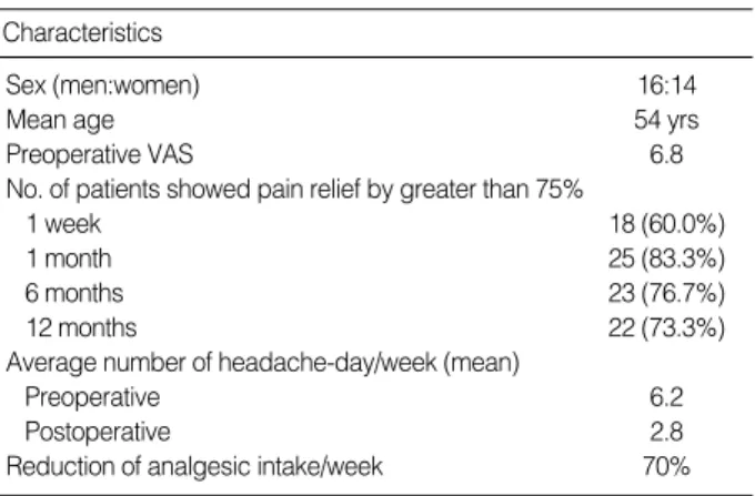
All patients well tolerated the procedure without additional analgesics or anesthetics. Eighteen (60%), 25 (83.3%), 23 (76.7%), and 22 (73.3%) patients showed pain relief by greater than 75% at 1 week, 1 month, 6 months, and at 12 months, respectively (Table 2). The average headache-days per week decreased from 6.2 days to 2.8 days, and the average analgesic intake per week showed a 70% reduction. There were no major complications related to the procedures. However, atax- ia was seen in 4 patients immediately after the procedure, which was resolved completely within a few hours to 2 or 3 days. Three patients obtained short-lasting relief (3 months).
None of the patients reported cutaneous numbness, dyses- thesia, or worsening of pain. However, 12 patients compla- ined of soreness on the posterior neck for 2-7 days following the procedures. These possible side effects were preoperatively explained to all patients and no specific measures were nec- essary except temporary oral analgesics for 2-5 days postop- eratively. All such symptoms disappeared within a week.
Interestingly, most of the patients showed definitive improve- ment after at least 2-4 weeks following the procedures. There were no instances of postoperative infection or other compli- cations related to the procedures.
DISCUSSION
The term ‘‘cervicogenic headache’’ is denoted for the hea- dache that originates from disorders of the neck (1). How-
I. Unilateral headache without side shift II. Symptoms and signs of neck involvement:
a. Provocation of attacks:
1. Pain triggered by neck movement and/or sustained awkward head position
2. Pain similar in distribution and character to the spontaneously occurring pain elicited by external pressure over ipsilateral upper, posterior neck region or occipital region
b. Ipsilateral neck, shoulder and arm pain of a rather vague, non-ra- dicular nature
c. Reduced range of motion in the cervical spine
Table 1.Major criteria for diagnosis of cervicogenic headache
Characteristics
Sex (men:women) 16:14
Mean age 54 yrs
Preoperative VAS 6.8
No. of patients showed pain relief by greater than 75%
1 week 18 (60.0%)
1 month 25 (83.3%)
6 months 23 (76.7%)
12 months 22 (73.3%)
Average number of headache-day/week (mean)
Preoperative 6.2
Postoperative 2.8
Reduction of analgesic intake/week 70%
Table 2.Characteristics of study subjects
Fig. 1.The target point and C3-C4 lesioning. A point of intersec- tion of 2 lines diagonally drawn from supero-anterior and supero- posterior to infero-posterior and infero-anterior articular pillar.
VAS, Visual Analogue Scale.
328 J.B. Lee, J.Y. Park, J. Park, et al.
ever, it is not a detailed description and is still controversial whether use of this term is appropriate as two currently major international organizations relevant to head pain, namely, the International Headache Society (IHS) and International Association for the Study of Pain (IASP), disagree with the view point (7, 8).
Symptoms and signs of cervicogenic headache such as me- chanical precipitation of attacks imply involvement of the neck. A reliable diagnosis of cervicogenic headache can be made based on the criteria established in 1990 and 1998 by the Cervicogenic Headache International Study Group (3, 4).
Currently, positive response after an appropriate nerve block is an essential diagnostic feature of the disorder.
Pathologic conditions such as fractures, congenital abnor- malities, bone tumors, rheumatoid arthritis, whiplash injury, and/or other distinct pathologies all may be related to the development of cervicogenic headache, but most authors be- lieve that these conditions should be described as underlying pathologies, and those without definitive structural patholo- gies should only be regarded as primary cervicogenic heada- che. The authors used the diagnostic criteria from 1990 (3) as the clinical diagnostic tool, and confirmed the diagnosis by C3-C4 cervical medial branch block. Some authors rec- ommend that when the diagnosis of cervicogenic headache is suspected, attempts should be made to confirm the diag- nosis with the objective tests of cervical facet block and C2 nerve diagnostic blocks (4, 8, 9). Although many cervical structures are suggested as the cause of cervicogenic headache, most authors agree that upper cervical nerves are the main cause. Among the upper cervical structures, we believe that C3-C4 cervical medial branches are the main culprit. Based on anatomical innervation of these branches to the upper cervical and occipital region, these may cause neck pain and radiating pain to the posterior and temporal areas of the head.
Bovim et al. reported that C2 nerve block seemed to be the most informative procedure and C4 and C5 nerve block was of little value in the work-up of cervicogenic headaches (9).
Stovner et al. reported that RF neurotomy of facet joints C2- C6 was not beneficial in cervicogenic headache in a random- ized, double-blind, sham-controlled study (10). Other stud- ies have shown what can be achieved if patients are carefully selected, using objective criteria under controlled conditions, and if meticulous surgical technique is used (6, 11-14). In a recent report of randomized controlled trial of treatment for cervicogenic headache (15), the authors did not find evidence that RF treatment of cervical facet joints and upper dorsal root ganglions was a better treatment than other conserva- tive management. And in a recent review, Martelletti and van Suijlekom (16) concluded that a consensus on a standard treatment for cervicogenic headache does not exist because of the great variability in patient selection and clinical effects.
Anatomically, the cervical zygapophyseal joints are inner- vated by articular branches derived from the medial branch- es of the cervical dorsal rami (17). The C4-C8 dorsal rami
arise from their respective spinal nerves just outside the inter- vertebral foramina and pass dorsally over the root of the ipsi- segmental transverse process. The medial branches of the typ- ical cervical dorsal rami are small (diameter, approximately 1 mm) and curve medially, hugging the waists of their ipsi- segmental articular pillars, covered by the tendinous slips of the origin of the semispinalis capitis. Each typical cervical zygapophyseal joint receives a dual innervation, from the medial branch above and from the medial branch below its location. The medial branches of the C3 dorsal ramus differ in their anatomy from those of lower cervical levels. A deep medial branch passes around the waist of the C3 articular pillar in a manner analogous to that of typical medial branch- es; this branch participates in the innervation of the C3-C4 zygapophyseal joint. The superficial medial branch of C3 is large (diameter, approximately 1.5-2.0 mm) and is known as the third occipital nerve. It winds around the lateral aspect and then the posterior aspect of the C2-C3 zygapophyseal joint. Articular branches may also arise from a communicat- ing loop that crosses the back of the joint between the third occipital nerve and the C2 dorsal ramus (17). Beyond the C2- C3 zygapophyseal joint, the third occipital nerve furnishes muscle branches to the semispinalis capitis and becomes cuta- neous over the suboccipital region. In this respect, the C3 dor- sal ramus is the only cervical dorsal ramus below C2 that reg- ularly has a cutaneous distribution. Those from C4 to C7 typi- cally lack any cutaneous branches (17). Therefore, on anato- mical grounds, pain suspiciously resulting from the C2-C3 levels can be blocked by coagulating the ipsilateral third occipi- tal nerve as it crosses the lateral aspect of the joint, and pain stemming below C2-C3 can be blocked by coagulating the cervical medial branches as they pass around the waists of the articular pillars above and below (18).
During the last decade, percutaneous RF neurotomy has been increasingly used in the treatment of chronic cervical pain and cervicobrachial pain. The rationale for this technique is that nociceptive transmission from the cervical zygapophy- seal joint and muscles as well as ligaments can be blocked by coagulating the medial branches of the dorsal rami that innervate these structures (17, 18). At typical cervical levels, RF neurotomy achieves complete relief of pain, provided that patients have had complete relief of pain following diagnos- tic blocks for chronic cervical zygapophyseal joint pain (5, 17). Those blocks can be placebo-controlled or comparative local anesthetic blocks (11, 12). Similarly, for patients with headache mediated by the third occipital nerve, complete relief of pain can be achieved if meticulous and demanding techniques are followed (13). Because of many possible eti- ologies in cervicogenic headache, however, we believe that complete relief of pain is difficult to achieve, especially in chronic cases. Also, many authors agree that RF denervation of the C2-C3 facet joint procedure is technically difficult and does not guarantee good outcome (10, 17). Having these factors in mind, we selected the patients with C3-C4 cervi-
RF Neurotomy in Chronic Cervicogenic Headache 329
cal level lesion with good postoperative results.
Some authors reported a lengthy operation time to denervate just one joint and, to practice third occipital neurotomy, about 90 min (5, 13). They do not rely on a stimulation test for adjusting electrode tip. However, we believe that through a stimulation tests, both sensory and motor threshold, surgeons can confirm the exact location of the electrode tip and reduce operation time. Also, a lengthy operation not only gives rise to patient discomfort caused by the prone position, but also results in a higher dose of radiation exposure to both the pa- tient and surgeon. Moreover, during the long operation, much needle manipulation may be necessary. In these cases, pain relief can be derived from other mechanisms including myo- fascial trigger point theory (14). Even with these adequate physiologic monitorings, the average duration of operation in our series was 19 min. The plane of insertion of electrodes is a critical factor. Electrodes must lie parallel to the nerve for the nerve to be incorporated in the radial lesion. Under these circumstances, however, the electrode must be within 2 mm of the nerve for adequate coagulation (18).
With these clinical, anatomical, and technical considera- tions, we undertook an audit of our experience with this pro- cedure. The patients selected for the study were those who had significant pain relief without definitive complications or worsening of symptoms when diagnostic blocks were pre- operatively performed under stringent, controlled conditions.
The procedure was based on antecedent anatomical studies of the target nerves; electrodes were introduced parallel to the target nerves, and multiple lesions were made to incor- porate the nerves. The limitations of this study lie in the fact that it was not a controlled, randomized study, and the num- ber of subjects was small. Also, data from long-term follow- up evaluation was not included. However, this study included results from carefully selected patients with up to 12-month period of follow-up evaluation. Therefore, we believe it rep- resents a moderate duration of pain relief without any seri- ous side effects in the majority of these chronically disabling patients.
REFERENCES
1. Sjaastad O, Saunte C, Hovdhal H, Breivik H, Grinbaek E. ‘‘Cervico- genic’’ headache. A hypothesis. Cephalalgia 1983; 3: 249-56.
2. Fredriksen TA, Hovdal H, Sjaastad O. ‘‘Cervicogenic headache’’.
Clinical manifestation. Cephalalgia 1987; 7: 147-60.
3. Sjaastad O, Fredriksen TA, Pfaffenrath V. Cervicogenic headache:
diagnostic criteria. Headache 1990; 30: 725-6.
4. Sjaastad O, Fredriksen TA, Pfaffenrath V. Cervicogenic headache:
diagnostic criteria. The Cervicogenic Headache International Study Group. Headache 1998; 38: 442-5.
5. Lord SM, Barnsley L, Wallis BJ, McDornald GJ, Bogduk N. Percu- taneous radio-frequency neurotomy for chronic cervical zygapophy- seal-joint pain. N Engl J Med 1996; 335: 1721-6.
6. van Suijlekom HA, van Kleef M, Barendse GA, Sluijter ME, Sjaas- tad O, Weber WE. Radiofrequency cervical zygapophyseal joint neu- rotomy for cervicogenic headache. A prospective study of 15 patients.
Funct Neurol 1998; 13: 297-303.
7. Headache Classification Committee of the International Headache Society. Classification and diagnostic criteria for headache disor- ders, cranial neuralgias and facial pain. Cephalalgia 1998; 8 (Suppl 7): 1-96.
8. International Association for the Study of Pain (IASP). Cervicogenic Headache. In: Merskey H, Bogduk N, editors. Classification of Chron- ic Pain. Description of Chronic Pain Syndromes and Definitions of Pain Terms, 2nd ed. Seattle: IASP Press, 1994; 94-5.
9. Bovim G, Berg R, Dale LG. Cervicogenic headache: anesthetic block- ades of cervical nerves (C2-C5) and facet joint (C2/C3). Pain 1992;
49: 315-20.
10. Stovner LJ, Kolstad F, Helde G. Radiofrequency denervation of facet joints C2-C6 in cervicogenic headache: a randomized, double-blind, sham-controlled study. Cephalalgia 2004; 24: 821-30.
11. Lord SM, Barnsley L, Bogduk N. The utility of comparative local anaesthetic blocks versus placebo-controlled blocks for the diagnosis of cervical zygapophyseal joint pain. Clin J Pain 1995; 11: 208-13.
12. Barnsley L, Lord S, Bogduk N. Comparative local anaesthetic blocks in the diagnosis of cervical zygapophyseal joints pain. Pain 1993;
55: 99-106.
13. Govind J, King W, Bailey B, Bogduk N. Radiofrequency neurotomy for the treatment of third occipital headache. J Neurol Neurosurg Psychiat 2003; 74: 88-93.
14. Jaeger B. Are ‘‘cervicogenic’’ headaches due to myofascial pain and cervical spine dysfunction? Cephalalgia 1989; 9: 157-64.
15. Haspeslagh SR, Van Suijlekom HA, Lame IE, Kessels AG, van Kleef M, Weber WE. Randomised controlled trial of cervical radiofrequen- cy lesions as a treatment for cervicogenic headache [ISRCTN0744- 4684]. BMC Anesthesiol 2006; 6: 1-11.
16. Martelletti P, Van Suijlekom HA. Cervicogenic headache: practical approaches to therapy. CNS Drugs 2004; 18: 793-805.
17. Bogduk N. The clinical anatomy of the cervical dorsal rami. Spine 1982; 7: 319-30.
18. Lord SM, Barnsley L, Bogduk N. Percutaneous radiofrequency neu- rotomy in the treatment of cervical zygapophyseal joint pain: a cau- tion. Neurosurgery 1995; 36: 732-9.