© 2010 The Korean Academy of Medical Sciences.
This is an Open Access article distributed under the terms of the Creative Commons Attribution Non-Commercial License (http://creativecommons.org/licenses/by-nc/3.0) which permits unrestricted non-commercial use, distribution, and reproduction in any medium, provided the original work is properly cited.
pISSN 1011-8934 eISSN 1598-6357
Severe Hypertriglyceridemia in Diabetic Ketoacidosis Accompanied by Acute Pancreatitis: Case Report
We report a case of diabetic ketoacidosis (DKA) and hypertriglyceridemia (severely elevated to 15,240 mg/dL) complicated by acute pancreatitis, which was treated successfully with insulin therapy and conservative management. A 20-yr-old woman with a history of type 1 diabetes came to the emergency department 7 months after discontinuing insulin therapy. DKA, severe hypertriglyceridemia and acute pancreatitis were diagnosed, with DKA suspected of contributing to the development of the other conditions. In Korea, two cases of DKA-induced hypertriglyceridemia and 13 cases of hypertriglyceridemia-induced acute pancreatitis have been previously reported separately.
Key Words: Diabetes Mellitus, Type 1; Diabetic Ketoacidosis; Hypertriglyceridemia; Acute Pancreatitis
Suk Jae Hahn, Jung-hyun Park, Jong Ho Lee, Jun Kyu Lee, and Kyoung-Ah Kim
Department of Internal Medicine, Dongguk University Ilsan Hospital, Dongguk University School of Medicine, Goyang, Korea
Received: 22 July 2009 Accepted: 29 October 2009 Address for Correspondence:
Kyoung-Ah Kim, M.D.
Department of Internal Medicine, Dongguk University Ilsan Hospital, 29 Donggung-no, Ilsandong-gu, Goyang 410-773, Korea
Tel: +82.31-961-7136, Fax: +82.31-961-9469 E-mail: [email protected]
DOI: 10.3346/jkms.2010.25.9.1375 • J Korean Med Sci 2010; 25: 1375-1378
CASE REPORT
Endocrinology, Nutrition & Metabolism
INTRODUCTION
Diabetic ketoacidosis (DKA) is an acute metabolic complica- tion that occurs mainly in type 1 diabetes mellitus (1). The risk factors are omission of insulin, infection, trauma and acute pan- creatitis (2, 3). Insulin deficiency increase free fatty acid (FFA) and amino acids release from adipose tissue and muscle, respec- tively and increased counter-regulatory hormones causes in- creased gluconeogenesis and glycogenolysis in the liver (4, 5).
Elevated FFA taken up by liver leads to increased production of very low density lipoprotein (VLDL), which causes hypertriglyc- eridemia (2-4). Hypertriglyceridemia is an uncommon cause of acute pancreatitis accounting for 1-4% of cases, especially when the serum triglyceride (TG) level exceeds 1,000 mg/dL (5). In Korea, 13 cases of hypertriglyceridemia-induced acute pancre- atitis have been reported. In two cases, where hypertriglyceri- demia was noted along with DKA, the serum TG levels were se- verely elevated (12,864 and 11,929 mg/dL) but it did not cause acute pancreatitis (6, 7) (Tables 1, 2). The present case also in- volved DKA and an extremely high TG level (15,240 mg/dL), which ultimately culminated in the development of acute pan- creatitis. DKA, hypertriglyceridemia and acute pancreatitis were successfully resolved by insulin and hydration therapy.
CASE REPORT
A 20-yr-old female visited the emergency department because of a 1-day history of vomiting (10 times) and was experiencing
epigastric pain with diarrhea on March 23, 2009. The upper gas- tric pain was continuous without radiation. The patient had been drinking almost daily alcoholic beverages soju (alcohol concen- tration in the range of 19-22%) for 5 days prior to admission. The patient had a smoking history of one pack-year. Two years pre- viously, the patient experienced DKA accompanied by acute pancreatitis. At that time, the patient had been diagnosed with type 1 diabetes mellitus. Insulin treatment began at that time.
However, 7 months prior to the current admission, the patient ceased taking insulin.
Upon admission, the patient was determined to be 161 cm in height, 55 kg in weight with a body mass index of 21.2. On ad- mission, the patient was alert but appeared acutely ill. Initial vi- tal signs were blood pressure 90/60 mmHg, pulse rate of 88 beats/
min, respiratory rate of 20/min and body temperature of 36.5°C.
Physical examination revealed a dehydrated tongue and skin turgor. There was no evidence of xanthoma, xanthelasma or eruptive xanthoma. No palpable lymph node enlargement was apparent on head and neck examination, and no abdominal tenderness on abdominal examination. Bowel sound was nor- moactive.
Initial laboratory findings were ABGA (pH 7.148, pCO2 12.9 mmHg, pO2 126 mmHg, HCO3- 8.4 mM/L, SaO2 98.0%), glucose level 281 mg/dL, hemoglobin A1c 13.8% , C-peptide (premeal) 0.441 ng/mL (normal reference: 1.1–4.4 ng/mL), total cholester- ol 1,640 mg/dL, TG 15,240 mg/dL, measured low density lipo- protein cholesterol (LDL-C) 246 mg/dL (determined by homo- geneous enzymatic colorimetry method assay), high density li-
Hahn SJ, et al. • Severe Hypertriglyceridemia in Diabetic Ketoacidosis Accompanied by Acute Pancreatitis
1376 http://jkms.org DOI: 10.3346/jkms.2010.25.9.1375
poprotein cholesterol (HDL-C) 69 mg/dL, serum ketone body 3.1 mM/L (normal reference 0–0.05 mM/L), total bilirubin 1.2 mg/dL, AST 19 IU/L, ALT 14 IU/L, total protein 8.4 g/dL, serum albumin 4.1 g/dL, alkaline phosphatase 147 IU/L, serum amy- lase 81 U/L, serum lipase 108 U/L, WBC 13,310/μL, hemoglo- bin 13.9 g/dL, hsCRP 5.616 mg/dL, BUN 14.0 mg/dL, creatinine
0.6 mg/dL, sodium 125 mEq/L, potassium 4.4 mEq/L and chlo- ride 95 mEq/L. There were no abnormalities in the coagulation test. Serum sample was milky and turbid, which suggested a li- pemic state (Fig. 1). Anti-glutamic acid decarboxylase (GAD) antibody was 0.12 U/mL (normal reference 0–0.9 U/mL), and anti-islet antibody-2 (IA-2) antibody was <0.4 U/mL (normal reference 0–0.4 U/mL). Apolipoprotein E genotyping assessed by polymerase chain reaction revealed ε2/ε3.
The patient was aggressively hydrated and treated with intra- venous insulin in the intensive care unit. On admission, serum sodium was 125 mEq/L and serum osmolality was 317 mOsm/kg.
We suspected pseudohyponatremia caused by hyperlipidemia and hyperglycemia, and tried normal saline infusion. In spite of significant improvement in glucose level for the first 12 hr, sodi- um level dropped to 115 mEq/L. After correcting the hyponatre- mia with 3% NaCl solution, continuous insulin infusion and hy- dration for next 12 hr, serum sodium level rose to 121 mEq/L.
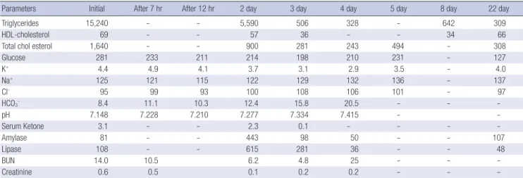
On the second day of hospitalization, the patient complained of aggravated epigastric pain. Follow-up laboratory analyses re- vealed a serum amylase level of 443 U/L and a serum lipase lev- el of 615 U/L. On abdominal computed tomography (CT) scan, the pancreas was diffusely swollen with peripancreatic fat infil- tration and fluid collection, which suggested acute pancreatitis Table 1. Sequential laboratory results
Parameters Initial After 7 hr After 12 hr 2 day 3 day 4 day 5 day 8 day 22 day
Triglycerides 15,240 - - 5,590 506 328 - 642 309
HDL-cholesterol 69 - - 57 36 - - 34 66
Total chol esterol 1,640 - - 900 281 243 494 - 308
Glucose 281 233 211 214 198 210 231 - 127
K+ 4.4 4.9 4.1 3.7 3.1 2.9 3.5 - 4.0
Na+ 125 121 115 122 129 132 136 - 137
Cl- 95 99 93 100 108 106 101 - 97
HCO3- 8.4 11.1 10.3 12.4 15.8 20.5 - - -
pH 7.148 7.228 7.210 7.277 7.334 7.415 - -
Serum Ketone 3.1 - - 2.3 0.1 - - - -
Amylase 81 - - 443 98 50 - - 107
Lipase 108 - - 615 281 36 - - 48
BUN 14.0 10.5 6.2 4.8 25 - - -
Creatinine 0.6 0.5 0.1 0.2 0.2 - - -
Triglycerides (mg/dL), HDL-cholesterol (mg/dL), Total cholesterol (mg/dL), Glucose (mg/dL), K+ (mEq/L), Na+ (mEq/L), Cl- (mEq/L), HCO3- (mEq/L), pH, Serum ketone (mmol/L), Amylase (U/L), Lipase (U/L), BUN (mg/dL), Creatinine (mg/dL).
Fig. 1. Photograph of hyperli- pidemic serum extracted by centrifugation from the pati- ent’s blood sample.
Table 2. Summary of severe hypertriglyceridemia in DKA in Korea
Ref. Year Sex/Age Past history Initial TG
(mg/dL) Follow-up TG (Time) Associated conditions Treatment Outcome
(5) 1999 F/25 T1D, without treatment 12,864 72 (after 72 hr)
83 (after 5 month) Non-specific Conservative with insulin Survival (20) 2002 M/38 T2D on insulin, Acromegaly 1,488 427 (after 72 hr)
80 (after 5 month)
Acute pancreatitis Conservative with insulin Survival (6) 2008 M/26 T1D on OHA (omit insulin) 11,929 1,316 (after 72 hr)
77 (after 45 days) Lipemia retinalis Conservative with insulin Survival
Present 2009 F/20 T1D, omit insulin 15,240 506 (after 72 hr)
309 (after 22 days) Acute pancreatits Conservative with insulin Survival OHA, oral hypoglycemic agent; TG, triglyceride; T1D, type 1 diabetes mellitus; T2D, type 2 diabetes mellitus.
Hahn SJ, et al. • Severe Hypertriglyceridemia in Diabetic Ketoacidosis Accompanied by Acute Pancreatitis
http://jkms.org 1377
DOI: 10.3346/jkms.2010.25.9.1375
grade D, according to the Balthazar CT severity index (Fig. 2).
The Ranson’s score was 4 at this point. There was no lipidemia retinalis on an ophthalmologic exam.
On the third day of hospitalization, the TG was 506 mg/dL, total cholesterol 281 mg/dL, LDL-C 101 mg/dL and HDL-C 36 mg/dL. The epigastric pain had diminished. The patient com- menced oral intake, multiple subcutaneous insulin injection and fibrate medication.
On the fourth day of hospitalization, the serum amylase level was 50 U/L and lipase level was 36 U/L. The patient was dis- charged without any complication on post-admission day 8.
Fourteen days after discharge, analyses revealed total choles- terol 308 mg/dL, TG 309 mg/dL, LDL-C 184 mg/dL, HDL-C 66 mg/dL, serum amylase level 107 U/L and serum lipase level 48 U/L. Lipoprotein electrophoresis performed after recovery showed a normal pattern.
DISCUSSION
In DKA, the deficiency of insulin activates lipolysis in adipose tissue releasing increased FFA, which accelerates formation of VLDL in the liver. In addition, reduced activity of lipoprotein li- pase in peripheral tissue decreases removal of VLDL from the plasma, resulting in hypertriglyceridemia (8). Moderate hyper- triglyceridemia is common during episodes of DKA (9). How- ever, severe hypertriglyceridemia, which is defined as a TG level
>2,000 mg/dL, is rare. Although morbidity is <1%, clinicians should be aware that devastating consequences such as acute pancreatitis or lipidemia retinalis are possible (8). In extreme
A B
Fig. 2. Contrast-enhanced pancreas CT scan (arterial phase). Initial pancreas CT scan shows diffuse swelling of pancreas body, tail, demonstrating CT grade D acute pancreatitis with peripancreatic fat infiltration (arrow), fluid collection suggesting inflammation (A) and edematous change of pancreatic head (openarrow) (B).
cases, co-existence of genetic mutations in lipoprotein lipase should be suspected (10).
During treatment of DKA with severe hypertriglyceridemia, pseudohyponatremia or pseudonormoglycemia due to labora- tory interference may lead to delay of proper management. Frier et al. suggested that if serum triglyceride concentration exceeds 2,500 mg/dL, measured electrolyte can decrease by over 5% be- cause of the intracellular movement of serum lipid components.
Therefore, in the hyponatremic state, the clinician should con- sider the possibility of pseudohyponatremia and avoid overtreat- ment with hypertonic saline (11). We presently observed a de- creased sodium level of 125 mEq/L to 115 mEq/L during the re- covery phase, which necessitate temporary correction with 3%
hypertonic saline.
In severe hypertriglyceridemia, there is an increased risk of developing acute pancreatitis. The mechanism is related to high plasma chylomicrons or TGs, which are hydrolyzed by lipase in the pancreatic capillaries and subsequently trigger FFA release (12) that, in turn, causes activation of trypsinogen and commenc- es pancreatic capillary damage by free radical damage (13, 14).
The common clinical scenario of hypertriglyceridemia-induced acute pancreatitis involves poorly-controlled diabetes mellitus with type IV hyperlipidemia (5), or chronic alcoholism (15). In contrast, moderate hyperlipidemia (usually <400 mg/dL) can be observed secondary to acute pancreatitis and should not be con- fused with the marked hypertriglyceridemia that causes acute pancreatitis (16), as in the present case.
Of note, normoamylasemia is possible in about 50% of pa- tients with hypertriglyceridemia-induced pancreatitis. The mech-
Hahn SJ, et al. • Severe Hypertriglyceridemia in Diabetic Ketoacidosis Accompanied by Acute Pancreatitis
1378 http://jkms.org DOI: 10.3346/jkms.2010.25.9.1375
anism is believed to be the interference with in vitro determina- tion of the actual amylase level by disturbance of the calorimet- ric method. Serial dilutions of the sample could reduce interfer- ence of light transmission by hyperlipidemic serum (17). In the present case, increased amylase occurred parallel to the de- creased TG level, which might have delayed the diagnosis of acute pancreatitis at initial presentation.
Although acute pancreatitis can initiate DKA, DKA itself may mask a co-existing acute pancreatitis that occurs in 10–15% of cases due to ambiguous clinical presentations (16). Even worse, nonspecific elevations of amylase and/or lipase without clinical evidence of pancreatitis have been reported in 24.7–79.0% of DKA cases (17). At least in those patients with continuous ab- dominal pain, it is prudent to seek further laboratory evaluation or a CT scan of the abdomen. In our case, not surprisingly, be- cause of the severe dehydration secondary to DKA, the initial and 48 h Ranson’s score was 4 points each, which is relatively high. In a previous study, the clinical course of acute pancreatitis with DKA seemed to be mild, although the mean Ranson’s score was 1.36±0.5 and the mean CT severity index (total 20 point) score was 4.6±1.2; none of the patients developed systemic complica- tion (17).
In the present case, hypertriglyceridemia was controlled with insulin without lipid lowering agents. However, in severe hyper- triglyceridemia, physicians should consider the application of plasma exchange to avoid complications (18). The present pa- tient has since been administered fibrate, which is the first-line drug of hypertriglyceridemia, and insulin treatment, in order to prevent recurrence of pancreatitis (19).
To our knowledge, this is the first report of a case in Korea of DKA and severe hypertriglyceridemia, complicated by acute pancreatitis. Although moderate hypertriglyceridemia in DKA is common, if the TG level exceeds 1,000 mg/dL, the clinician should consider the devastating consequences such as acute pancreatitis or lipemic retinalis, which might benefited from insulin administration and conservative management, unless otherwise necessitating the plasma exchange.
REFERENCES
1. Kitabchi AE, Nyenwe EA. Hyperglycemic crises in diabetes mellitus: dia- betic ketoacidosis and hyperglycemic hyperosmolar state. Endocrinol Metab Clin North Am 2006; 35: 725-51.
2. Chiasson JL, Aris-Jilwan N, Belanger R, Bertrand S, Beauregard H, Ekoe JM, Fournier H, Havrankova J. Diagnosis and treatment of diabetic keto- acidosis and the hyperglycemic hyperosmolar state. CMAJ 2003; 168:
859-66.
3. Kitabchi AE, Umpierrez GE, Murphy MB, Barrett EJ, Kreisberg RA, Malo- ne JI, Wall BM. Hyperglycemic crises in diabetes. Diabetes Care 2004; 27 Suppl 1: S94-102.
4. Exton JH. Mechanisms of hormonal regulation of hepatic glucose me- tabolism. Diabetes Metab Rev 1987; 3: 163-83.
5. Fortson MR, Freedman SN, Webster PD 3rd. Clinical assessment of hy- perlipidemic pancreatitis. Am J Gastroenterol 1995; 90: 2134-9.
6. Choi DS, Oh JH, Park IB, Kim JW, Choi KM, Kim YH, Kim NH, Kim SJ, Baik SH. A case of severe hypertriglyceridemia with diabetic ketoacido- sis. J Korean Diabetes Assoc 1999; 23: 715-21.
7. Choi SH, Sohn TS, Lee JI, Kim ES, Choi WH, Shin J, Son HS. A case of se- vere hypertriglyceridemia in diabetic ketoacidosis. Korean Clin Diabetes 2008; 9: 336-40.
8. Fulop M, Eder H. Severe hypertriglyceridemia in diabetic ketosis. Am J Med Sci 1990; 300: 361-5.
9. Fulop M, Eder HA. Plasma triglycerides and cholesterol in diabetic keto- sis. Arch Intern Med 1989; 149: 1997-2002.
10. Karagianni C, Stabouli S, Roumeliotou K, Traeger-Synodinos J, Kavaz- arakis E, Gourgiotis D, Lambrou J, Kanavakis E. Severe hypertriglyceri- daemia in diabetic ketoacidosis: clinical and genetic study. Diabet Med 2004; 21: 380-2.
11. Frier BM, Steer CR, Baird JD, Bloomfield S. Misleading plasma electro- lytes in diabetic children with severe hyperlipidaemia. Arch Dis Child 1980; 55: 771-5.
12. Havel RJ. Pathogenesis, differentiation and management of hypertriglyc- eridemia. Adv Intern Med 1969; 15: 117-54.
13. Havel RJ. Approach to the patient with hyperlipidemia. Med Clin North Am 1982; 66: 319-33.
14. Tsuang W, Navaneethan U, Ruiz L, Palascak JB, Gelrud A. Hypertriglyc- eridemic pancreatitis: presentation and management. Am J Gastroen- terol 2009; 104: 984-91.
15. Yadav D, Pitchumoni CS. Issues in hyperlipidemic pancreatitis. J Clin Gastroenterol 2003; 36: 54-62.
16. Nair S, Yadav D, Pitchumoni CS. Association of diabetic ketoacidosis and acute pancreatitis: observations in 100 consecutive episodes of DKA. Am J Gastroenterol 2000; 95: 2795-800.
17. Yadav D, Nair S, Norkus EP, Pitchumoni CS. Nonspecific hyperamylase- mia and hyperlipasemia in diabetic ketoacidosis: incidence and correla- tion with biochemical abnormalities. Am J Gastroenterol 2000; 95: 3123-8.
18. Furuya T, Komatsu M, Takahashi K, Hashimoto N, Hashizume T, Waji- ma N, Kubota M, Itoh S, Soeno T, Suzuki K, Enzan K, Matsuo S. Plasma exchange for hypertriglyceridemic acute necrotizing pancreatitis: report of two cases. Ther Apher 2002; 6: 454-8.
19. Huang DB, Raskin P. Diabetic hypertriglyceridemia-induced acute pan- creatitis masquerading as biliary pancreatitis. J Diabetes Complications 2002; 16: 180-2.
20. Lee CY, Lee MK, Lee SY, Hong SN, Kim HH, Kang BH, Kang HW, Lee BW, Park YJ, Min YK, Lee MS, Kim KW, Kim JH. A case of acromegaly with diabetic detoacidosis and hypertriglyceridemia-induced acute pan- creatitis. J Korean Soc Endocrinol 2002; 17: 110-6.