© 2011 The Korean Academy of Medical Sciences.
This is an Open Access article distributed under the terms of the Creative Commons Attribution Non-Commercial License (http://creativecommons.org/licenses/by-nc/3.0) which permits unrestricted non-commercial use, distribution, and reproduction in any medium, provided the original work is properly cited.
pISSN 1011-8934 eISSN 1598-6357
Recurrent Bilateral Branch Retinal Artery Occlusion with Hearing Loss and Encephalopathy: The First Case Report of Susac
Syndrome in Korea
We report the first case of Susac syndrome in Koreans, in a 23-yr-old female patient who presented with sudden visual loss and associated neurological symptoms. Ophthalmic examination and fluorescein angiography showed multiple areas of branch retinal artery occlusion, which tended to recur in both eyes. Magnetic resonance imaging showed dot- like, diffusion-restricted lesions in the corpus callosum and left fornix, and audiometry showed low-frequency sensory hearing loss, compatible with Susac syndrome. She received immunosuppressive therapy with oral steroid and azathioprine. Three months later all the symptoms disappeared but obstructive vasculitis have been relapsing. This patient demonstrated the entire clinical triad of Susac syndrome, which tends to occur in young females. Although this disorder has rarely been reported in Asian populations, a high index of suspicion is warranted for early diagnosis and timely treatment.
Key Words: Susac Syndrome; Korean; Branch Retinal Artery Occlusion; Hearing Loss;
Encephalopathy Soo Geun Joe1, June-Gone Kim1,
Sun Uck Kwon2, Choong Wook Lee3, Hyun Woo Lim4 and Young Hee Yoon1 Departments of 1Ophthalmology, 2Neurology,
3Radiology and 4Otolaryngology, Asan Medical Center, University of Ulsan, College of Medicine, Seoul, Korea
Received: 26 July 2011 Accepted: 21 September 2011 Address for Correspondence:
Young Hee Yoon, MD
Department of Ophthalmology, Asan Medical Center, University of Ulsan, College of Medicine, 88 Olympic-ro 43-gil, Songpa-gu, Seoul 138-736, Korea
Tel: +82.2-3010-3675, Fax: +82.2-470-6440 E-mail: [email protected]
http://dx.doi.org/10.3346/jkms.2011.26.11.1518 • J Korean Med Sci 2011; 26: 1518-1521
CASE REPORT
Ophthalmology
INTRODUCTION
Susac syndrome (SS) consists of the clinical triad of encephalop- athy, branch retinal artery obstruction (BRAO) and hearing loss without prominent systemic manifestations (1-4). It is thought to be caused by pre-capillary arteriole obstruction of the brain, retina and inner ear due to damage from circulating anti-endo- thelial cell antibodies (3-5). These symptoms may not always occur, and disease presentation is often insidious (2, 4, 6), such that patients with initial presenting symptoms may be misdiag- nosed. Although many patients with SS have been reported in Western countries, none has yet been reported in Korea.
CASE DESCRIPTION
A 23-yr-old woman visited the emergency clinic on November 23, 2011 with visual disturbance and a visual field defect in her left eye which had developed 1 day earlier. She had started to experience recurrent visual field defects in both eyes and head- ache 6 months earlier, and these symptoms became aggravated 1 month earlier. She denied having diabetes, hypertension, con- nective tissue disease, tuberculosis, hematologic disease or car- diovascular disease. She also stated that she had not used oral contraceptives and was not pregnant.
On initial examination, her pupils were reactive to light with-
out relative afferent papillary defect. Corrected visual acuity was 20/20 in her right eye and 20/25 in her left eye. IOP was 18 mmHg in both eyes. Slit lamp examination revealed no abnormal find- ings in the anterior segment of both eyes. Fundoscopic exami- nation showed an edematous lesion in the supra-temporal area of her left eye (Fig. 1A), and normal findings in her right eye. Flu- orescein angiography (FA) revealed obstructive vasculitis in the supra-temporal branch of the retinal artery in her left eye (Fig.
1B). A visual field test showed an infra-nasal field defect in her left eye and a small field defect in her right eye (Fig. 2).
Two days later, the patient again visited the emergency clinic due to sudden hearing loss in her left ear, which had developed that morning. Pure tone audiometry showed sensory neuronal hearing loss at low-frequency in her left ear (Fig. 3). Three weeks later, she complained of a visual field defect in her right eye. A repeat FA showed BRAO in her right eye, but the signs of obstruc- tive vasculitis in her left eye had disappeared. Two days later, she was admitted to the neurologic ward for tinnitus and severe headache with arm numbness.
Laboratory tests, including for lupus anticoagulant, anticar- diolipin antibody, erythrocyte sedimentation rate, and C-reac- tive protein, were all normal. Cerebrospinal fluid (CSF) analysis showed a slight increase in protein concentration (74.4 mg/dL), without evidence of oligoclonal bands. There were no specific hematologic or rheumatologic abnormalities. Magnetic reso-
Joe SG, et al. • Susac Syndrome in Korea
http://jkms.org 1519
http://dx.doi.org/10.3346/jkms.2011.26.11.1518
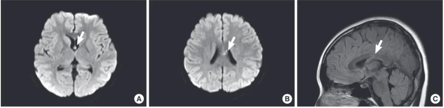
nance (MR) imaging showed dot-like, diffusion-restricted lesions with T2 signal change in the corpus callosum and left fornix (Fig. 4A, B).
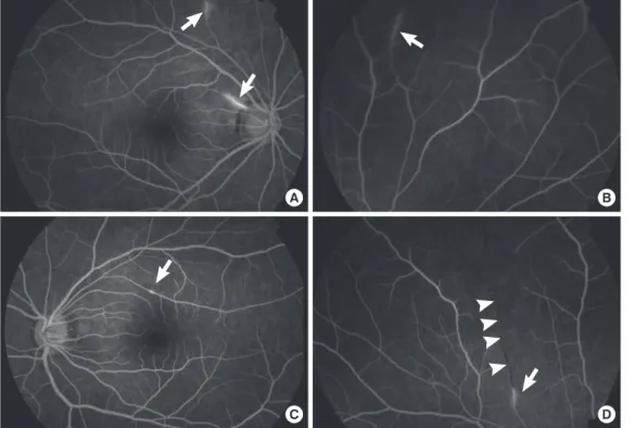
After considering all these findings, she was diagnosed with SS and started on high dose steroid therapy. Two months later, FA detected newly developed occlusive vasculitis in both eyes (Fig. 5). Follow-up sagittal Fluid-attenuated inversion recovery (FLAIR) MR imaging showed a signal change in the area corre- sponding to that of the previous infarction in the corpus callo- sum (Fig. 4C), but there was no evidence of newly developed ischemic brain lesions. The dosage of oral steroids was main- tained, and azathioprine (50 mg bid) was added. Three months
later, her symptoms of headache, tinnitus and visual field defect had all resolved. However, FA showed new vasculitis and obstruc- tion of the branch retinal artery, although all previously affected lesions had normalized.
DISCUSSION
We have described here a young female patient who showed the clinical triad of SS: BRAO, hearing loss and encephalopathy. To our knowledge, this is the first case report of SS in Korea. Since the first description of SS in 1977, hundreds of patients with SS have been reported, mainly in Western countries (3). Women
A B
Fig. 1. Fundus photo showing edematous lesions in the supra-temporal area (A). Fluorescein angiography, show- ing a hyperfluorescent arterial wall proximal to the ob- structed branch retinal artery (B).
A B C
Fig. 4. Magnetic resonance image of the brain. (A, B) Diffusion weighted images, showing a diffusion restricted lesion in the corpus callosum and left fornix. (C) Sagittal Fluid- attenuated inversion recovery (FLAIR) image showing a focal signal change in the corpus callosum.
LEFT EYE
TOTAL DEVIATION PUPIL DIAMETER:
VISUAL ACUITY:
RX:
TEST DURATION: 1:59 FIXATION TARGET: Central FIXATION ERRS: 0/10 (0%) FALSE POS ERRS: 0/10 (0%)
TEST DURATION: 1:59 FIXATION TARGET: Central FIXATION ERRS: 0/10 (0%) FALSE POS ERRS: 0/10 (0%) PUPIL DIAMETER:
VISUAL ACUITY:
RX:
RIGHT EYE
Fig. 2. Visual field test results, showing an infranasal field defect of the left eye cor- responding to the obstructed lesion and a small area of visual field defect in the right eye.
Fig. 3. Pure tone audiometry, showing decreased sensitivity to low-frequency sounds in the left ear.
Right Left
Frequency (Hz) Frequency (Hz)
Hearing level (dB) Hearing level (dB)
125 125
0 0
20 20
60 60
40 40
80 80
10 10
30 30
70 70
50 50
90 90
100 100
110 110
250 500 1000 200030004000 8000 250 500 1000 200030004000 8000
Joe SG, et al. • Susac Syndrome in Korea
1520 http://jkms.org http://dx.doi.org/10.3346/jkms.2011.26.11.1518
are more frequently affected than men, with young white wom- en between the age of 20 and 40 yr being most susceptible (3, 6).
A MEDLINE search for SS in Asian populations showed only one case report, in a Japanese male (1).
Retinal arterial occlusion is rare in younger individuals (7), with nearly 75% of these patients being older individuals with retinal arterial occlusion caused by embolization of atheroma- tous plaques of the carotid artery (7, 8). Retinal arterial occlusion in younger patients is usually due to migraine, a coagulation ab- normality, cardiac disease, trauma, drug abuse or connective tissue disease (7, 9). In patients with SS, localized retinal vascu- litis rather than embolism is believed to cause arteriolar occlu- sions, which usually do not occur at bifurcations (10). BRAO tends to be bilateral, and to be multiple, widely disseminated in the retina, and temporally separated by as long as several months.
The white material in the arterial lumen may represent aggre- gations of immune complexes or debris from damaged endo- thelium (11). A characteristic feature on FA is arterial wall hyper- fluorescence, often proximal to sites of occlusion, as shown in our patient, and suggesting endothelial dysfunction (11).
Common neurological features of SS include severe headache, which may occur several months prior to other symptoms, and personality disorder (12). Due to the multifocal vasculitis, neu- rological manifestations are extremely variable and include atax- ia, vertigo, corticospinal tract signs, and even seizure. On MRI, microinfarcts are best seen on T2 weighted images as hyperin- tense lesions (13). These microinfarcts are usually multifocal and located in the area of the corpus callosum, as seen in our patient (3, 4). CSF may contain a normal or high protein concen-
A B
C D
Fig. 5. Fluorescein angiography showing hyperfluorescent vasculi- tis lesions (arrows) (A-D) and an obstructed branch retinal artery (arrowheads) (D).
tration. High protein concentration, coupled with an absence of oligoclonal bands, is typical, as in our patient (14).
Characteristic audiological findings in SS include low-frequen- cy hearing loss, vertigo, and tinnitus. Our patient had both low- frequency hearing loss and tinnitus, which were likely due to mi- croangiopathic lesions of the apical cochlea end arterioles (2).
The diagnosis of SS is often difficult because its characteristic signs often do not occur simultaneously or may be too subtle for the patient to notice (1, 15). In one case report, the patient pre- sented with encephalopathy 10 yr before hearing loss, with recur- rence of encephalopathy 18 yr later (2). Another patient experi- enced multiple BRAO episodes over 30 yr, followed by a Swiss- cheese like corpus callosum on MR imaging without signs of encephalopathy (1). Therefore, patients who manifest one of the clinical signs of SS should be suspected of this disorder. Al- though SS has been considered rare, it may be more common than reported (1, 13, 14).
As in many other autoimmune diseases, steroid therapy is the mainstay of treatment for SS (2-4). Initial high dose cortico- steroid therapy, followed by tapering, has been recommended (2, 4). Additional immune suppressive treatment, with cyclo- phosphamide, mycophenolate mofetil, methotrexate, or cyclo- sporine, may be helpful (2). Many patients have improved after receiving intravenous immunoglobulin (2).
To avoid disastrous consequences, aggressive and timely treat- ment is needed, for a sufficient period of time (2). However, pro- gression of SS may be insidious and the symptoms do not always occur together.
Joe SG, et al. • Susac Syndrome in Korea
http://jkms.org 1521
http://dx.doi.org/10.3346/jkms.2011.26.11.1518
REFERENCES
1. Murata Y, Inada K, Negi A. Susac syndrome. Am J Ophthalmol 2000; 129:
682-4.
2. Rennebohm RM, Egan RA, Susac JO. Treatment of Susac’s syndrome.
Curr Treat Options Neurol 2008; 10: 67-74.
3. Susac JO, Egan RA, Rennebohm RM, Lubow M. Susac’s syndrome: 1975- 2005 microangiopathy/autoimmune endotheliopathy. J Neurol Sci 2007;
257: 270-2.
4. Rennebohm R, Susac JO, Egan RA, Daroff RB. Susac’s syndrome: update.
J Neurol Sci 2010; 299: 86-91.
5. Jarius S, Neumayer B, Wandinger KP, Hartmann M, Wildemann B. Anti- endothelial serum antibodies in a patient with Susac’s syndrome. J Neu- rol Sci 2009; 285: 259-61.
6. Martinet N, Fardeau C, Adam R, Bodaghi B, Papo T, Piette JC, Lehoang P.
Fluorescein and indocyanine green angiographies in Susac syndrome.
Retina 2007; 27: 1238-42.
7. Trevino R, Pearlman R. Idiopathic recurrent branch retinal arterial oc- clusion in a young adult. Optom Vis Sci 1998; 75: 11-6.
8. Beatty S, Au Eong KG. Acute occlusion of the retinal arteries: current con-
cepts and recent advances in diagnosis and management. J Accid Emerg Med 2000; 17: 324-9.
9. Recchia FM, Brown GC. Systemic disorders associated with retinal vas- cular occlusion. Curr Opin Ophthalmol 2000; 11: 462-7.
10. Gass JD, Tiedeman J, Thomas MA. Idiopathic recurrent branch retinal arterial occlusion. Ophthalmology 1986; 93: 1148-57.
11. Notis CM, Kitei RA, Cafferty MS, Odel JG, Mitchell JP. Microangiopathy of brain, retina, and inner ear. J Neuroophthalmol 1995; 15: 1-8.
12. Susac JO, Hardman JM, Selhorst JB. Microangiopathy of the brain and retina. Neurology 1979; 29: 313-6.
13. O’Halloran HS, Pearson PA, Lee WB, Susac JO, Berger JR. Microangiop- athy of the brain, retina, and cochlea (Susac syndrome). A report of five cases and a review of the literature. Ophthalmology 1998; 105: 1038-44.
14. Susac JO. Susac’s syndrome: the triad of microangiopathy of the brain and retina with hearing loss in young women. Neurology 1994; 44: 591-3.
15. Dörr J, Radbruch H, Bock M, Wuerfel J, Bruggemann A, Wandinger KP, Zeise D, Pfueller CF, Zipp F, Paul F. Encephalopathy, visual disturbance and hearing loss-recognizing the symptoms of Susac syndrome. Nat Rev Neurol 2009; 5: 683-8.