• 검색 결과가 없습니다.

Neuroimaging-Based Deep Learning in Autism Spectrum Disorder and Attention-Deficit/Hyperactivity Disorder

N/A
N/A
Protected

Academic year: 2021

Share "Neuroimaging-Based Deep Learning in Autism Spectrum Disorder and Attention-Deficit/Hyperactivity Disorder"

Copied!
8
0
0

로드 중.... (전체 텍스트 보기)

전체 글

(1)

INTRODUCTION

During the first 12 months of life, various factors such as heredity and environment affect the progression of neuro- developmental disorders such as autism spectrum disorder (ASD) and attention-deficit/hyperactivity disorder (ADHD) [1]. Several studies have pointed out that in the case of ASD, behavioral signs may appear during the age of 6–12 months [2], but in clinical settings, on average, clear diagnosis is only possible after 4 years of age [3]. In ADHD, diagnostic accura- cy in children older than 7 years of age is limited due to dif- ficulties in distinguishing them from children with normal development [4-7]. Therefore, in longitudinal studies, the sta- bility of ADHD diagnosis before 7 years of age has been a prob- lem; up to 50 other studies have concluded that diagnostic sta- bility rate is below 50% for children under this age [8-11].

To facilitate better course and prognosis of neurodevelop- mental disorders, it is important for clinical interventions to precede certain critical periods of brain plasticity develop- ment through early and accurate diagnosis. Although early detection of neurodevelopmental disorders is vital, the de- velopment of non-invasive, quantitative, and sensitive bio- markers has been insufficient. It would, thus, be valuable to

detect biomarkers (e.g., brain images, blood, genes, pupil- lometry, etc.) early in development that can differentiate chil- dren with neurodevelopmental disorders and those with normal development pathways.

The several potential biomarkers found thus far exist only as sporadic information, and it is difficult to derive mean- ingful information on diagnosis by collecting them. In order to solve this problem, deep learning (DL) technology can be used to collect biomarker information, allowing early detec- tion of neurodevelopmental disorders in a simple, accurate, timely, and cost-efficient manner.

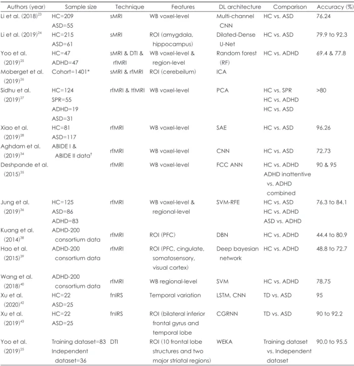
This review summarizes the latest papers that use DL in the field of neuroimaging in ASD and ADHD research (Table 1).

Through our review, the merits and limitations, as well as fu- ture directions of neuroimaging-based DL of neurodevelop- mental disorders will be brought to light.

DEEP LEARNING

DL involves training and testing a multi-layered neural net- work using artificial intelligence (AI) to learn complex struc- tures and achieve a high level of abstraction [12]. It is a ma- chine learning technique which is different from existing techniques in that it can acquire optimal representation through consecutive nonlinear transformation of large amounts of raw data. This feature has strengths in terms of abstraction

This is an Open Access article distributed under the terms of the Creative Commons Attribution Non-Commercial License (https://creativecommons.org/licenses/by-nc/4.0) which permits unrestricted non-commercial use, distribution, and reproduction in any medium, provided the original work is properly cited.

Neuroimaging-Based Deep Learning in Autism Spectrum Disorder and Attention-Deficit/Hyperactivity Disorder

Jae-Won Song

1

, Na-Rae Yoon

1

, Soo-Min Jang

1

, Ga-Young Lee

2

, and Bung-Nyun Kim

1

1

Department of Child and Adolescent Psychiatry, Seoul National University Hospital, Seoul, Korea

2

Seoul National University Hospital, Autism and Developmental Disorder Center, Seoul, Korea

Deep learning (DL) is a kind of machine learning technique that uses artificial intelligence to identify the characteristics of given data and efficiently analyze large amounts of information to perform tasks such as classification and prediction. In the field of neuroimaging of neurodevelopmental disorders, various biomarkers for diagnosis, classification, prognosis prediction, and treatment response prediction have been examined; however, they have not been efficiently combined to produce meaningful results. DL can be applied to overcome these limitations and produce clinically helpful results. Here, we review studies that combine neurodevelopmental disorder neuroimag- ing and DL techniques to explore the strengths, limitations, and future directions of this research area.

Key Words: Neuroimaging; Neurodevelopmental disorder; Autism spectrum disorder; Attention-deficit/hyperactivity disorder;

Deep learning; Review.

Received: May 13, 2020 / Revision: June 8, 2020 / Accepted: June 11, 2020

Address for correspondence: Bung-Nyun Kim, Department of Child and Adolescent Psychiatry, Seoul National University Hospital, 101 Daehak-ro, Jongno-gu, Seoul 03080, Korea

Tel: +82-2-2072-3647, Fax: +82-2-747-2471, E-mail: [email protected]

(2)

and complexity, enabling accurate visual recognition. In the medical field, it has gained a spotlight in the field of radiolo- gy, and is used for classification, diagnosis, risk factor analy- sis, prognosis, and prediction of treatment response. In the field of neuroimaging, it is used for classification and diag-

nosis of neurologic conditions such as stroke, neurodegen- erative disorders, and psychiatric disorders [13-18].

DL uses artificial neural networks (ANN), which are mod- eled on the structure of human neural networks to perform cognitive functions [19]. In a feedforward network, informa-

Table 1. Summary of neuroimaging based deep learning studies

Authors (year) Sample size Technique Features DL architecture Comparison Accuracy (%) Li et al. (2018)

23

HC=209

ASD=55

sMRI WB voxel-level Multi-channel CNN

HC vs. ASD 76.24

Li et al. (2019)

24

HC=215 ASD=61

sMRI ROI (amygdala,

hippocampus)

Dilated-Dense U-Net

HC vs. ASD 79.9 to 92.3

Yoo et al.

(2019)

25

HC=47 ADHD=47

sMRI & DTI &

rfMRI

WB voxel-level &

region-level

Random forest (RF)

HC vs. ADHD 69.4 & 77.8

Moberget et al.

(2019)

26

Cohort=1401* sMRI & rfMRI ROI (cerebeilum) ICA

Sidhu et al.

(2019)

27

HC=124 SPR=55 ADHD=19 ASD=31

rfMRI & tfMRI WB voxel-level PCA HC vs. SPR HC vs. ADHD HC vs. ASD

>80

Xiao et al.

(2019)

28

HC=81 ASD=117

rfMRI WB voxel-level SAE HC vs. ASD 96.26

Aghdam et al.

(2019)

34

ABIDE I &

ABIDE II data

rfMRI WB voxel-level CNN HC vs. ASD 72.73

Deshpande et al.

(2015)

35

rfMRI WB voxel-level FCC ANN HC vs. ADHD

ADHD inattentive vs. ADHD combined

90 & 95

Jung et al.

(2019)

36

HC=125 ASD=86 ADHD=83

rfMRI WB voxel-level &

regional-level

SVM-RFE HC vs. ASD HC vs. ADHD ASD vs. ADHD

76.3 to 84.1

Kuang et al.

(2014)

38

ADHD-200

consortium data rfMRI ROI (PFC) DBN HC vs. ADHD 44.4 to 80.9

Hao et al.

(2015)

39

ADHD-200 consortium data

rfMRI ROI (PFC, cingulate, somatosensory, visual cortex)

Deep bayesian network

HC vs. ADHD 48.8 to 72.7

Wang et al.

(2018)

40

ADHD-200

consortium data rfMRI WB regional-level SVM HC vs. ADHD 78.75

Xu et al.

(2020)

42

HC=22 ASD=25

fnIRS Temporal variation LSTM, CNN TD vs. ASD 95

Xu et al.

(2019)

43

HC=22 ASD=25

fnIRS ROI (bilateral inferior frontal gyrus and temporal lobe

CGRNN TD vs. ASD 90 to 92.2

Yoo et al.

(2019)

25

Training dataset=83 Independent dataset=36

DTI ROI (10 frontal lobe structures and two major striatal regions)

WEKA Training dataset vs. Independent dataset

90.0 to 95.5

*philadelphia neurodevelopmental cohort, age 8–23 years,

ABIDE I and II: autism brain imaging data exchange I and II. HC: healthy control, ASD: autism sepectrum disorder, ADHD: attention-deficit/hyperactivity disorder, SPR: schizophrenia, MRI: magnetic reso- nance imaging, sMRI: structural MRI, DTI: diffuse tensor imaging, rfMRI: resting state functional MRI, tfMRI: task-based function MRI, fNIRS: functional near-infrared spectroscopy, WB: whole brain, ROI: region of interest, PFC: prefrontal cortex, ICA: independent component analysis, PCA: principal component analysis, SAE: sparse auto-encoder, CNN: convolutional neural network, FCC ANN:

fully connected cascade artificial neural network, SVM-RFE: support vector machine-recursive feature elimination, DBN: deep belief

network, SVM: support vector machine, LSTM: long short-term memory, CGRNN: consisting of CNN and GRU, WEKA: waikato envi-

ronment for knowledge analysis

(3)

tion is propagated in one direction from the input to the out- put layer, whereas in a recurrent network, past information makes feedback links that can affect the output. This con- nection allows information to be retained in a neural network in a manner similar to memory, and a model capable of pro- cessing data can be constructed.

There are two main stages of DL. The first (training) stage acquires the optimal algorithm to perform the desired task (e.g., classification, diagnosis, prediction, etc.) by learning the available data. In the second (test) stage, an evaluation of whether the trained model can accurately distinguish the newly observed data is conducted [20].

Convolutional neural networks (CNN), which are most commonly used in neuroimaging, were developed as mod- els of the visual cortex to process visual information and are a type of feedforward neural network. CNN consists of three layers: input layer, convolutional layer, and pooling layer. The convolutional layer consists of several feature maps. All ar- tificial neurons in a given feature map are connected to the previous layer of the feature map in the same region (local connectivity), and the locality is distinguished by connec- tions with different weights from feature maps in other re- gions. Therefore, a feature map can be used as a feature de- tector to scan a pattern type, called a kernel. The process of making local connections with different weights to form nu- merous kernels is done automatically within the network and without any interventions. When multiple convolutional lay- ers exist, increasing amounts of abstract visual information can be processed [12].

Currently, in addition to CNN, various models such as Fully Convolutional Network and Dilated-Dense U-Net are used in research [12].

NEUROIMAGING-BASED DEEP LEARNING IN PSYCHIATRY

In the field of neuroimaging, the interest in generalization of classification using machine learning is on the rise. DL tech- nology is mainly used in psychiatry as a diagnostic model for distinguishing between patient and normal groups, a model for predicting the risk of developing disorders in a high-risk group, and a model for predicting response to a treatment [12]. A number of studies on diagnostic models have been conducted on Alzheimer’s disease and mild cognitive impair- ment, but the number of studies on neurodevelopmental dis- orders is small.

In one study, classification using machine learning was performed to evaluate large heterogeneous groups of four diseases: ASD (n=988), ADHD (n=930), post-traumatic stress disorder (n=87), and Alzheimer’s disease (n=132) [21]. In this

study, 18 machine learning classifications were applied, and the predictive powers of these classifiers were combined to form one consensus-classifier proposal. The identified func- tional connection pattern was firmly applied to differences in classification algorithms, age, and data acquisition sites, and was confirmed to have diagnostic predictive ability along with the ability to distinguish statistically significant differences between groups.

Research on neuroimaging-based DL of neurodevelop- mental disorders is mainly conducted using magnetic reso- nance imaging (MRI), functional MRI (fMRI), and functional near-infrared spectroscopy (fNIRS). Most studies focus on evaluating diagnostic accuracy through DL algorithms, and several MRI studies utilize DL to analyze structural differenc- es between brain regions of patient groups and normal devel- opment groups to develop biomarkers for early detection.

MAGNETIC RESONANCE IMAGING

In a 2019 study by Moon et al. [22], 40 machine learning algorithm studies for ASD published between 2007 and 2018 were systematically reviewed and meta-analysis was con- ducted. Among them, meta-analysis of 12 samples using struc- tural MRI revealed that the integrated sensitivity was 0.83, specificity was 0.84, and area under the curve/partial area under the curve (AUC/pAUC) was 0.90/0.83. Based on these results, the authors deemed the accuracy of the machine learning algorithm using structural MRI in ASD diagnosis to be acceptable.

Li et al. [23] applied multi-channel CNN using a patch- level data-expanding strategy to brain MRI data to predict ASD early in life. A total of 264 brain MRI scans [55 ASD groups, 209 normal control (NC) groups] registered in the National Database for Autism Research (NDAR) were col- lected and areas of brain structures showing statistically sig- nificant differences between the ASD and NC groups were selected (data-driven anatomical landmark identification).

When the corresponding region was extracted from MRI scans and subjected to DL using a multi-channel CNN, the diagnostic accuracy improved by 24% over a three-dimen- sional CNN.

Li et al. [24] also used a new DL approach, the Dilated-

Dense U-Net, to find ASD neurobiological anomalies and

confirm critical timing of the progression through longitu-

dinal studies of amygdala and hippocampus subfield devel-

opment. They collected 276 longitudinal brain MRI scans (30

ASD, 31 mild autism spectrum, 215 NC groups) registered

in the NDAR. Cross-analysis revealed that the ASD group

showed bilateral amygdala and bilateral CA1-3 size expan-

sion at 6 months after birth and bilateral amygdala, left CA1-

(4)

3, left subiculum size expansion at 24 months after birth. Lon- gitudinal analysis showed that at about 6 months of age, there was a size expansion of bilateral amygdala and bilateral CA1-3 in the ASD group with an increase in the size difference be- tween the groups at around 24 months of age. They suggest- ed that overgrowth of amygdala and CA1-3 starting at 6 months of age may be associated with the expression of ASD, and highlighted the possibility of early ASD detection from 6 months of age onwards.

Yoo et al. [25] confirmed that machine learning using multi- mode neurological imaging and genetic data could distin- guish typically developing children from an ADHD group.

Polygenetic risk scores calculated from cortical morphology, diffuse scalar and resting state functional connectivity, and norepinephrine/dopamine/glutamate genes were extracted from 47 ADHD children and 47 matched NC children. A mod- el consisting of cortical thickness and volume characteristics achieved a maximum accuracy of 85.1%. Morphological chang- es across insula, sensory/motor cortex, and ventral frontal cortex were also identified as key predictors, accounting for 18% of the ADHD measurement scale. Dynamic zone ho- mogeneity within the default network accounted for 6.4% of the missing errors in the continuous performance test (CPT) and validity testing achieved an accuracy of 69.4%. The re- sults of this study indicate that structural abnormalities re- lated to salience detection, sensory processing and response suppression can be powerful classifiers for ADHD diagnosis and symptoms.

Evidence is accumulating on the role of the cerebellum in psychiatric diseases such as schizophrenia, bipolar disorder, depression, anxiety disorder, and ADHD; however, little is known about its role during the development of these diseas- es. Currently, it is unclear whether the morphological aspects of the cerebellum are associated with the expression of spe- cific symptoms. In one study, analysis using machine learn- ing was conducted to determine whether morphological char- acteristics of the cerebellum could reliably predict general cognitive function and psychiatric symptoms in a large-scale developmental community composed of adolescents [26]. The morphological characteristics of the cerebellum were corre- lated with both general cognitive function and general psy- chopathology (mean correlations between predicted and ob- served values: r=0.20 and r=0.13, respectively; p<0.001).

Analysis of specific symptom areas revealed that norm-vio- lating behaviors (r=0.17; p<0.001), psychotic symptoms (r=

0.12; p<0.001) and anxiety symptoms (r=0.09; p=0.012) were significantly associated with cerebellar morphology. On the contrary, there were no associations with ADHD, depressive disorder, mania, and obsessive-compulsive disorder symp- toms. Cerebellar features appeared to be most important in

predicting general psychopathology, psychotic symptoms, and norm-violating behaviors.

FUNCTIONAL MAGNETIC RESONANCE IMAGING

fMRI measures neural activity using blood oxygenation- level dependent (BOLD) differences. A 2019 meta-analysis study conducted on fMRI and machine learning algorithm studies on ASD published between 2007 and 2018 [22] found the integrated sensitivity to be 0.69, specificity to be 0.66, and AUC/pAUC to be 0.71.

In one study, nonlinear dimensionality reduction (Locally Linear Embedding) was introduced to extract neural activi- ty information using BOLD time-series [27]. The methodolo- gy was verified by leave-one-out-cross-validation (LOOCV) accuracy for classification of psychiatric diagnosis using rest- ing state and task-related fMRI. Embedded fMRI showed high diagnostic performance (>80%) for 11 datasets includ- ing patients with schizophrenia, ADHD, ASD, and NC. Un- like the original fMRI data, embedded fMRI showed signifi- cantly better results than chance classification in ten out of 11 datasets.

Xiao et al. [28] attempted to validate diagnostic classifica- tion using DL on the full brain frequency from resting-state fMRI data of 198 school-aged ASD children. The average di- agnostic accuracy was found to be 96.26%, sensitivity to be 98.03%, and specificity to be 93.62%. These results show high- er diagnostic accuracy of up to 15% when compared with pre- vious studies that classified diagnosis using only one or two frequency bands [29-33].

Aghdam et al. [34] examined whether diagnostic discrim- ination is possible when resting-state fMRI data of 5–10 year old ASD children is subjected to DL with CNN [34]. Analy- sis using various methods showed that the maximum diag- nostic accuracy was 0.7273, sensitivity was 0.712, and speci- ficity was 0.7348.

In a 2015 study, Deshpande et al. [35] applied a fully con- nected cascade ANN to compare functional connectivity be- tween ADHD and NC children. Using this method, ADHD children of inattentive and combined subtype and an NC group were classified with an accuracy of 90%, and two sub- types with an accuracy of 95%.

In another fMRI study using machine learning technolo-

gy, functional connection patterns were examined in ADHD,

ASD, and matched typically developing children (7–15 year

old males) [36]. In the ASD group, functional connectivity

within the limbic system and somatomotor network was in-

creased as compared to the NC group. In addition, it was con-

firmed that the ADHD group showed an increase in func-

(5)

tional connectivity within the limbic system, visual, default mode, somatomotor, dorsal attention, fronto-parietal, and ventral cognitive networks as compared to the ASD group.

Using machine learning, ASD and normal groups, ADHD and normal groups, ADHD and ASD groups were classified with accuracies of 76.3%, 84.1%, and 79.3%, respectively.

The ADHD-200 consortium provides open-data sharing of ADHD patients with 776 resting-state fMRI and anatomi- cal datasets aggregated across eight independent imaging sites for better understanding of the neural basis of ADHD.

Several studies have been conducted using the ADHD-200 consortium data. Milham et al. used DL to analyze the brain areas that distinguish NC groups from ADHD groups, and found that the prefrontal, cingulate, and visual cortices are the most informative [37]. In a study using the same data, it was found that the prefrontal cortex region could be used to distinguish between the ADHD and NC groups of three dif- ferent samples with accuracies of 44.4%, 55.6%, and 80.9%, respectively [38]. In a 2015 study, the dataset of prefrontal, cingulate, somatosensory, and visual cortices were combined to distinguish between the ADHD and NC groups of three different samples with accuracies of 48.8%, 54%, and 72.7%, respectively [39]. In a 2018 study by Wang and Jiao [40], data from the ADHD-200 consortium was used to construct a di- agnostic model based on support vector machines (SVMs).

The performance of the diagnostic model was analyzed us- ing LOOCV and 10-fold cross-validations (CV). LOOCV re- vealed that the model had 78.75% accuracy, 76% sensitivity, and 80.71% specificity. The 10-fold CV average prediction ac- curacy was 75.54±1.34%, average sensitivity was 70.5±2.34%, and mean specificity was 77.44±1.47%. The differential pat- tern of ADHD was investigated using SVM, and the result was consistent with those of the previous studies, highlight- ing its potential as a biomarker.

FUNCTIONAL NEAR-INFRARED SPECTROSCOPY

It is known that the ASD patient and normal development groups differ in terms of spontaneous hemodynamic fluctu- ation in oxygenated hemoglobin (HbO2) and deoxyhemo- globin (Hb) [41].

According to a study by Xu et al. [42], hemodynamic chang- es in the temporal cortex of ASD children show weaker sta- tionarity and stronger memory and persistence for wireless shock than typically developing children. When analyzing whether diagnostic differentiation can be accomplished by applying a DL model to the spontaneous hemodynamic fluc- tuation of the brain over time, the temporal change of HbO2 showed 91.4% specificity, 98.6% sensitivity, and 95% accuracy.

The temporal change of Hb was able to discriminate between the groups with 94.3% specificity and 97.1% sensitivity.

In a study by Xu et al. [43], fNIRS was used to measure HbO2, Hb, and total hemoglobin in the bilateral inferior frontal gyrus and bilateral temporal cortex for 480 seconds.

The record was divided into 7-second units that were par- tially overlapped and they examined whether ASD and typ- ically developing children could be differentiated using a convolutional gated recurrent neural network (CGRNN), a multi-layer neural network that combines CNN and gate re- current unit. As a result, Hb of channel 10 showed 92.2% ac- curacy, 85% sensitivity, and 99.4% specificity and HbO2 of channel 43 showed 90.8% accuracy, 87.9% sensitivity, 93.8%

specificity. The total hemoglobin of channel 25 was able to discriminate between the ASD and NC groups with 90% ac- curacy, 81.6% sensitivity, and 98.5% specificity. Xu et al. sug- gested that the CGRNN model used in the study can be ap- plied clinically in a mere 7 seconds.

MULTIMODAL APPROACHES

In a study that used DL, Yoo et al. [44] sought to determine if pre-treatment demographics, clinical symptoms, environ- mental factors, neuropsychological characteristics, genetic factors, and brain imaging characteristics could predict sleep side effects after methylphenidate (MPH) administration.

They collected information on 83 ADHD patients, such as demographics, ADHD Rating Scale-IV (ADHD-RS) and Dis- ruptive Behavior Disorder rating scales, neuropsychological tests [CPT, Stroop color word test], genetic and environmen- tal variables (dopamine and norepinephrine receptor gene [DAT1, DRD4, ADRA2A, SLC6A2] polymorphisms, blood lead, and urine cotinine levels), and structural connectivity of fronto-striatal circuits. The resulting data were analyzed using three machine learning algorithms [logistic ridge re- gression (LR), SVM, J48]. The accuracy of predicting sleep problems was 95.5% for LR (AUC 0.99), 91% for SVM (AUC 0.85), and 90% for J48 (AUC 0.87). Inattentive symptoms in ADHD-RS, CPT response time variability, DAT1, ADRA2A, DRD4, SLC6A2 A3081T polymorphism and fronto-striatal structural connectivity were identified as the most differen- tial features. Validation analysis showed an accuracy of 86.1%

(AUC 0.92). This study suggested that multimodal classifiers, especially neuroimaging features, are helpful in predicting MPH side effects in ADHD children.

DISCUSSION

Machine learning and DL are among the most popular tech-

nologies of recent years, and meaningful results are constant-

(6)

ly being derived by combining them with neuroimaging. They have the advantage of overcoming limitations of traditional analytical methods by using previous mass-univariate statis- tics and enabling analysis of multivariate brain patterns and inter-regional correlations [45]. As described above, DL tech- niques provide significant diagnostic accuracy and results in detecting brain regions associated with disease, moving a step forward from traditional neuroimaging techniques. As multimodal approaches become easier with DL, opportuni- ties to better implement the bio-psycho-social model of psy- chiatric disorders in clinical situations have opened up.

The combination of neuroimaging and DL is still a field that requires much caution and improvement. The first is- sue is the problem of overfitting. Overfitting refers to the problem of failing to form an appropriate algorithm due to excessive generalization that results from fitting to various types of data [46-48]. This is especially troublesome when the dataset is small [49], causing overestimations of accuracy [50-52]. The more features one needs to analyze, the more training data is required to avoid overfitting, further exacer- bating the “curse of dimensionality.” Thus, there is a need to apply dimensionality reduction techniques such as principal components analysis or factor analysis in many machine learning studies [52]. In an attempt to overcome this diffi- culty, some studies have used methods such as regulariza- tion [53], early stopping [54], and drop out. [55]

The second issue is that, when using DL, bigger datasets result in more accurate algorithms, implying that the entire dataset will include all the images acquired in different ways (e.g., imaging tool, sites, etc.). In such cases, inadequate stan- dardization can lead to inaccurate processing [56]. As a po- tential solution to this problem, a field called “brittle AI” is being studied and implemented.

The third problem is that of psychiatric classification. The diagnosis of psychiatric disorders is based on observed symp- toms and signs, which can be an obstacle to prediction through DL. The training criteria for evaluating the accuracy of re- sults obtained through DL also follow the current diagnostic classification system. Therefore, a new approach called the re- search domain criteria initiative [57] was devised to overcome the heterogeneity of psychiatric disorder. This is a method used to detect the brain circuits associated with shared symp- toms and derive meaningful results for pathophysiology and etiology of psychiatric illnesses.

Finally, there is an overall lack of transparency and conse- quent interpretability from such experiments. Unlike general statistical analysis, it is difficult to intuitively recognize the results of analysis from the original data [58]. For example, it may be more difficult to detect abnormalities in specific brain regions of images from individuals using DL. This may be

due to a focus on features that are not clinically meaningful (e.g., artifacts) or a lack of information on treatment and/or developmental stages. Input modification methods [59] and deconvolution methods [60] are typically used to address this issue.Research that applies DL to neuroimaging analysis has been mainly conducted over the past 5 years and the field is still at an early stage. There are many research methods, such as targeting the entire brain area or a specific brain re- gion, and each study utilizes various ANNs. Hence, there is great potential for development of the technique. In the fu- ture, after overcoming the above-mentioned limitations, DL can be used to discover a biomarker that distinguishes vari- ous psychiatric disorders and not just to distinguish between patient and NC groups. In addition, studies on the prediction of prognosis and treatment effects for neurodevelopmental disorders would be beneficial.

CONCLUSION

Neuroimaging studies of neurodevelopmental disorders have suggested various biomarkers; however, there are limi- tations in comprehensively analyzing such biomarkers to de- rive clinically significant results. Considering that the comor- bidities of neurodevelopmental disorders are high, a new approach called DL can be applied to the clinical information extracted from currently available standardized diagnostic evaluations to improve the accuracy of diagnosis and better help clinicians in determining diagnosis and treatment.

Acknowledgments

This work was supported by the National Research Foundation of Korea (NRF), funded by the Ministry of Science, ICT (NRF-2015M3 C7A1028926 to B.-N.K).

Conflicts of Interest

The authors have no potential conflicts of interest to disclose.

Author Contributions

Conceptualization: Jae-Won Song, Na-Rae Yoon, Bung-Nyun Kim.

Data curation: all authors. Investigation: all authors. Project administration:

Bung-Nyun Kim. Supervision: Bung-Nyun Kim. Validation: Bung-Nyun Kim. Writing—original draft: Jae-Won Song, Na-Rae Yoon, Soo-Min Jang, Ga-Young Lee. Writing—review & editing: Bungnyun Kim.

ORCID iDs

Jae-Won Song https://orcid.org/0000-0002-3109-3794 Na-Rae Yoon https://orcid.org/0000-0002-5415-4617 Soo-Min Jang https://orcid.org/0000-0003-3515-7211 Ga-Young Lee https://orcid.org/0000-0002-6687-5737 Bung-Nyun Kim https://orcid.org/0000-0002-2403-3291

REFERENCES

1) Lai MC, Lombardo MV, Baron-Cohen S. Autism. Lancet 2014;383:

896-910.

(7)

2) Filipek PA, Accardo PJ, Baranek GT, Cook EH Jr, Dawson G, Gor- don B, et al. The screening and diagnosis of autistic spectrum dis- orders. J Autism Dev Disord 1999;29:439-484.

3) Baird G, Cass H, Slonims V. Diagnosis of autism. BMJ 2003;327:

488-493.

4) Kadesjö C, Kadesjö B, Hägglöf B, Gillberg C. ADHD in Swedish 3- to 7-year-old children. J Am Acad Child Adolesc Psychiatry 2001;

40:1021-1028.

5) Pineda D, Ardila A, Rosselli M, Arias BE, Henao GC, Gomez LF, et al. Prevalence of attention-deficit/hyperactivity disorder symp- toms in 4- to 17-year-old children in the general population. J Ab- norm Child Psychol 1999;27:455-462.

6) Smidts DP, Oosterlaan J. How common are symptoms of ADHD in typically developing preschoolers? A study on prevalence rates and prenatal/demographic risk factors. Cortex 2007;43:710-717.

7) Campbell SB. Behavior problems in preschool children: a review of recent research. J Child Psychol Psychiatry 1995;36:113-149.

8) Harvey EA, Youngwirth SD, Thakar DA, Errazuriz PA. Predicting attention-deficit/hyperactivity disorder and oppositional defiant disorder from preschool diagnostic assessments. J Consult Clin Psy- chol 2009;77:349-354.

9) Tandon M, Si X, Luby J. Preschool onset attention-deficit/hyperac- tivity disorder: course and predictors of stability over 24 months. J Child Adolesc Psychopharmacol 2011;21:321-330.

10) Pierce EW, Ewing LJ, Campbell SB. Diagnostic status and symp- tomatic behavior of hard-to-manage preschool children in middle childhood and early adolescence. J Clin Child Psychol 1999;28:44- 11) McGee R, Partridge F, Williams S, Silva PA. A twelve-year follow- 57.

up of preschool hyperactive children. J Am Acad Child Adolesc Psychiatry 1991;30:224-232.

12) Vieira S, Pinaya WH, Mechelli A. Using deep learning to investi- gate the neuroimaging correlates of psychiatric and neurological disorders: methods and applications. Neurosci Biobehav Rev 2017;

74(Pt A):58-75.

13) Kamal H, Lopez V, Sheth SA. Machine learning in acute ischemic stroke neuroimaging. Front Neurol 2018;9:945.

14) Mateos-Pérez JM, Dadar M, Lacalle-Aurioles M, Iturria-Medina Y, Zeighami Y, Evans AC. Structural neuroimaging as clinical predic- tor: a review of machine learning applications. Neuroimage Clin 2018;20:506-522.

15) Feng R, Badgeley M, Mocco J, Oermann EK. Deep learning guided stroke management: a review of clinical applications. J Neurointerv Surg 2018;10:358-362.

16) Davatzikos C. Machine learning in neuroimaging: progress and challenges. Neuroimage 2019;197:652-656.

17) Zaharchuk G, Gong E, Wintermark M, Rubin D, Langlotz CP.

Deep learning in neuroradiology. AJNR Am J Neuroradiol 2018;39:

1776-1784.

18) Plis SM, Hjelm DR, Salakhutdinov R, Allen EA, Bockholt HJ, Long JD, et al. Deep learning for neuroimaging: a validation study. Front Neurosci 2014;8:229.

19) Zhu G, Jiang B, Tong L, Xie Y, Zaharchuk G, Wintermark M. Ap- plications of deep learning to neuro-imaging techniques. Front Neurol 2019;10:869.

20) Chartrand G, Cheng PM, Vorontsov E, Drozdzal M, Turcotte S, Pal CJ, et al. Deep learning: a primer for radiologists. Radiograph- ics 2017;37:2113-2131.

21) Lanka P, Rangaprakash D, Dretsch MN, Katz JS, Denney TS Jr, Deshpande G. Supervised machine learning for diagnostic classi- fication from large-scale neuroimaging datasets. Brain Imaging Behav 2019 Nov 5 [Epub]. https://doi.org/10.1007/s11682-019- 00191-8.

22) Moon SJ, Hwang J, Kana R, Torous J, Kim JW. Accuracy of ma- chine learning algorithms for the diagnosis of autism spectrum

disorder: systematic review and meta-analysis of brain magnetic resonance imaging studies. JMIR Ment Health 2019;6:e14108.

23) Li G, Liu M, Sun Q, Shen D, Wang L. Early diagnosis of autism disease by multi-channel CNNs. Mach Learn Med Imaging 2018;

11046:303-309.

24) Li G, Chen MH, Li G, Wu D, Lian C, Sun Q, et al. A longitudinal MRI study of amygdala and hippocampal subfields for infants with risk of autism. Graph Learn Med Imaging (2019) 2019;11849:164- 25) Yoo JH, Kim JI, Kim BN, Jeong B. Exploring characteristic fea- 171.

tures of attention-deficit/hyperactivity disorder: findings from multi-modal MRI and candidate genetic data. Brain Imaging Be- hav 2019 Jul 18 [Epub]. https://doi.org/10.1007/s11682-019-00164-x.

26) Moberget T, Alnæs D, Kaufmann T, Doan NT, Córdova-Palomera A, Norbom LB, et al. Cerebellar gray matter volume is associated with cognitive function and psychopathology in adolescence. Biol Psychiatry 2019;86:65-75.

27) Sidhu G. Locally linear embedding and fMRI feature selection in psychiatric classification. IEEE J Transl Eng Health Med 2019;

7:2200211.

28) Xiao Z, Wu J, Wang C, Jia N, Yang X. Computer-aided diagnosis of school-aged children with ASD using full frequency bands and enhanced SAE: a multi-institution study. Exp Ther Med 2019;17:

4055-4063.

29) Chen H, Duan X, Liu F, Lu F, Ma X, Zhang Y, et al. Multivariate classification of autism spectrum disorder using frequency-specif- ic resting-state functional connectivity--a multi-center study. Prog Neuropsychopharmacol Biol Psychiatry 2016;64:1-9.

30) Uddin LQ, Supekar K, Lynch CJ, Khouzam A, Phillips J, Feinstein C, et al. Salience network-based classification and prediction of symptom severity in children with autism. JAMA Psychiatry 2013;

70:869-879.

31) Anderson JS, Nielsen JA, Froehlich AL, DuBray MB, Druzgal TJ, Cariello AN, et al. Functional connectivity magnetic resonance im- aging classification of autism. Brain 2011;134:3742-3754.

32) Murdaugh DL, Shinkareva SV, Deshpande HR, Wang J, Pennick MR, Kana RK. Differential deactivation during mentalizing and classification of autism based on default mode network connectiv- ity. PLoS One 2012;7:e50064.

33) Iidaka T. Resting state functional magnetic resonance imaging and neural network classified autism and control. Cortex 2015;63:55-67.

34) Aghdam MA, Sharifi A, Pedram MM. Diagnosis of autism spec- trum disorders in young children based on resting-state functional magnetic resonance imaging data using convolutional neural net- works. J Digit Imaging 2019;32:899-918.

35) Deshpande G, Wang P, Rangaprakash D, Wilamowski B. Fully connected cascade artificial neural network architecture for atten- tion deficit hyperactivity disorder classification from functional magnetic resonance imaging data. IEEE Trans Cybern 2015;45:

2668-2679.

36) Jung M, Tu Y, Park J, Jorgenson K, Lang C, Song W, et al. Surface- based shared and distinct resting functional connectivity in atten- tion-deficit hyperactivity disorder and autism spectrum disorder.

Br J Psychiatry 2019;214:339-344.

37) The ADHD-200 Consortium. The ADHD-200 Consortium: a mod- el to advance the translational potential of neuroimaging in clini- cal neuroscience. Front Syst Neurosci 2012;6:62.

38) Kuang D, Guo X, An X, Zhao Y, He L. Discrimination of ADHD based on fMRI data with deep belief network. Proceedings of 10th International Conference; 2014 Aug 3-6; Taiyuan, China.

Cham, Switzerland: Springer;2014.

39) Hao AJ, He BL, Yin CH. Discrimination of ADHD children based

on deep Bayesian network. Proceedings of 2015 IET International

Conference on Biomedical Image and Signal Processing; 2015

Nov 19; Beijing, China.

(8)

40) Wang XH, Jiao Y, Li L. Identifying individuals with attention defi- cit hyperactivity disorder based on temporal variability of dynamic functional connectivity. Sci Rep 2018;8:11789.

41) Zhang F, Roeyers H. Exploring brain functions in autism spectrum disorder: a systematic review on functional near-infrared spectros- copy (fNIRS) studies. Int J Psychophysiol 2019;137:41-53.

42) Xu L, Liu Y, Yu J, Li X, Yu X, Cheng H, et al. Characterizing autism spectrum disorder by deep learning spontaneous brain activity from functional near-infrared spectroscopy. J Neurosci Methods 2020;331:

108538.

43) Xu L, Geng X, He X, Li J, Yu J. Prediction in autism by deep learn- ing short-time spontaneous hemodynamic fluctuations. Front Neu- rosci 2019;13:1120.

44) Yoo JH, Sharma V, Kim JW, McMakin DL, Hong SB, Zalesky A, et al. Prediction of sleep side effects following methylphenidate treatment in ADHD youth. Neuroimage Clin 2020;26:102030.

45) Calhoun VD, Adali T. Feature-based fusion of medical imaging data. IEEE Trans Inf Technol Biomed 2009;13:711-720.

46) Todd MT, Nystrom LE, Cohen JD. Confounds in multivariate pat- tern analysis: theory and rule representation case study. Neuroim- age 2013;77:157-165.

47) Woolgar A, Golland P, Bode S. Coping with confounds in multi- voxel pattern analysis: what should we do about reaction time dif- ferences? A comment on Todd, Nystrom & Cohen 2013. Neuroim- age 2014;98:506-512.

48) Alizadeh S, Jamalabadi H, Schönauer M, Leibold C, Gais S. De- coding cognitive concepts from neuroimaging data using multivari- ate pattern analysis. Neuroimage 2017;159:449-458.

49) Schnack HG, Kahn RS. Detecting neuroimaging biomarkers for psychiatric disorders: sample size matters. Front Psychiatry 2016;

7:50.

50) Kambeitz J, Kambeitz-Ilankovic L, Leucht S, Wood S, Davatzikos C, Malchow B, et al. Detecting neuroimaging biomarkers for schizophrenia: a meta-analysis of multivariate pattern recognition studies. Neuropsychopharmacology 2015;40:1742-1751.

51) Kambeitz J, Cabral C, Sacchet MD, Gotlib IH, Zahn R, Serpa MH, et al. Detecting neuroimaging biomarkers for depression: a meta- analysis of multivariate pattern recognition studies. Biol Psychia- try 2017;82:330-338.

52) Librenza-Garcia D, Kotzian BJ, Yang J, Mwangi B, Cao B, Pereira Lima LN, et al. The impact of machine learning techniques in the study of bipolar disorder: a systematic review. Neurosci Biobehav Rev 2017;80:538-554.

53) Kolbak M, Lauria K, Lee I, Mohan S, Phan HP, Salisbury J. Regu- larization for deep learning. Deep Learning 2016;221-261.

54) Prechelt L. Early stopping — but when? In: Montavon G, Orr GB, Müller KR, editors. Neural networks: tricks of the trade. 2nd ed.

Heidelberg: Springer Berlin Heidelberg;2012. p.53-67.

55) Srivastava N, Hinton G, Krizhevsky A, Sutskever I, Salakhutdinov R. Dropout: a simple way to prevent neural networks from overfit- ting. J Mach Learn Res 2014;15:1929-1958.

56) Pesapane F, Volonté C, Codari M, Sardanelli F. Artificial intelli- gence as a medical device in radiology: ethical and regulatory is- sues in Europe and the United States. Insights Imaging 2018;9:745- 753.

57) Insel T, Cuthbert B, Garvey M, Heinssen R, Pine DS, Quinn K, et al. Research domain criteria (RDoC): toward a new classification framework for research on mental disorders. Am J Psychiatry 2010;

167:748-751.

58) Suk HI, Lee SW, Shen D; Alzheimer’s Disease Neuroimaging Ini- tiative. Latent feature representation with stacked auto-encoder for AD/MCI diagnosis. Brain Struct Funct 2015;220:841-859.

59) Zeiler MD, Fergus R. Visualizing and understanding convolutional networks. Proceeding of 13th European Conference on Computer Vision; 2014 Sep 6-12; Zurich, Switzerland. Cham, Switzerland:

Springer;2014.

60) Springenberg JT, Dosovitskiy A, Brox T, Riedmiller MA. Striving

for simplicity: the all convolutional net [cited 2020 May 1]. Avail-

able from URL: https://arxiv.org/abs/1412.6806.

수치

Table 1. Summary of neuroimaging based deep learning studies

참조

관련 문서

Objective: The purpose of this study was to report improvement in patients with autism spectrum disorder (ASD) treated with herbal medication described

action time; ASD, autism spectrum disorder; TDC, typically developing children; N, neutral; M, matched; MM, mismatched; KASLAT, Korean Autism Social Language Task.. 2A, only

Review of Traditional Chinese Medicine Clinical Studies on Tuina Treatment of Children's Autism Spectrum Disorder.. Park

The purpose of this study is to report a case of one autism spectrum disorder child who was treated by Korean

Objective: The purpose of this study was to report improvement in patients with autism spectrum disorder (ASD) treated with herbal medication described

Objectives: This study identified changes in social communication and interaction among children with autism spectrum disorder (ASD) through Theraplay.. Methods: This study

Identification of unique gene expression profile in children with regressive autism spectrum disorder (ASD) and ileocolitis. 33) Santocchi E, Guiducci L, Fulceri F, Billeci

Table 8. ASD: autism spectrum disorder, DSM: Diagnostic and Statistical Manual of Mental Disorders.. Second, the study of the discriminatory power of the K- CBCL 1.5-5