INTRODUCTION
Neurodevelopmental disorders, including attention-deficit/
hyperactivity disorder (ADHD) and autism spectrum disor- der (ASD), represent a broad spectrum of pathologies char- acterized by disruption of brain development [1]. With a prev- alence of up to 5% [2] and 2% [3], respectively, ADHD and ASD appear to be the most prevalent neurodevelopmental disorders with a higher incidence in males [4]. Despite a dis- tinct clinical appearance, ADHD and ASD are characterized by certain similarities including impaired sensory process- ing, sleep disorders, and anxiety [5]. Moreover, ADHD fre- quently co-occurs in ASD [6]. It is proposed that ADHD and ASD may represent a single pathogenetic continuum [5].
Environmental factors [7], including nutrition [8], play a significant role in neurodevelopmental disorders. Particu- larly, children with ADHD [9] and ASD [10] are characterized by an impaired nutritional status. Existing data demonstrate a potential role of trace element, mineral, vitamin, and other micronutrient deficiencies in neurodevelopmental disorders due to their function in brain development [11].
Magnesium (Mg) is an essential element for child growth, and the recommended daily allowances for children aged 1–3, 4–8, and 9–13 years old are estimated at 80, 130, and 240 mg, respectively. Mg regulation occurs through a balance between absorption, excretion, and redistribution from bones, and the element is involved in multiple processes [12] including brain development and functioning [13]. Multiple studies have as- sessed Mg status in ADHD, although with conflicting results at times [14,15]. However, data on Mg levels in children with ASD are limited. Mg content in the hair has been shown to
This is an Open Access article distributed under the terms of the Creative Commons Attribution Non-Commercial License (https://creativecommons.org/licenses/by-nc/4.0) which permits unrestricted non-commercial use, distribution, and reproduction in any medium, provided the original work is properly cited.
Magnesium Status in Children
with Attention-Deficit/Hyperactivity Disorder and/or Autism Spectrum Disorder
Anatoly V. Skalny
1,2,3, Anna L. Mazaletskaya
1, Olga P. Ajsuvakova
1,2,3, Geir Bjørklund
4, Margarita G. Skalnaya
2,3, Lyubov N. Chernova
3, Andrey A. Skalny
1,3, and Alexey A. Tinkov
1,2,31
Laboratory of Biotechnology and Applied Bioelementology, Yaroslavl State University, Yaroslavl, Russia
2
Laboratory of Molecular Dietetics, IM Sechenov First Moscow State Medical University, Moscow, Russia
3
Department of Medical Elementology, RUDN University, Moscow, Russia
4
Council for Nutritional and Environmental Medicine, Mo i Rana, Norway
Objective: The objective of this study was to assess serum, hair, and urinary magnesium (Mg) levels in children with attention-deficit/
hyperactivity disorder (ADHD), autism spectrum disorder (ASD), and both ASD and ADHD to reveal potential interactive effects.
Methods: A total of 148 boys aged 4–9 years old were enrolled in this study, including 44 children with ADHD, 40 pediatric patients with ASD, 32 patients with both ADHD and ASD, as well as 32 healthy neurotypical children. Hair, serum, and urinary Mg levels were assessed using inductively-coupled plasma mass spectrometry (ICP-MS). Laboratory quality control was performed using certified ref- erence materials of human hair, plasma, and urine.
Results: No significant group difference in serum Mg levels was observed. Mg content in hair was found to be reduced in children with ADHD and ADHD+ASD compared to that in healthy controls by 11% and 15%, respectively. Urinary Mg levels in children with ADHD+ASD exceeded the control, ADHD, and ASD values by 51, 76, and 65%, respectively. Factorial analysis revealed significant con- tribution of ADHD to hair and urinary Mg levels. Multiple regression analysis demonstrated that hair and urinary Mg levels were con- sidered as significant predictors of neurodevelopmental disorder complexity.
Conclusion: We propose that impaired Mg status may provide a link between ADHD and ASD.
Key Words: Neurodevelopmental disorders; Magnesium; Comorbidity; Urine; Hair.
Received: October 28, 2019 / Revision: December 4, 2019 / Accepted: December 16, 2019
Address for correspondence: Alexey A. Tinkov, Laboratory of Biotechnology and Applied Bioelementology, Yaroslavl State University, 14 Sovetskaya, Yaroslavl 150003, Russia
Tel: +7-961-9378198, Fax: +7-4852-797751, E-mail: [email protected]
J Korean Acad Child Adolesc Psychiatry 2020;31(1):41-45
https://doi.org/10.5765/jkacap.190036
be inversely associated with autism severity in an Indian co- hort of children with ASD [16]. It is proposed that impaired Mg status may be associated with both ADHD and ASD [17].
Moreover, Mg supplements are also used as a complementa- ry therapy in children with both ADHD [18] and ASD [19].
However, the potential interactive effects of ADHD and ASD on Mg status have not been studied.
In view of ADHD and ASD comorbidity, as well as the role of Mg in both ADHD and ASD, it is hypothesized that im- paired Mg status may provide a link between these neuro- developmental disorders, explaining the occurrence of hy- peractivity in ASD. Therefore, the objective of the study was to assess serum, hair, and urinary Mg levels in children with ADHD, ASD, and both ADHD and ASD.
METHODS
The protocol of the study was approved by the Local In- stitutional Ethics Committee at Yaroslavl State University (Yaroslavl, Russia; approval no. 12/3-21.12.2018) in agree- ment with the principles of the Declaration of Helsinki (1964-2013). All children and their parents participated in the investigation on a voluntary basis. The informed consent form was signed by the parents. All procedures including anthropometric hair and blood sampling were performed in the presence of one of the parents.
A total of 148 boys aged 4–9 years old were enrolled in the present study including 44 children with ADHD, 40 pediatric patients with ASD, 32 patients with both ADHD and ASD, as well as 32 healthy neurotypical children. No significant group difference in age, weight, height, and body mass index (BMI) was observed between the study groups (Table 1). The data on ADHD (ICD-10: F90.0) and ASD (ICD-10: F84.0) diagnosis were extracted from the children’s clinical records.
ADHD was diagnosed using ICD-10 recommendations in- cluding inattention, hyperactivity, and impulsivity (not less than 3 symptoms of each). The Childhood Autism Rating Scale (CARS) was also used for validation of ASD diagnosis.
ASD was also diagnosed using the following ICD-10 crite- ria: impaired development before the age of 3 years; impaired
social interaction; abnormalities in communication; restrict- ed, repetitive, and stereotyped behavior; and interests not at- tributable to other pervasive developmental disorders.
Blood serum was obtained through blood centrifugation at 1,600×g for 10 minutes with an EBA 200 (Hettich, Kirch- lengern, Germany) centrifuge. Urine samples were collected by the parents using Vacuette
®urine collection cups (Grein- er Bio-One International AG, Kremsmünster, Austria). The proximal parts of hair strands (0.05–0.10 g) were collected using ethanol-precleaned stainless steel scissors from the occipital region with subsequent acetone-water washing.
Serum and urine samples were diluted with a diluent (1:15 v/v) containing (v/v) 1% 1-butanol (Merck KGaA, Darmstadt, Germany) and 0.1% Triton X-100 (Sigma-Aldrich, Co., St. Lou- is, MO, USA) in 18.2 MΩ distilled deionized water (Lab- conco Corp., Kansas City, MO, USA) acidified with 0.07%
HNO3 (Sigma-Aldrich, Co.) (pH=2). The hair samples were subjected to microwave digestion in Berghof SpeedWave-4 (Berghof Products & Instruments, Eningen, Germany).
Hair, serum, and urinary Mg levels were assessed using inductively-coupled plasma mass spectrometry (ICP-MS) at NexION 300D (PerkinElmer Inc., Waltham, MA, USA) equipped with ESI SC-2 DX4 autosampler (Elemental Scien- tific Inc., Omaha, NE, USA). The certified reference materi- als of human hair GBW09101 (SINR, Shanghai, China), plasma (ClinChek
®Plasma Control, RECIPE Chemicals+
Instruments GmbH, Munich, Germany) and urine (Clin- Chek
®Urine Control) were used for laboratory quality control.
Statistical analysis was performed using Statistica 10.0 soft- ware (StatSoft, Tulsa, OK, USA). Distribution normality was assessed using Shapiro-Wilk and Kolmogorov-Smirnov tests.
Data on hair, serum, and urine Mg levels were expressed as median and the respective interquartile range (IQR) due to skewed distribution. The initial values were log-transformed for further processing with repeated distribution analysis.
Group comparisons were performed using analysis of cova- riance (ANCOVA) adjusted for age and anthropometric pa- rameters (weight, height, BMI) with subsequent Bonferroni correction for multiple testing for the group mean compari- sons of log-transformed serum and hair Mg levels. As the log-
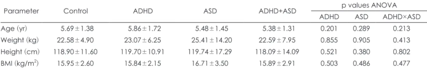
Table 1. Age and anthropometric parameters of the examined children
Parameter Control ADHD ASD ADHD+ASD p values ANOVA
ADHD ASD ADHD×ASD
Age (yr) 5.69±1.38 5.86±1.72 5.48±1.45 5.38±1.31 0.201 0.289 0.213
Weight (kg) 22.58±4.90 23.07±6.25 25.41±14.20 22.59±7.95 0.855 0.905 0.413
Height (cm) 118.90±11.60 119.70±10.91 119.74±17.29 118.09±14.09 0.521 0.380 0.802
BMI (kg/m
2) 15.95±2.60 15.84±2.15 16.71±3.50 15.89±2.91 0.503 0.486 0.477
Data are expressed as mean±standard deviation; no significant group difference was observed (p>0.05). ADHD: attention-defi-
cit/hyperactivity disorder, ASD: autism spectrum disorder, BMI: body mass index, ANOVA: analysis of variance
transformed data on urinary Mg levels did not reach log-nor- mal distribution, group comparison was performed using nonparametric Kruskal-Wallis test with adjustment for mul- tiple comparisons. Factorial analysis was performed using two-way ANCOVA using log-transformed data. The results of the tests were considered significant at p<0.05.
RESULTS
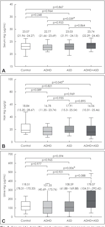
The obtained data showed that the patterns of Mg status depended significantly on the studied samples (Fig. 1). No significant group difference in serum Mg levels was observed when compared to that in the control group. Hair Mg con- tent was found to be reduced in children with ADHD and ADHD+ASD compared to the controls by 11% and 15%, re- spectively. It is notable that no significant group difference in hair Mg content was observed between children with ADHD or ASD. Urinary Mg levels in children with ADHD+ASD ex- ceeded the control, ADHD, and ASD values by 51, 76, and 65%, respectively.
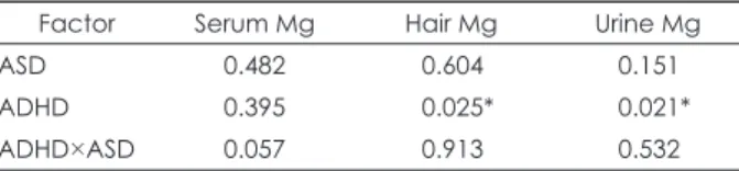
Factorial analysis using two-way ANCOVA (Table 2) dem- onstrated that although ADHD and ASD did not affect se- rum Mg levels, factorial interaction (ADHD×ASD) had a nearly significant effect (p<0.10). In contrast, ADHD had a signifi- cant effect on hair and urinary Mg levels, whereas ASD did not have any factorial influence.
Correlation analysis demonstrated that only hair Mg con- tent was weakly but significantly associated with age values (r=0.163; p=0.026), whereas no other significant associations with anthropometric parameters were revealed (data not shown).
DISCUSSION
This study provides unique data on comparative analysis of Mg status in children with ADHD and/or ASD. Generally, the revealed associations between hair (inverse) and urinary (direct) Mg levels and neurodevelopmental disorders may be indicative of increased Mg excretion in children with ADHD and ASD, ultimately leading to a reduced Mg burden in the body. The lack of significant alterations in serum Mg levels may occur due to homeostatic regulation aimed at mainte- nance of circulating Mg levels through modulation of ab- sorbance, excretion, and tissue redistribution (especially bones) [20].
The current findings generally agree with the earlier indi- cations of altered Mg status in ADHD and ASD. Particularly, Mg deficiency was found to be prevalent in 65% of examined Egyptian children with ADHD and also correlated with im- pulsivity, hyperactivity, and inattention [21]. Moreover, the
first detailed study of Mg status in ADHD also revealed Mg deficiency in 95% of children with ADHD [22]. Results of the most recent meta-analysis have demonstrated that children with ADHD have 0.105 mmol/L lower serum Mg levels than in neurotypical controls [15]. Furthermore, hair Mg content
40
35 30
25 20
15
Control
p=0.867 p=0.964
p=0.039
*
p=0.24823.07 (21.96-24.27)
22.77 (21.66-23.69)
23.03 (21.91-24.13)
23.74 (22.29-24.40) p=0.953
p=0.864
ADHD ASD ADHD+ASD
Serum Mg
(μg/mL
)
A
100
80
60
40
20
0
Control
p=0.043
*
p=0.821p=0.969 p=0.089
18.84 (15.20-28.67)
16.78 (11.35-23.74)
17.91 (15.3-25.54)
16.06 (10.31-25.46) p=0.935
p=0.895
ADHD ASD ADHD+ASD
Hair Mg
(μg/g
)
B
700 600 500 400 300 200 100 0
Control
p=0.594 p=0.965
p=0.006
*
p=0.977p=0.921
p=0.088
ADHD ASD ADHD+ASD
Urine Mg
(μg/mL
)
C
118.51
(78.31-170.33) 101.35 (40.69-173.76)
108.59 (61.88-169.88)
178.07 (104.51-292.42)