INTRODUCTION
Angiogenesis is essential for tumor growth and metasta- sis. Tumors cannot exceed 1-2 L in volume without devel- oping new blood vessels (1). These new vessels may allow tumor dissemination by providing tumor cells with a portal of entry into the circulation (2), and decreased angiogenesis is associated with a decreased rate of metastasis (3).
Angiogenetic capacity, that is, the ability to induce neo- vascularization, is characteristic of most neoplastic cells.
Among various angiogenic factors, vascular endothelial growth factor (VEGF) is known as a powerful endothelial cell-specific mitogen that is involved in tumor neovascular- ization (4). VEGF is also a highly potent direct mediator of microvascular permeability, and it is thought to be respon- sible for the characteristic leakiness of tumor blood vessels (5). VEGF expression has been demonstrated in a number of human cancer cell lines (6), as well as in clinical speci- mens of breast, brain, ovarian, esophageal, and colon cancer, suggesting a trophic role of VEGF in supporting tumor growth via host angiogenesis (7). In previous studies, VEGF
expression was significantly associated with the degree of vas- cularization in non-small-cell lung cancer (NSCLC) (8, 9).
Although hypoxia is a strong inducer of VEGF (10), muta- tionally activated ras or p53 oncogenes act synergistically with hypoxia to induce VEGF expression (11, 12).
The wild-type (wt) p53 oncogene has recently been shown to inhibit angiogenesis via regulation of thrombospondin-1, an inhibitor of angiogenesis (13), and to down-regulate VEGF-promoter activity (14). Mutant ras genes up-regulate the expression of a variety of other growth factors thought to have direct or indirect stimulating effects on angiogenesis, e.g., transforming growth factor (TGF)- , TGF- , and VEGF (15). Although experimental studies have shown that mutant p53 and ras oncogenes contribute to angiogenesis (11-15), there are few reports on the relationship between these oncogenes and angiogenesis in NSCLC (16, 17).
Here, we investigated the relationship and correlation between tumor angiogenesis and VEGF, mutant p53, and K-ras protein expression by immunohistochemistry. In addition, we evaluated the relationship between tumor angiogenesis and overall survival.
Yu-Ho Kang, Kyu-Sik Kim, Young-Kwon Yu, Sung-Chul Lim, Young-Chul Kim, Kyung-Ok Park
Department of Internal Medicine, Chonnam National University Medical School, Research Institute of Medical Science, Kwangju, Korea
Received : 6 February 2001 Accepted : 9 May 2001
Address for correspondence Young-Chul Kim, M.D.
Department of Internal Medicine, Chonnam National University Hospital, 8 Hak-dong, Dong-gu, Kwangju 501-757, Korea
Tel : +82.62-220-6573, Fax : +82.62-225-8578 E-mail : [email protected]
417
The Relationship between Microvessel Count and the Expression of Vascular Endothelial Growth Factor, p53, and K-ras in Non-Small Cell Lung Cancer
Using immunohistochemical staining, we studied the relationship between the microvessel count (MC) and the expression of K-ras, mutant p53 protein, and vas- cular endothelial growth factor (VEGF) in 61 surgically resected non-small cell lung cancers (NSCLC) (42 squamous cell carcinoma, 14 adenocarcinoma, 2 large cell carcinoma, 2 adenosquamous carcinoma, and 1 mucoepidermoid carcino- ma). MC of the tumors with lymph node (LN) metastasis was significantly higher than that of tumors without LN metastasis (66.1±23.1 vs. 33.8±13.1, p<0.05).
VEGF was positive in 54 patients (88.5%). MC was 58.1±25.2 (mean±S.D.) in a
×200 field, and it was significantly higher in VEGF(+) tumors than in VEGF(-) tumors (61.4±23.7 vs. 32.9±23.8, p<0.05). VEGF expression was higher in K- ras-positive or mutant p53-positive tumors than in negative tumors (p<0.05). MC was significantly higher in K-ras(+) tumors than in K-ras(-) tumors, although it did not differ according to the level of mutant p53 protein expression. Survival did not differ with VEGF, mutant p53, or K-ras expression, or the level of MC. In conclu- sion, there is a flow of molecular alterations from K-ras and p53, to VEGF expres- sion, leading to angiogenesis and ultimately lymph node metastasis. Correlations between variables in close approximation and the lack of prognostic significance of individual molecular alterations suggest that tumorigenesis and metastasis are multifactorial processes.
Key Words: Vascular Endothelial Growth Factor; K-ras; Protein p53; Carcinoma, Non-Small-Cell Lung
MATERIALS AND METHODS
Paraffin-embedded tumor specimens from 61 patients with NSCLC who had undergone surgery at the Chonnam National University Hospital were studied using immuno- histochemistry. Pathologic and clinical data at the time of surgery were reviewed to provide accurate staging. The clinical follow-up of the patients (46 men and 15 women) ranged from 1.5 to 75.7 months (average 18.5 months).
Tumors were staged and classified using the TNM staging system (18) and the World Health Organization Histologi- cal Classification (19) after a complete mediastinal lymph node dissection in all cases. The mean age of patients was 56.3 yr (range, 31 to 71 yr). The characteristics of the pa- tients are listed in Table 1. All the patients had an Eastern Cooperative Oncology Group performance status of 0 or 1, and normal findings on abdominal computed tomograms and radionuclide bone scans. The histologic types included 42 squamous cell carcinoma, 14 adenocarcinoma, 2 large cell carcinoma, 2 adenosquamous carcinoma, and 1 muco- epidermoid carcinoma. TNM staging consisted of 1 Stage Ia, 8 Ib, 13 IIb, 30 IIIa, and 9 IIIb. Patients who had died within 1 month of the surgery were excluded.
Vascular endothelial growth factor (VEGF) expression
Immunohistochemical staining for VEGF was performed using a Microprobe immuno/DNA staining system (Fisher Scientific, Pittsburgh, PA, U.S.A.), which is based on capil- lary action (20). Three-micrometer sections were cut from paraffin blocks and mounted on Probe on Plus slides (Fisher Scientific). Dewaxed sections were incubated with Pepsin solution (Research Genetics, Huntsville, AL, U.S.A.) for 1 min at 45℃. After washing with Universal Buffer (Research Genetics), sections from each tumor were incubated with primary VEGF antibody (Santa Cruz Biotechnology, Santa
Cruz, CA, U.S.A.) at a 1:50 dilution for 2 hr at room tem- perature. The slides were washed with Universal Buffer and incubated in biotinylated anti-mouse IgG (Sigma, St. Louis, MO, U.S.A.) for 7 min at 45℃.
Endogenous peroxidase activity was blocked with Auto- blocker (Research Genetics) for 5 min at 45℃. After wash- ing with Universal Buffer, the sections were incubated with streptavidin horseradish peroxidase (STR-HRP) (Research Genetics) for 7 min at 45℃. The reaction products were again washed in Universal Buffer, and then developed with the chromogen 3-amino-9-ethylcarbazole (AEC) for 7 min at 45℃. Sections were washed in running tap water and lightly counterstained with hematoxylin. To evaluate VEGF expression, a score corresponding to the percentage of positive cells (0=0% positive cells, 1=≤10%, 2=10 to 25%, 3=26 to 50%, 4=≥50%) was established. Negative controls were stained similarly, but without the primary antibody. VEGF expression was considered positive if stain- ing of the cytoplasm was seen in more than 25% of the tumor cells in the slide containing the largest section of the tumor.
Determination of microvessel count
Microvessels were identified by immunohistochemistry using the CD34 mouse monoclonal antibody (QBEnd/10, BioGenex, San Ramon, CA, U.S.A.) in a 1:50 dilution at 45℃for 15 min. The microvessel count (MC) was assessed by light microscopy in the areas of the tumor containing the highest number of capillaries and small venules at the invasive edge, as described previously (21). The three highly vascular areas were identified by scanning tumor sections at low power×40 and×100). After the area of highest neo- vascularization was identified on a×200 field×20 objec- tive and (10 ocular, 0.739 mm2per field), the highest value was taken as the MC.
p53 and K-ras protein expression
Three-micrometer sections were cut from paraffin blocks and mounted on Probe on Plus slides. Dewaxed sections, were immersed in citric acid buffer (0.01 M, pH 6.0), and then placed in a microwave oven at 100℃for 10 min (for p53). Sections from each tumor were incubated with prima- ry antibodies for p53 (BP53.12, Zymed, South San Francis- co, CA, U.S.A.) and K-ras (F234, Santa Cruz Biotechnolo- gy, Santa Cruz, CA, U.S.A.) for 15 to 25 min. The slides were washed with Universal Buffer and incubated with biotinylated anti-mouse IgG for 7 min.
Endogenous peroxidase activity (for p53 and K-ras) was blocked with Autoblocker for 5 min. After washing with Universal Buffer, the sections were incubated with STR- HRP (for p53 and K-ras) for 7 min. The reaction products were again washed in Universal Buffer, then developed
Mean age (yr) 56.3±9.3
Male : Female 46 : 15
Smoking (pack-years) 24.1±17.7
Histology
Squamous cell carcinoma 42
Adenocarcinoma 14
Large cell carcinoma 2
Adenosquamous cell carcinoma 2
Mucoepidermoid carcinoma 1
N status
N0 15
N1-3 46
TNM stage
≤ IIB 22
≥ IIIA 39
n=61 Table 1.Clinical characteristics of subjects
using 3-amino-9-ethylcarbazole (AEC, for p53 and K-ras, 7 min) as a chromogen. All steps in the staining procedure were done at 45℃. Sections were washed in running tap water and lightly counterstained with hematoxylin. All pro- cedures were performed using a Microprobe immuno/DNA staining system, which is based on capillary action (23). Pos- itive controls consisted of colon cancer for p53 and gastric cancer for K-ras. For negative controls, the same positive control slides were stained without the primary antibody.
The immunoreactivity of the slides was examined by two investigators using standard light microscopy, and scored in quartiles as the percentage of positive tumor cells: 0=0%, 1=≤10%, 2=10 to 50% and 3=≥50%. The intensity of the staining reaction was not quantified.
K-ras immunoreactivity was defined as a diffuse cytoplas- mic stain in neoplastic cells. p53 immunoreactivity was defined as nuclear reactivity in neoplastic cells. The expres- sion of p53 and K-ras was considered to be positive if respective staining of the cytoplasm or nuclei was seen in more than 10% of the tumor cells in the slide containing the largest section of the tumor.
Statistical analyses
The statistical analyses were performed with the SPSS for Windows program package. The survival time was recorded in months (m) from the day of surgery. Univariate and mul- tivariate analyses of survival data were undertaken, using survival curves and applying the Kaplan and Meyer method with log rank analysis, and the Cox regression model. Data were expressed as the mean±standard deviation (S.D.). The significance of associations was determined by using the 2 test, Fisher’s exact probability test, or a two-tailed Student’s t-test. p<0.05 was considered significant.
RESULTS Microvessel count in tumors
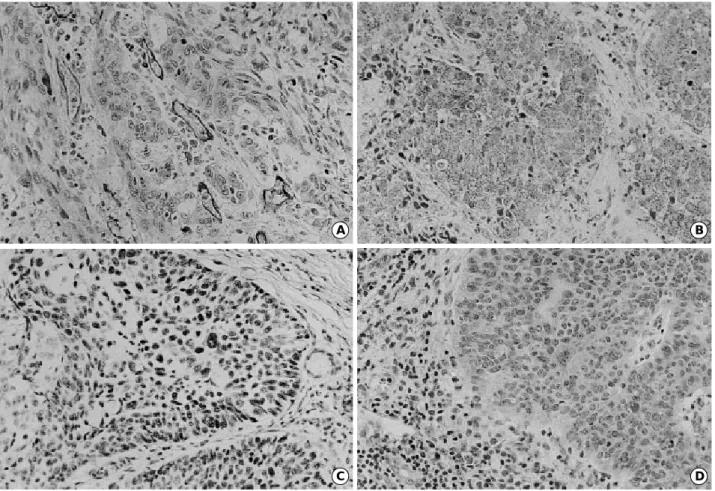
Blood vessels were heterogeneously distributed in the stroma of the tumors (Fig. 1A). The mean MC was 58.1± 25.2 and the median was 58 (range, 14-124) in a ×200 field. MC according to the pathologic state, and VEGF,
C D
A B
Fig. 1. A:Tumor area with high microvessel count in squamous cell carcinoma (ABC method, ×200). B:cytoplasmic VEGF expression in squamous cell carcinoma (×200). C:p53 protein accumulation in the nuclei of neoplastic cells in squamous cell carcinoma (×200).
D:Cytoplasmic K-ras expression in squamous cell carcinoma (×200).
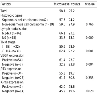
mutant p53, and K-ras protein expression are shown in Table 2. There was no significant difference in MC among the different histological subtypes of NSCLC. In addition, MC was not significantly different between low stage (≤
Stage IIb) and advanced stage (≥ Stage IIIa). The patients with lymph node metastasis had a higher MC than those without metastasis (66.1±23.1 vs. 33.8±13.1, p<0.001).
VEGF expression and microvessel count
VEGF expression was mainly identified in the cytoplasm of tumor cells (Fig. 1B). Positive staining for VEGF was seen in 54 out of 61 cases (88.5%). MC of VEGF-positive tumors was significantly higher than that of VEGF-negative tumors (61.4±23.7 vs. 32.9±23.8, p=0.004) (Table 2). VEGF expression did not differ according to lymph node metastasis or stage (low stage vs. advanced stage) (Table 3).
p53 staining, microvessel count, and VEGF
Mutant p53 was stained in the nuclei of neoplastic cells (Fig. 1C). Of 61 specimens, 34 (55.7%) stained positive for mutant p53 protein. p53 expression did not differ according to lymph node metastasis or stage. MC did not differ between p53-positive and p53-negative tumors (61.7±30.8 vs. 55.3
±19.7, p=0.353) (Table 2). However, VEGF expression in p53-positive tumors (33/34, 97.1%) was greater than that in p53-negative tumors (21/27, 77.8%) (p=0.037) (Table 3).
K-ras staining, microvessel count, and VEGF
K-ras was stained in the cytoplasm of neoplastic cells (Fig. 1D). Forty-seven specimens stained positive for K-ras protein (77%). K-ras expression did not differ according to lymph node metastasis or stage (data not shown). MC in K- ras-positive tumors was significantly higher than that in K- ras-negative tumors (62.0±25.6 vs. 45.2±19.6, p= 0.028) (Table 2). VEGF expression in K-ras-positive tumors (45/
47, 95.7%) was greater than that in K-ras-negative tumors (9/14, 64.3%) (p=0.005) (Table 3).
Overall survival analysis
The three-year survival rate (YSR) was higher in low stages (≤IIb) than in advanced stages (≥IIIa) (3YSR:
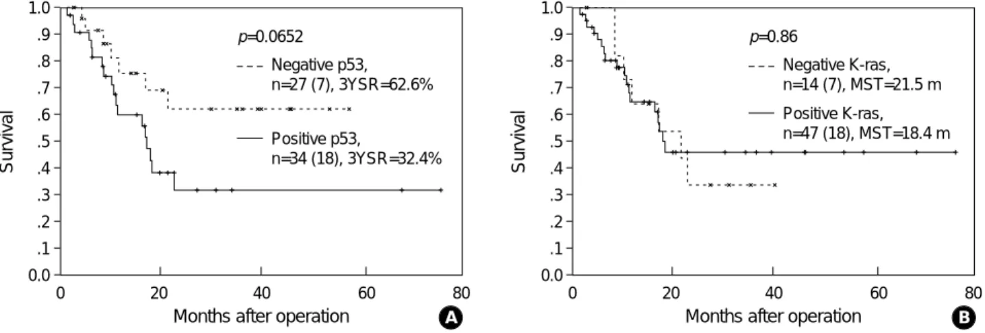
79.8% vs. 28.8%, p=0.0043) (Fig. 2). The patients were divided into two groups: those with low MC (<58) or high MC (≥58: median of MC). The overall survival did not dif- fer between the low and high MC groups (Fig. 3A). In addition, the overall survival did not differ according to VEGF, p53, or K-ras expression (Fig. 3B, 4). The univariate analysis using a Cox regression model (shown in Table 4) underlines the strong prognostic relevance of TNM stage (p=0.0098).
Total 58.1±25.2
Histologic types
Squamous cell carcinoma (n=42) 57.5±24.2
Non-squamous cell carcinoma (n=19) 59.6±27.9 0.766 Lymph nodal status
N1-N3 (n=46) 66.1±23.1
N0 (n=15) 33.8±13.1 0.000
TNM stage
≤ IIB (n=22) 50.6±28.9
≥ IIIA (n=39) 62.4±22.2 0.081
VEGF expression
Positive (n=54) 61.4±23.7
Negative (n=7) 32.9±23.8 0.004
P53 expression
Positive (n=34) 55.3±19.7
Negative (n=27) 61.7±30.8 0.353
K-ras expression
Positive (n=47) 62.0±25.6
Negative (n=14) 45.2±19.6 0.028
Factors Microvessel counts p value
Table 2. Correlation between the microvessel counts and VEGF expression, mutant p53 protein, K-ras protein expres- sion in tumor tissue
Age (yr) 0.987 0.450-2.167 0.97
Sex 2.267 0.775-6.636 0.14
Lymph node status 2.617 0.781-8.769 0.12
TNM stage 4.938 1.471-16.581 0.0098
Microvessel counts 1.124 0.512-2.469 0.77
VEGF 1.083 0.324-3.626 0.90
Mutant p53 2.233 0.930-5.365 0.07
K-ras 0.924 0.385-2.218 0.86
Factors Hazard ratio 95% Confidence interval p value Table 4.Results of Cox regression analysis in 61 patients with NSCLC; univariate analysis of the various prognostic factors Lymph node status
Negative 4 11
Positive 3 43 0.055
TNM stage
≤ IIB 4 18
≥ IIIA 3 36 0.240
Mutant p53
Negative 6 21
Positive 1 33 0.037
K-ras
Negative 5 9
Positive 2 45 0.005
VEGF expression
Factors p-value
Negative (n=7) Positive (n=54)
Table 3.VEGF expression according to the expression of mutant p53, K-ras protein, lymph nodal status, and TNM stage
Age; 0: <58, 1: ≥58, Sex: 0: Female, 1: Male. Lymph node status; 0:
Negative, 1: Positive. TNM stage; 0: ≤IIB, 1: ≥IIIA. Microvessel count;
0: less than median, 1: equal or greater than median. Other factors; 0:
negative, 1: positive.
DISCUSSION
Various growth factors stimulate angiogenesis, including acidic fibroblast growth factor (aFGF), basic fibroblastic growth factor (bFGF), TGF- , TGF- , platelet-derived endothelial cell growth factor, hepatocyte growth factor, and VEGF (1, 7). In this study we focused on the role of VEGF.
As a result of alternative splicing of messenger RNA, VEGF exists as four different homodimeric molecular species; the monomers have 121, 165, 189, or 206 amino acids (VEGF121, VEGF165, VEGF189, VEGF206, respectively) (4). VEGF121 and VEGF165 are soluble pro- teins, whereas VEGF189 and VEGF206 are bound to hep- arin-containing proteoglycans on the cell surface or in the basement membrane (22). Experiments utilizing the gene transfection method (23, 24) have established strong evi- dence for a contribution of VEGF to the progressive growth
Survival
1.0 .9 .8 .7 .6 .5 .4 .3 .2 .1 0.0
0 20 40 60 80
p=0.004
≤IIB, n=22 (3), 3YSR: 79.8%
≥IIIA, n=39 (22), 3YSR: 28.8%
Months after operation
Fig. 2. Suvival difference according to the TNM stage. n repre- sents the number of subjects with the number of deaths in parentheses. 3YSR, three year survival rate.
Survival
1.0 .9 .8 .7 .6 .5 .4 .3 .2 .1 0.0
0 20 40 60 80
p=0.7706
Lower than median MC, n=30 (12), MST=21.5 m Higher than median MC, n=31 (13), MST=22.7 m
Fig. 3. Survival difference according to the microvessel count (A) and VEGF expression (B). n represents the number of subjects with the number of deaths in parentheses. MC, microvessel count; MST, median survival time (month).
Survival
1.0 .9 .8 .7 .6 .5 .4 .3 .2 .1 0.0
0 20 40 60 80
p=0.897
Negative VEGF, n=7(3), MST=11.8 m Positive VEGF, n=54 (22), MST=21.5 m
Survival
1.0 .9 .8 .7 .6 .5 .4 .3 .2 .1 0.0
0 20 40 60 80
p=0.0652 Negative p53, n=27 (7), 3YSR=62.6%
Positive p53,
n=34 (18), 3YSR=32.4%
Fig. 4. Survival difference according to the p53 protein (A) and K-ras protein expression (B). n represents the number of subjects with the number of deaths in parentheses. 3YSR, three year survival rate; MST, median survival time (month).
Survival
1.0 .9 .8 .7 .6 .5 .4 .3 .2 .1 0.0
0 20 40 60 80
p=0.86
Negative K-ras, n=14 (7), MST=21.5 m Positive K-ras, n=47 (18), MST=18.4 m
A Months after operation B
Months after operation
A B
Months after operation Months after operation
of solid tumors through its effects in promoting tumor angiogenesis.
Numerous cytokines and growth factors produced by tumor and normal cells affect VEGF expression. It has been reported that VEGF expression is enhanced by epidermal growth factor, keratinocyte growth factor, TGF- , TGF- , insulin-like growth factor I, PGE2, IL-1 , IL-1 and IL-6 (7). In addition, oncogenes, such as p53 and ras, affect VEGF expression. VEGF overexpression was correlated with nucle- ar accumulation of p53 in human lung and colon cancer (12, 16). Likewise, vessel counts in p53-positive tumors were sig- nificantly higher than in p53-negative tumors (15). Besides mutant p53, mutation of the ras gene also has been shown to up-regulate VEGF expression (11, 15).
As in other reports (8, 16, 25, 26), our results showed that lymph node (LN) metastasis was associated with the microvessel count. In addition, our data are in agreement with previous studies, in that VEGF expression was signifi- cantly associated with the degree of vascularization in NSCLC (8, 9). However, there were no direct correlations between LN metastasis and VEGF expression. These find- ings suggest that there are many other factors controlling the degree of LN metastasis that cannot be explained by VEGF expression alone.
In this study, p53 and K-ras were shown to have no direct effect on lymph node metastasis. However, both oncopro- teins were correlated with VEGF expression. VEGF expres- sion is correlated with the onset of mutations in both ras (11) and p53 (12), and these mutations act synergistically with hypoxia to induce VEGF expression. Our data also showed that K-ras expression was correlated with MC, while p53 expression was not. These results suggest that K- ras induces angiogenesis via VEGF expression.
If we postulate a sequential pathway of angiogenesis, oncogenes (p53 and K-ras), VEGF expression, microvessel count (angiogenesis), and finally degree of metastasis can be lined up. We observed a direct relationship between the closely approximated variables; however, direct relation- ships between distant variables are hard to prove. These findings suggest that there are many other factors control- ling the dependent variable, since it cannot be explained with only one or two independent variables.
Although the role of VEGF in tumor metastasis has been demonstrated by several experiments that showed that VEGF induces neovascularization (1), the association of VEGF expression with the prognosis of NSCLC is controversial.
Imoto et al. reported poor survival in VEGF-immunopositive NSCLC (27). However, Decaussin et al. (28) argued against this finding by demonstrating that positive staining for VEGF did not indicate a poor prognosis. Our data showed that survival did not differ according to VEGF expression.
Some studies have found that patients with p53-positive tumors have a significantly poorer prognosis in lung cancers (29), whereas others have found no differences in survival
between p53-positive and p53-negative lung cancers (30).
Moreover, a recent study by Lee et al. (31) showed that p53 expression implied a favorable prognosis in a series of 156 resected primary NSCLC. According to our data, p53 expres- sion did not affect patient survival.
VEGF expression can be induced by the mutant ras onco- gene (15), and mutant K-ras knockout sublines were incapable of forming tumors in nude mice (11). Furthermore, in serum- starved NIH 3T3 cells, a ras-transformed cell line expresses enhanced levels of VEGF transcripts (32). Although the K-ras mutation is known to be a poor prognostic factor in pul- monary adenocarcinoma (33), our data did not show any survival difference according to K-ras expression.
The prognostic significance of angiogenesis in NSCLC has not been established. Giatromanolaki et al. (25) and Harpole et al. (34) reported that vascularity was a signifi- cant factor in predicting poor survival. However, this study and others (10, 35) have found no differences in survival according to MC in lung cancer.
In conclusion, there is a flow of molecular alterations from K-ras and p53, and VEGF expression, that lead to angiogenesis and finally to lymph node metastasis. Correla- tions between variables in close approximation and the lack of prognostic significance of individual molecular alter- ations suggest that tumorigenesis and metastasis are multi- factorial processes.
As shown in the report by Kim et al. (36), however, anti- VEGF monoclonal antibody inhibits the growth of rhab- domyosarcoma (A673) and leiomyosarcoma (SK-LMS-1) cell lines, and the magnitude of the response is greater in rhabdomyosarcoma, which proliferates more rapidly and is therefore more angiogenesis-dependent. The important role of VEGF expression in angiogenesis in NSCLC suggests a potential therapeutic use of anti-VEGF monoclonal anti- body for blocking VEGF action in this type of tumor.
REFERENCES
1. Folkman J, Shing Y. Angiogenesis: minireview. J Biol Chem 1992;
267: 10931-4.
2. Liotta LA, Kleinerman J, Saidel GM. Quantitative relationships of intravascular tumor cells, tumor vessels, and pulmonary metastases following tumor implantation. Cancer Res 1974; 34: 997-1004.
3. Starkey JR, Crowle PK, Taubenberger S. Mast cell-deficient W/Wvmice exhibit a decreased rate of tumor angiogenesis. Int J Cancer 1988; 42: 48-52.
4. Leung DW, Cachianes G, Kuang WJ, Goeddel DV, Ferrara N. Vas- cular endothelial growth factor is a secreted angiogenic mitogen.
Science 1989; 246: 1306-9.
5. Senger DR, Brown LF, Claffey KP, Dvorak HF. Vascular perme- ability factor, tumor angiogenesis and stroma generation. Invasion Metastasis 1994; 14: 385-94.
6. Senger DR, Perruzzi CA, Feder J, Dvorak HF. A highly conserved
vascular permeability factor secreted by a variety of human and rodent tumor cell lines. Cancer Res 1986; 46: 5629-33.
7. Ferrara N, Davis-Smyth T. The biology of vascular endothelial growth factor. Endocr Rev 1997; 18: 4-25.
8. Giatromanolaki A, Koukourakis MI, Kakolyris S, Turley H, O’Byrne K, Scott PAE, Pezzella F, Georgoulias V, Harris AL, Gat- ter KC. Vascular endothelial growth factor, wild-type p53, and angiogenesis in early operable non-small cell lung cancer. Clin Cancer Res 1998; 4: 3017-24.
9. Mattern J, Koomagi R, Volm M. Association of vascular endothe- lial growth factor expression with intratumoral microvessel density and tumor cell proliferation in human epidermoid lung carcinoma.
Br J Cancer 1996; 931-4.
10. Shweiki D, Itin A, Soffer D, Keshet E. Vascular endothelial growth factor induced by hypoxia may mediate hypoxia-initiated angiogen- esis. Nature 1992; 359: 843-5.
11. Rak J, Mitsuhashi Y, Bakyo L, Filmus J, Shirasawa S, Sasazuki T, Kerbel RS. Mutant ras oncogenes up-regulate VEGF/VPF expres- sion: implications for induction and inhibition of tumor angiogene- sis. Cancer Res 1995; 55: 4575-80.
12. Takahashi Y, Bucana CD, Cleary KR, Ellis LM. p53, vessel count, and vascular endothelial growth factor expression in human colon cancer. Int J Cancer 1998; 79: 34-8.
13. Dameron KM, Volpert OV, Tainsky MA, Bouck N. Control of angiogenesis in fibroblasts by p53 regulation of thrombospondin-1.
Science 1994; 265: 1582-4.
14. Mukhopadhyay D, Tsiokas L, Sukhatme VP. Wild-type p53 and v- Src exert opposing influences on human vascular endothelial growth factor gene expression. Cancer Res 1995; 55: 6161-5.
15. Rak J, Filmus J, Finkenzeller G, Grugel S, Marme D, Kerbel RS.
Oncogenes as inducers of tumor angiogenesis. Cancer Metastasis Rev 1995; 14: 263-77.
16. Fontanini G, Vignati S, Lucchi M, Mussi A, Calcinai A, Boldrini L, Chine S, Silvestri V, Angeletti CA, Basolo F, Bevilacqua G.
Neoangiogenesis and p53 protein in lung cancer: their prognostic role and their relation with vascular endothelial growth factor (VEGF) expression. Br J Cancer 1997; 75: 1295-301.
17. Tsao MS, Liu N, Nicklee T, Shepherd F, Viallet J. Angiogenesis correlates with vascular endothelial growth factor expression but not with Ki-ras oncogene activation in non-small cell lung carcino- ma. Clin Cancer Res 1997; 3: 1807-14.
18. Mountain CF. Revisions in the international system for staging lung cancer. Chest 1997; 111: 1710-7.
19. World Health Organization. The World Health Organization. Histo- logical typing of lung tumors. Am J Clin Pathol 1982; 77: 123-36.
20. Reed JA, Manahan LJ, Park CS, Brigati DJ. Complete one-hour immunohistochemisty based on capillary action. Biotechniques 1992; 13: 434-43.
21. Weidner N, Semple JP, Welch WR, Folkman J. Tumor angiogene- sis and metastasis-correlation in invasive breast carcinoma. N Engl J Med 1991; 324: 1-8.
22. Houck KA, Leung DW, Rowland AM, Winer J, Ferrara N. Dual regulation of vascular endothelial growth factor bioavailability by
genetic and proteolytic mechanisms. J Biol Chem 1992; 267:
26031-7.
23. Millauer B, Shawver LK, Plate KH, Risau W, Ullrich A. Glioblas- toma growth inhibited in vivo by a dominant-negative Flk-1 mutant.
Nature 1994; 367: 576-9.
24. Zhang HT, Craft P, Scott PA, Ziche M, Weich HA, Harris AL, Bicknell R. Enhancement of tumor growth and vascular density by transfection of vascular endothelial cell growth factor into MCF-7 human breast carcinoma cells. J Natl Cancer Inst 1995; 87: 213-9.
25. Giatromanolaki A, Koukourakis M, O’Byrne K, Fox S, Whitehouse R, Talbot DC, Harris AL, Gatter KC. Prognostic value of angiogene- sis in operable non-small cell lung cancer. J Pathol 1996; 179: 80-8.
26. Yuan A, Yang PC, Yu CJ, Lee YC, Yao YT, Chen CL, Lee LN, Kuo SH, Luh KT. Tumor angiogenesis correlates with histologic type and metastasis in non-small cell lung cancer. Am J Respir Crit Care Med 1995; 152: 2157-62.
27. Imoto H, Osaki T, Taga S, Ohgami A, Ichiyoshi Y, Yasumoto K.
Vascular endothelial growth factor expression in non-small-cell lung cancer: prognostic significance in squamous cell carcinoma. J Thorac Cardiovasc Surg 1998; 115: 1007-14.
28. Decaussin M, Sartelet H, Robert C, Moro D, Claraz C, Brambilla C, Brambilla E. Expression of vascular endothelial growth factor (VEGF) and its two receptors (VEGF-R1-Flt1 and VEGF-R2-Flk1/
KDR) in non-small cell lung carcinomas (NSCLCs): correlation with angiogenesis and survival. J Pathol 1999; 188: 369-77.
29. Quinlan DC, Davidson AG, Summers CL, Warden HE, Doshi HM.
Accumulation of p53 protein correlates with a poor prognosis in human lung cancer. Cancer Res 1992; 52: 4828-31.
30. McLaren R, Kuzu I, Dunnill M, Harris A, Lane D, Gatter KC. The relationship of p53 immunostaining to survival in carcinoma of the lung. Br J Cancer 1992; 66: 735-8.
31. Lee JS, Yoon A, Kalapurakal SK, Ro JY, Lee J, Tu N, Hittelman WN, Hong WK. Expression of p53 oncoprotein in non-small cell lung cancer: a favorable prognostic factor. J Clin Oncol 1995; 13:
1893-903.
32. Grugel S, Finkenzeller G, Weindel K, Barleon B, Marme D. Both v- Ha-Ras and v-Raf stimulate expression of the vascular endothelial growth factor in NIH 3T3 cells. J Biol Chem 1995; 270: 25915-9.
33. Kern JA, Slebos RJ, Top B, Rodenhuis S, Lager D, Robinson RA, Weiner D, Schwartz DA. C-erbB-2 expression and codon 12 K-ras mutations both predict shortened survival for patients with pul- monary adenocarcinomas. J Clin Invest 1994; 93: 516-20.
34. Harpole DH Jr, Richards WG, Herndon JE II, Sugarbaker DJ.
Angiogenesis and molecular biologic substaging in patients with stage I non-small cell lung cancer. Ann Thorac Surg 1996; 61:
1470-6.
35. Macchiarini P, Fontanini G, Dulmet E, de Montpreville V, Chape- lier AR, Cerrina J, Ladurie FL, Dartevelle PG. Angiogenesis: an indicator of metastasis in non-small cell lung cancer invading the thoracic inlet. Ann Thorac Surg 1994; 57: 1534-9.
36. Kim KJ, Li B, Winer J, Armanini M, Gillett N, Phillips HS, Ferrara N. Inhibition of vascular endothelial growth factor-induced angio- genesis suppresses tumor growth in vivo. Nature 1993; 362: 841-4.