키토산-비타민 C 나노입자
Aarti R. Deshmukh, 김범수*
Chitosan-Vitamin C Nanoparticles
Aarti R. Deshmukh and Beom Soo Kim*
Received: 4 September 2019 / Accepted: 24 October 2019
© 2019 The Korean Society for Biotechnology and Bioengineering
Abstract: Vitamin C is a very important module related to many biochemical functions of human body. Vitamin C is highly sensitive and can be lost if exposed to high temperature, humid air, light, and alkaline pH. In recent decades, use of chi- tosan nanoparticles as nanocarriers has received much attention due to their biocompatibility, biodegradability, and non-toxicity.
Chitosan nanoparticles containing vitamins, flavors, enzymes, and antimicrobial agents can maintain their activity. Chitosan nanoparticles can stimulate the stabilization of vitamin C and improve controlled release. This review provides a summary of the preparation and characterization of chitosan nanoparticles and potential benefits of vitamin C-containing chitosan nanopar- ticles. In addition, biological functions, dietary restrictions, and mechanisms of vitamin C are reviewed. Various challenges and future scope for improving the properties of vitamin C using chitosan nanoparticles are also discussed.
Keywords: chitosan, vitamin C, anoparticles, ionic gelation
1. INTRODUCTION
Vitamin C, also known as ascorbic acid or L-ascorbic acid, is a water-soluble vitamin. Vitamin C is a nutritional indicator for overall health. It is essential to strengthen the immune system deficiency and minimize the risk of cardiovascular diseases,
eye diseases, cancer and skin wrinkling [1,2]. Because human and animal bodies do not have L-gulonolactone oxidase, they cannot synthesize vitamin C from the digestive tract. To restock this valuable vitamin, it must be supplied through exogenous sources [2,3]. Fig. 1 represents mg vitamin C per 100 g fruits, vegetables, and nuts. It is naturally found in red peppers, kiwi fruit, papaya, cabbage, broccoli, strawberries, cauliflower, brussels sprouts, potatoes, peas, grapefruits, oranges, potatoes, cantaloupe, lemons and limes, mangos, carambola, peppers, parsley, nuts, chestnuts, and many others [4]. Vitamin C is a cofactor required for many enzyme reactions. Studies show that insufficient intake of vitamin C can have a negative impact on human health and normal life span and can lead to cancer in humans.
Vitamin C is easily broken down into biologi-cally inactive com- pounds and loses its function when exposed to high temperature, humid air, light, and alkaline pH [5]. Therefore, development of a more stable form of vitamin C is a prerequisite for human and animal nutrition. Encapsulation of vitamin C with chitosan nanoparticles can be used to increase stability [5,6]. Because vitamin C deficiency causes strepto-zotocin-induced renal scar- ceness, vitamin C is essential for maintaining kidney function in diabetes [7].
Chitosan is a cationic polyelectrolyte of N-acetyl gluco- samine. Next to cellulose, it is second abundant biopolymer that exists in nature [8]. Chitosan is mostly identified by its molecular weight and its degree of deacetylation. Chitosan is obtained from deacetylation of natural polymer chitin. Chitosan is edible and has excellent properties such as biocompatibility, biodegradability, bioadhesiveness, non-toxicity, in situ gelling transfection enhancement, bioactive compound/drug release control, permeation enhancement, and mucoadhesion [8-10].
충북대학교 화학공학과
Department of Chemical Engineering, Chungbuk National University, Cheongju, Chungbuk 28644, Korea
Tel.: +82-43-261-2372, Fax: +82-43 -269-2370 e-mail: [email protected]
Review Paper
222 Korean Society for Biotechnology and Bioengineering Journal 34(4): 221-232 (2019)
These properties make chitosan a versatile compound that can be used for a variety of applications. It is insoluble in water because of free amino groups. It can be dissolved in water at acidic pH because of the protonation of amino groups. Chitosan has been reported to be soluble in acid-free water through a physical dissolution-precipitation process. Water-soluble chitosan obtained by this method can be a promising source for food packing or carriers for drug controlled release system [10].
Chitosan has been successfully applied in drug delivery systems to enhance the absorption and bioavailability of the drug, bioactive compound, vitamin carrier, membrane dressing, DNA delivery, biofilm formation, and tissue engineering [11-21]. It is also a commonly used membrane material for nanofiltration, ultrafiltration and reverse osmosis for wastewater treatment, desalination, and antifouling agent [22-26]. Chitosan nano- particles and nanocomposites were found to be excellent adsorbents for the removal of heavy metal ions [10,27].
This review highlights an overall discussion of the prepara- tion and characterization of chitosan nanoparticles, effect of different parameters on the size of nanoparticles, and modified techniques. This article furthered reviews the biological functions, sources, dietary allowance, and mechanism of vitamin C. The interaction between vitamin C and chitosan nanoparticles, in vivo and in vitro release studies of vitamin C, challenges and future prospects for vitamin C-containing chitosan nanoparticles are also discussed. Although some review articles are available for preparation and drug delivery applications of chitosan nanoparticles [28-31], this is the first review showing the insights into chitosan-vitamin C nanoparticles.
2. Vitamin C
2.1. Biological Function of Vitamin C
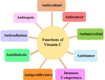
Vitamin C is considered a very powerful reducing agent and
radical scavenger. Many in vitro and in vivo studies have shown that intake of vitamin C is multifunctional in human health (Fig. 2). Vitamin C has proven to be an effective antioxidant. It can protect plasma lipids against peroxidation of cellular membranes surrounding cells as well as intracellular organelles. In addition, it reinstates the activity of fat-soluble vitamin E. Vitamin C plays an important role as an antioxidant for extracellular fluids surrounding lens, retina, and lungs. It protects against pathogen invasion [32]. Ascorbate has been shown to be the most active aqueous antioxidant in human plasma. It gives protection against diseases and deterioration processes caused by oxidative stress [33]. It plays an important role in the synthesis of catecholamine, cortisol, and vasopressin which are key mediators in the sepsis process [34]. When fucoxanthin and vitamin C were used together, lymphocytes showed more antioxidant and anti-inflammatory effects. It also reduces the proliferation rate of lymphocytes [35]. It significantly reduces total mortality and breast cancer risk [36].
According to in vitro studies, vitamin C generates hydrogen peroxide-dependent cytotoxicity in a variety of cancer cells [37]. In vivo studies in mice bearing glioblastoma xenografts showed that a single dose of ascorbate produced persistent ascorbate radical and hydrogen peroxide selectively inside the interstitial fluids of tumors and significantly decreased the growth rates of ovarian glioblastoma and pancreatic tumors [38]. It has been suggested that ascorbate as a prodrug has an advantage in the treatment of cancer with poor prognosis [39].
Vitamin C stimulates apoptosis in leukemia cells by oxidative stress mechanisms associated with superoxide anion radicals and H
2O
2generation [40]. It has been suggested that vitamin C can be useful in the treatment of leukemia [41]. 2-O-α-D- Glucopyranosyl-L-ascorbic acid as well as ascorbic acid signi- ficantly inhibited tumor growth in colon-26 tumor-bearing Fig. 1. Vitamin C content of various food.
Fig. 2. Biological function of vitamin C.
mice [42]
Vitamin C supplements reduce oxidative stress and platelet biochemical function in diabetes patients [43]. Vitamin C maintains an intracellular antioxidant network because it can act as an anti-inflammatory agent by eliminating reactive oxygen species responsible for proinflammatory cytokines in many inflamma- tory diseases [44-45]. Vitamin C has been reported to have a hepatoprotective effect against cholestasis liver injury [46]. In vivo and in vitro studies showed that the rate of blastocyst development in vitamin C-treated somatic cell nuclear transfer embryos was significantly higher than that of untreated embryos [12]. In vivo administration of vitamin C regulates T cell proliferation and cytokine secretion and improves hepato- renal and testicular toxicity as antioxidants [7,47]. Recent studies indicate that vitamin C can be a new, safe, and useful trigger for external activation of reduction sensitive nanothera- peutics [48]. In tumor therapy, slow release of drugs and poor tumor penetration are two major problems. Sequential admin- istration of paclitaxel-loaded shell-sheddable disulfide-linked poly(ethylene glycol)-b-poly(ε-caprolactone) micelles and vitamin C showed significantly higher tumor inhibition effects and improved survival rates. This enhanced antitumor effect is probably due to rapid extracellular paclitaxel release in tumors stimulated by vitamin C. Vitamin C promotes the uptake of paclitaxel by tumor cells with deep penetration of paclitaxel in tumor [49].
2.2. Sources and Dietary Allowance for Vitamin C Because vitamin C cannot be produced by humans and animal bodies, they must be replenished with diet or dietary supplements.
The recommended intake of vitamin C and other nutrients is shown in the dietary reference [50]. Table 1 shows the intake levels of vitamin C by age.
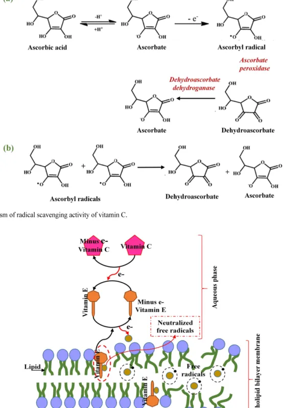
2.3. Role of Vitamin C in Tocopherol Regeneration One of the OH groups of vitamin C donates electron to quench radicals. Fig. 3 shows the mechanism of radical scavenging activity of vitamin C. The peroxidase initiates reaction and
delivers one electron from ascorbate derivative, converting the ascorbate to ascorbyl radical and ascorbyl radical to dehydroascorbic acid (Fig. 3(a)). This is one electron transfer reaction. Dehydroascor- bate dehydrogenase (reductase) converts dehydroascorbate back to ascorbate and makes two electron transfers. If ascorbyl radical is not immediately oxidized by ascorbate peroxidase, the ascorbyl radical pairs spontaneously become disproportionate to one another to form ascorbate and dehydroascorbate (Fig.
3(b)) [51]. Ascorbate is the major free radical scavenger in blood after uric acid.
Vitamin C supplies electrons to free radicals and neutralizes them. The tocopheroxyl radical is scavenged to pheroxyl radical and reduced by ascorbate to regenerate tocopherol. This type of synergistic inhibition can occur in homogeneous solution as well as heterogeneous membrane system. When oxidation proceeds in the lipid domain, ascorbic acid acts as an antioxi- dant. Although tocopherol and vitamin C are present in two different phases, lipid and aqueous phase, respectively, interaction occurs between them [19]. Because vitamin C is water soluble, it cannot enter the phospholipid bilayer and must interact with tocopheroxyl radical near the membrane surface. Tocopherol is regenerated by reducing tocopheroxyl radical with the help of glutathione and cysteine. Fig. 4 shows the schematic of vitamin E (tocopherol) regeneration mechanism by vitamin C. Ascorbic acid is much more active and efficient than cysteine and glutathione in reducing pheroxyl radical [33].
2.4. Influence of Temperature and Humidity on the Degradation Process of Vitamin C
Vitamin C is easily degraded into biologically inactive compounds and loses its functionality when exposed to high temperature, humid air, light, and alkaline pH [5]. Increased temperature and moisture content causes oxidation and degradation of ascorbic acid [52]. Its degradation was the fastest at high temperature [53]. No significant instability of vitamin C was observed at 22
oC and 35
oC, and there was a great chemical instability above 40
oC to 60
oC [54,55].
Color loss was significantly higher in indoor environmental
Table 1. Intake levels of vitamin C
Age Recommended dietary allowances for vitamin C (mg/day) Tolerable upper intake for vitamin C (mg/day)
Male Female Pregnancy Lactation Male Female
0–6 months 40 40 - -
Not possible to establish
7–12 months 50 50 - -
1–3 years 15 15 - - 400 400
4–8 years 25 25 - - 650 650
9–13 years 45 45 - - 1,200 1,200
14–18 years 75 65 80 115 1,800 1,800 (Same for pregnancy and lactation)
19+ years 90 75 85 120 2,000 2,000 (Same for pregnancy and lactation)
Smokers Additional 35 mg vitamin C/day than nonsmokers.
224 Korean Society for Biotechnology and Bioengineering Journal 34(4): 221-232 (2019)
conditions than in a cold chamber environment. The color of vitamin C decreased linearly with the initial moisture content and storage time of the product. The moisture content of vitamin C in the room-preserved samples did not change
during the same storage period and varied in the order of polyethylene bag>plastic container>glass container. The color difference increased with the storage time and varied in the order of glass container > plastic container > polyethylene bag [56].
Fig. 4. Regeneration of vitamin E (tocopherol) by vitamin C.
Fig. 3. Mechanism of radical scavenging activity of vitamin C.
3. Effect of Different Parameters on the Preparation of Chitosan Nanoparticles
It has been reported that tripolyphosphate (TPP) is widely used as an ionic crosslinking agent due to its non-toxic property in the preparation of chitosan nanoparticles [57,58]. TPP has been used in the synthesis of films, beads, gels, and nanoparticles [59]. The use of TPP improves stability and antibacterial properties of chitosan-TPP nanoparticles. Chitosan-TPP composites support biomineralization with phosphate as nucleation site and improves cellular response [60-63].
Many drug delivery systems have been developed to improve drug efficacy and minimize side effects. Nanoparticles are capable of delivering a wide range of active molecules to the different levels of the body for prolonged periods [64]. Chitosan has been widely used for nanoparticle synthesis due to its biodegradability, biocompatibility, and low toxicity. Various methods such as sieving method, emulsification, ionic gelation, spray drying, reverse micelle method, crosslinking, coacervation/
precipitation, emulsion droplet coalescence method, and mem- brane contactor have been reported for the preparation of chitosan nanoparticles. Fig. 5 shows a method of preparing chitosan-TPP nanoparticles.
Different methods for synthesizing chitosan nanoparticles have been reported. Table 2 summarizes studies on the preparation of chitosan-TPP nanoparticles.
Britto et al. [13] analyzed the effect of molecular weight of chitosan and the addition of vitamin C on chitosan nanoparticles.
Chitosan-vitamin C nanoparticles of 185-585 nm size were prepared by ionic gelation method with chitosan of molecular weight of 65 kDa and 450 kDa using sodium tripolyphosphate as crosslinking agent. In controlled in vivo experiments on trout fish, release of vitamin C was observed for 48 hours. The release of vitamin C was pH dependent and low molecular weight chitosan preferred higher encapsulation efficiency of vitamin C. Chitosan encapsulated vitamin C improved the
lifespan and release of vitamin C. The effect of vitamin C was analyzed in chitosan nanoparticles and observed a significant increase in particle size from 198 to 535 nm.
Jang and Lee [8] reported the fabrication of chitosan-TPP nanoparticles by ionic gelation of chitosan in the range of 170 nm. They demonstrated that nanoparticle size increases with chitosan concentration and decreases with TPP concentration.
The zeta potential of chitosan nanoparticles increased linearly with decreasing TPP concentration. Nanoparticles containing vitamin C with particle sizes ranging from 186 to 201 nm with zeta potential values of nanoparticles in the range of 2.43 and 19.27 mV exhibited adequate stability. Nanoparticles were stable during heat treatment. The crosslinking structure related to the formation of layer was affected by heat treatment. The particle structure consisted of two parts, surface and inner part.
Heat treatment mostly affects the adsorbed ascorbic acid on the surface and reduces particle size and surface charge. Further heat treatment did not affect particle size and zeta potential.
Since the matrix structure of ascorbic acid and chitosan combined through ionic gelation inside the particles was more stable than surface, they remained within a certain range.
Therefore, stability of inner part obtained by heat transfer was higher than that of particle surface, and stability of the surface was highly influenced by high temperature. Costa et al. [20]
prepared low molecular weight chitosan nanoparticles by ionic gelation between chitosan and TPP. The resulting nanoparticles had an average size of 244 ± 12 nm and showed biological activity against Staphylococcus aureus.
Sacco et al. [64] presented the contribution of the non-
traditional crosslinking agent pyropolyphosphate (PPi) to
chitosan nanoparticles. The size of the obtained nanoparticles
was 14.7-22.3 nm. They demonstrated that TPP and PPi favors
the formation of homogeneous and inhomogeneous systems,
respectively. The presence of an extra negative charge in case
of TPP than PPi clearly demonstrated the higher binding
affinity to chitosan chains compared to PPi. The effect of
Fig. 5. Schematic representation of the preparation of chitosan-TPP nanoparticles and vitamin C-loaded chitosan-TPP nanoparticles.
226 K o rean S o c iet y fo r Bi otec h nology and Bioe ngineer ing J our nal 34( 4) : 221-2 32 (2019) Table 2. Summary of preparation and characterization of chitosan nanoparticles
Preparation method Source of chitosan
Degree of deacetylation of
chitosan (DA) (%), molecularity
Crosslinking agent pH Ratio of
chitosan/TPP Particle size (nm) polydispersity
index (PDI) Zeta potential (mV) References
Ionic gelation N,N,N-trimethyl chitosan 80 TPP
7.54 196 ± 8 - +31.6 ± 0.6
[13]
10 302 ± 15 - +24.7 ± 1.0
5 606 ± 25 - +12.7 ± 0.4
Ionic gelation Chitosan 86.6, LMW TPP 1.67 171.70 ± 1.18 0.343 ± 0.005 +29.23 ± 0.85 [8]
Ionic gelation Chitosan 75-85, LMW TPP 7 244 ± 12 0.358 +17.3 ± 1.41 [20]
Ionic gelation Chitosan LMW
TPP
2.6 3.8 11.0 ± 0.1 - -
[64]
2.6 5.2 10.5 ± 0.2 - -
2.6 7.0 14.0 ± 0.7 - -
Pyrophosphate
2.6 3.8 22.3 ± 0.9 - -
2.6 5.2 14.9 ± 5.2 - -
2.6 7.0 14.7 ± 0.8 - -
Ionic gelation Chitosan Seacure ® 123 - TPP and polyethylene
oxide-poly lactic acid 4.5-5 1.75 200-1000 +20 to 60 [65]
Ionic gelation Chitosan Very LMW TPP 5.5 5 151 ± 10 0.185 24 ± 4
LMW TPP 5.5 6 200 ± 24 0.227 25 ± 3 [59]
Ionic gelation Chitosan - TPP - 8 293 ± 9 0.34 +37.5 ± 0.9 [66]
Pre-gelation ionotropic
method Chitosan 91.23 Sodium alginate - - 119.5 ± 49.9 0.454 ± 0.066 − 30.9 ± 0.5 [67]
Ionic gelation with
membrane Chitosan, 75-85 TPP - 2 90-100 0.22 +31 [68]
Ionic gelation
Chitosan hydrochloride (Protasan, UP CL 113,
Mw 110 kDa)
86
Sulfobutylether-b- cyclodextrin sodium
salt
- 3 503 ± 64 0.32-0.23 +33.8 ± 0.2 [69]
Ultrasonic ionotropic
gelation 0.25% Protasan - TPP - 2 283 ± 3 0.30 ± 0.01 +45.51 ± 0.29 [70]
Ultra-sonication Chitosan 98 TPP - 3.75 < 300 nm - - [71]
Ultrasonic degradation Chitosan 90 - - - 5 - -
95 - - - 12-13 - - [72]
Self-aggregation method N-acyl chitosan 84.9
Propionic anhydride - - 444.2 ± 12.2 - +26.0 ± 4.6
[14]
Hexanoic anhydride - - 469.3 ± 15.6 - +10.2 ± 3.2
Nonanoyl chloride - - 481.2 ± 3.8 - +12.5 ± 3.8
Lauroyl chloride - - 463.3 ± 0.4 - +14.6 ± 2.4
Pentadecanoyl
chloride - - 469.8 ± 9.6 - +22.0 ± 5.5
Stearoyl chloride - - 486.6 ± 0.1 - +28.9 ± 7.4
Ionotropic gelation Chitosan 86 TPP 2.53 100-900 0.1 to 0.45 - [73]
Ionic Gelation Chitosan 91.5, LMW TPP - - 138 0.026 +35 [74]
Ionic crosslinking Chitosan ≥95 TPP - 5 104 ± 6.52 0.221 ± 0.024 +22.7 ± 4.92 [75]
Modified ionic gelation Chitosan 75-85 TPP 2.5 100-300 - [76]
scattering ability confirmed higher ability of TPP to interact with chitosan than PPi. Calvo et al. [65] analyzed the effect of diblock copolymer of ethylene oxide and propylene oxide (PEO-PPO) and chitosan concentration on chitosan nanoparticles.
Chitosan/PEO-PPO nanoparticles exhibited a compact core surrounded by a thick, fluffy PEO-PPO coating, whereas CS- TPP nanoparticles had a solid and consistent structure. The core sizes of two nanoparticles were similar. The CS/PEO-PPO nanoparticles were more stable than CS-TPP nanoparticles.
The size of chitosan nanoparticles depended on the concen- tration of chitosan and TPP and formation of nanoparticles was only possible at certain concentrations. Particle size increased with increasing concentration of copolymer. These nano- particles had excellent capacity for entrapment of proteins and continuous protein release. Chitosan/alginate nanoparticles were prepared for encapsulation and controlled release of vitamin B2 [67]. Average size of nanoparticles was 119.5 ± 49.9 nm with zeta potential of −30.9 ± 0.5. Synthesized nanoparticles were more sensitive to alginate protonation and pH changes, leading to higher values of size and polydispersity index.
In vivo tests on chick embryos confirmed biocompatibility of both chitosan and nanostructures [59]. Smaller nanoparticles were formed by using very LMW chitosan. The size of nano- particles increased with TPP concentration and addition time.
De Campos et al. [66] prepared chitosan nanoparticles from a mixture of acetonitrile and water. The incorporation of acetonitrile affected entanglement of chitosan chains and gelation process. The prepared chitosan nanoparticles were effective to deliver immunosuppressive peptide CyA specifically to the ocular mucosa. Aresta et al. [69] prepared chitosan nanoparticles by ionic gelation using sulfobutylether-b- cyclodextrin sodium salt as a crosslinking agent and evaluated potential of vitamin loaded chitosan nanoparticles for food packing application. The average size of chitosan nanoparticles was 375-503 nm and the zeta potential was +16.0 to +33.8 mV.
Ionic gelation was further modified by application of membranes and ultrasound. Hassani et al. [68] used nickel micro-engineered membranes with uniform cylindrical pores in a perfectly ordered hexagonal array with pores in the center of each cell. Size of the nanoparticles prepared by ionic gelation using membranes ranged from 90 to 100 nm. Ultrasound processing was effective in decomposing aggregates, reducing particle size and polydispersity of nanoparticles, and improving storage stability at the same time [70,71]. Souza et al. [72] also reported ultrasound degradation of chitosan. It was possible to control nanoparticle size and shape according to sonication time. Cho et al. [15] prepared chitosan nanoparticles modified by anhydrides and halides with an average size of 216-218 nm through self-aggregation method. Li and Huang [71] reported
293 nm chitosan nanoparticles by ultrasonication. Chitosan composition (source, degree of acetylation, and molecular weight), crosslinking agent, ratio of chitosan and crosslinking agent, method of fabrication, and parameters related to fabri- cation greatly influenced size, biological and physicochemical properties of nanoparticles (Table 2). Low molecular weight chitosan and ultrasonication treatment have been shown to be effective for very small size (5, 12, and 93 nm) nanoparticle formation [15,73-76].
4. Chitosan Nanoparticles as Enhancer and Carrier for Vitamin C
Vitamin C has a low molecular weight and relatively simple structure. Due to the presence of carbonyl and acid groups, it has a high density of negative charges. When added to a chitosan solution, electrostatic interactions and ionic bonds start between these two molecules [3]. Chitosan and alginate nanoliposomes prepared by electron deposition suggested that shell modifies the surface properties from oxidation and hydrolysis by surface barrier, resulting in long term stability of vitamin C [11].
Fig. 5 shows a schematic view of vitamin C-loaded chitosan-
TPP nanoparticles. Different methods for the fabrication of
nanoparticles with vitamin C are summarized in Table 3. The
chitosan nanoparticles carrying vitamin C showed lower
resistance time compared to controlled release profiles in the
gastrointestinal tract of rainbow trout [77]. This controlled
release mainly occurs due to the bioadhesive properties of
chitosan nanoparticles [79]. Nanoparticles surround vitamin C
and prevent direct contact with environmental variables such
as light, heat, and oxygen [77]. At a pH lower than 6.5, amino
moieties on the surface of chitosan nanoparticles becomes
positively charged, which connects negative compounds
present in epithelial cell membrane [61]. This characteristic of
nanoparticles has resulted in enhanced vitamin C uptake
through the opening of intercellular and intracellular tight
junctions in epithelial cell membrane [78]. Due to bioadhesion
to epithelial cell membrane, chitosan nanoparticles can
significantly improve stability and catechin release. It has been
reported that chitosan nanoparticles are suitable for encapsulating
vitamin C. It maintains and enhances the immune competence
of vitamin C. In vitro studies indicate that plain chitosan can be
completely dissolved in acidic conditions, limiting its application
in controlled release delivery systems. However, N-acyl modified
chitosan showed significant resistance under acidic conditions
due to the hydrophobic N-acyl side chains. In addition, vitamin
C in the N-acyl chitosan nanoparticles can crosslink to attract
228 K o rean S o c iet y fo r Bi otec h nology and Bioe ngineer ing J our nal 34( 4) : 221-2 32 (2019) Table 3. Summary of chitosan-based vitamin C nanoparticles
Preparation Method
Chitosan source and DA (%)
Crosslinking
agent Source of vitamin C
Loading efficiency/
Release period
Particle size (nm)
polydispersity index (PDI)
Zeta potential
(mV) Research findings References
Ionotropic
gelation Chitosan, 90 TPP Commercial 32-48%, 48 h 185.4 ± 2.1 - +49.3 ± 1.6
Chitosan extends the shelf life and delivery of vitamin C.
[77]
Ionic gelation N,N,N-trimethyl
chitosan, 80 TPP
White powder, assay 99%, heavy metal <10 ppm, ash 0.03%, oxalic acid
<0.3%
No data 534 ± 20 - +36.3 ± 0.7
The cooperative effect of negatively charged groups and the anion TPP favored the nanoparticled size in- creasing for vitamin C en- capsulation.
[13]
Self-aggregation
Propionyl chitosan
Without cross-
inker L-ascorbic acid >99%
55%, 168 h 215.6 ± 18.1
-
+18.4 ± 2.7 In vitro release test of vitamin C-loaded N-acyl chitosan nanoparticles veri- fied that N-acyl modified chitosan nanoparticles was not dissolved in acidic condition and controlled released the core vitamin C over the releasing time.
[15]
Hexanoyl chitosan 59%, 168 h 233.9 ± 13.5 +5.9 ± 2.0
Nonanoyl chitosan 64%, 168 h 239.3 ± 11.4 +6.3 ± 1.9
Lauroyl chitosan 67%, 168 h 245.4 ± 14.5 +6.9 ± 3.1
Pentadecanoyl
chitosan 66%, 168 h 275.6 ± 12.8 +13.2 ± 3.3
Stearoyl chitosan 67%, 168 h 288.2 ± 10.2 +18.1 ± 2.5
Ionic gelation Chitosan (LMW),75 TPP Sodium
L-ascorbate
4.47 ± 1.5%,
24 h 255.3 ± 22.9 0.34 ± 0.04 +34.1 ± 1.6
In vivo and in vitro study verified that vitamin C can penetrate through the intestinal epithelium. Sta- bility of vitamin C in intestinal fluid and enhan- cement of antioxidant acti- vity of vitamin C.
[78]
Ionic gelation Chitosan (LMW),
86.6 TPP
Sodium L- ascorbate
(mg/ml)
0.1
-
186.67 ± 5.44 0.369 ± 0.007 +24.43 ± 0.35 Vitamin C-chitosan nano- particle found to be stable during heat treatment 0.2 195.60 ± 4.18 0.387 ± 0.004 +21.60 ± 0.36 [8]
0.3 201.83 ± 4.51 0.400 ± 0.003 +19.27 ± 0.30
Ionic gelation
Chitosan hydrochloride (Protasan, UP CL 113,
Mw 110 kDa), 86
Sulfobutylether- b-cyclodextrin
sodium salt
Vitamin C 52%
7 days 375 ± 13 0.26 to 0.20 +16.0 ± 2.0
In vitro release studies showed a slow and con- tinuous vitamin(s) release from chitosan nanoparti- cles. 7 days monitoring suggested the efficacy of the synthesized nano- particles as preservative agents in food.
[69]
particle walls and reduce particle size. N-acyl chitosan nano- particles containing vitamin C were controlled to release vitamin C without being dissolved in acidic conditions [15]. A new drug delivery system for in vitro and in vivo vitamin C administra- tion has been reported. The performance of vitamin C loaded chitosan nanoparticles was studied in the zebrafish liver cell line and post-metamorphic larvae of Solea senegalensis.
Vitamin C loaded chitosan nanoparticles were internalized in the intestinal epithelium of fish after 2 hours. Liver cells of zebrafish treated with vitamin C loaded chitosan nanoparticles after induction of oxidative stress showed antioxidant activity [80].
5. Challenges and Future Prospects
The stability of nanoparticles is influenced by a variety of factors such as ions, pH, and enzymes. When chitosan nanoparticles containing vitamin C enter the gastrointestinal tract, they can be exposed to various pH, different enzymes, and excessive ions, which can affect vitamin C delivery.
Interactions between chitosan nanoparticles and digestive enzymes in gastrointestinal tract can affect the properties of nanoparticles. Ionic interactions between chitosan nanoparticles and ions can increase the size of nanoparticles [81]. Therefore, developing stable nanoparticles in gastrointestinal tract is a future challenge.
In food processing and storage, the deterioration of vitamin C is a major cause of color and quality changes. Therefore, the most stable form of vitamin C is essential for human and animal nutrition. Encapsulation and controlled release are an appropriate technique to improve the stability of vitamin C to prevent oxidation or destruction of vitamin C and deliver it to a specific site [77]. It has been reported that the active com- pounds encapsulated in chitosan particles are protected from stringent conditions inside the gastrointestinal tract and the absorption is enhanced [82].
Bioavailability and permeability of active pharmaceutical ingredients (APIs) play a very important role in drug delivery.
A variety of systems have been developed and tested in vitro, in vivo, and in clinical trials to control these two properties.
Chitosan nanoparticles have been widely used as delivery carriers for bioactive compounds and APIs such as proteins, peptides, vitamins, plasmids, drugs, and oligonucleotides [42].
Chitosan nanoparticles have the ability to improve pharmaco- kinetic bioavailability, drug retention capability, enhanced mucoadhesion, and sustained release of active ingredient in vivo and during storage [84,85]. Therefore, chitosan can be used for the active delivery of vitamin C. More research is
needed to select well-coordinated chitosan nanoparticles to encapsulate vitamin C. Modification in chitosan nanoparticles can improve bioavailability and targeting. The physical properties of chitosan can be improved by modifying the hydroxyl and amine groups present in chitosan. Thermal stability, pH sensitivity, and targeting accuracy can be improved by chemical modification. However, before proceeding with in vivo studies, the toxicity of newly developed nanoparticle systems should be verified.
6. CONCLUSION
Vitamin C is very unstable in air, light, and humid conditions and can be easily broken down. Chitosan nanoparticles can protect vitamin C from degradation and improve the stability of vitamin C. Chitosan-based drug delivery systems can be improved by adopting different synthetic techniques and selecting appropriate process parameters and functional properties. New ultrasonication and ultrasonic ionotropic gelation have been developed to synthesize controlled nanosized chitosan-TPP nanoparticles. Using the properties of chitosan to improve shelf life, delivery and stability of vitamin C, modified chitosan nanoparticles can be used for effective drug delivery and release. Future research can focus on the development of chitosan nanoparticles containing vitamin C for better bioavaila- bility, stability, and release.
Acknowledgements
이 논문은 2019학년도 충북대학교 연구년제 사업의 연구비 지원에 의하여 연구되었음.
REFERENCES
1. Ohno, S, Y. Ohno, N. Suzuki, G. I. Soma, M. Inoue, et al. (2009) High-dose vitamin C (ascorbic acid) therapy in the treatment of patients with advanced cancer. Anticancer Res. 29: 809-815.
2. Estevinho, B. N., I. Carlan, A. Blaga and F. Rocha (2016) Soluble vitamins (vitamin B12 and vitamin C) microencapsulated with dif- ferent biopolymers by a spray drying process. Powder Technol., 289: 71-78.
3. Alishahi, A., A. Mirvaghefi, M. R. Tehrani, S. Koshio, et al. (2011) Chitosan nanoparticle to carry vitamin C through the gastrointesti- nal tract and induce the non-specific immunity system of rainbow trout (Oncorhynchus mykiss). Carbohydr. Polym. 86: 142-146.
4. Eitenmiller, R. R., W. O. Landen and L. Ye (2007) Vitamin analy- sis for the health and food sciences. CRC Press, Taylor & Francis Group.
230 Korean Society for Biotechnology and Bioengineering Journal 34(4): 221-232 (2019)
5. Desai, K. G. H. and H. J. Park (2005) Encapsulation of vitamin C in tripolyphosphate cross-linked chitosan microspheres by spray drying. J. Microencapsul. 22: 179-192.
6. Comunian, T. A., M. Thomazini, A. J. G. Alves, F. E. de Matos Junior, J. C. de Carvalho Balieiro, and C. S. Favaro-Trindade (2013) Microencapsulation of ascorbic acid by complex coacerva- tion: protection and controlled release. Food Res. Int. 52(1): 373- 379.
7. Maeng, H. G., H. Lim, Y. Jeong, A. Woo, J. S. Kang, et al. (2009) Vitamin C enters mouse T cells as dehydroascorbic acid in vitro and does not recapitulate in vivo vitamin C effects. Immunobiol- ogy 214: 311-320.
8. Jang, K. I. and H. G. Lee (2008) Stability of chitosan nanoparticles for L-ascorbic acid during heat treatment in aqueous solution. J.
Agric. Food Chem.56: 1936-1941.
9. Sung, I., J. Y. Song, and B. S. Kim (2011) Preparation of chitosan/
poly-γ-glutamic acid nanoparticles and their application to removal of heavy metals. Korean Chem. Eng. Res. 49: 475-479.
10. Fu, Y. and C. Xiao (2017) A facile physical approach to make chi- tosan soluble in acid-free water. Int. J. Biol. Macromol. 103: 575- 580.
11. Liu, N. and H. J. Park (2010) Factors effect on the loading effi- ciency of Vitamin C loaded chitosan-coated nanoliposomes. Col- loids Surf. B. 76: 16-19.
12. Huang, Y., X. Tang, W. Xie, Y. Zhou, D. Li, et al. (2011) Vitamin C enhances in vitro and in vivo development of porcine somatic cell nuclear transfer embryos. Biochem. Biophys. Res. Commun.
411: 397-401.
13. Britto, D., M. R. Moura, F. A. Aouada, L. H. C. Mattoso, and O.
B. G. Assis (2012) N,N,N-trimethyl chitosan nanoparticles as a vitamin carrier system. Food Hydrocolloid. 27, 487-493.
14. Cho, Y., J. T. Kim, and H. J. Park (2012) Preparation, characteriza- tion, and protein loading properties of N-acyl chitosan nanoparti- cles. J. Appl. Polym. Sci. 124: 1366-1371.
15. Cho, Y., J. T. Kim, and H. J. Park (2012) Size-controlled self- aggregated N-acyl chitosan nanoparticles as a vitamin C carrier.
Carbohydr. Polym. 88: 1087-1092.
16. Csaba, N., M. K. Hoggard, and M. J. Alonso (2009) Ionically crosslinked chitosan/tripolyphosphate nanoparticles for oligonu- cleotide and plasmid DNA delivery. Int. J. Pharm. 382: 205-214.
17. Souza, H. S. K., J. M. Campina, A. M. M. Sousa, F. Silva, and M.
P. Gonçalves (2013) Ultrasound-assisted preparation of size-con- trolled chitosan nanoparticles: Characterization and fabrication of transparent biofilms. Food Hydrocolloid. 31: 227–236.
18. Nie, J., Z. Wang, K. Zhang, and Q. Hu (2015) Biomimetic multi- layered hollow chitosan-tripolyphosphate rod with excellent mechanical performance. RSC Adv. 5: 37346-37352.
19. Niki, E. (1991) Action of ascorbic acid as a scavenger of active and stable oxygen radicals. Am. J. Clin. Nutr. 54: 1119S-1124S.
20. Costa, E. M., S. Silva, S. Vicente, C. Neto, P. M. Castro, et al.
(2017) Chitosan nanoparticles as alternative anti-staphylococci agents: Bactericidal, antibiofilm and antiadhesive effects. Mater.
Sci. Eng., C. 79: 221-226.
21. Ma, Y., L. Xina, H. Tan, M. Fan, J. Li, et al. (2017) Chitosan mem- brane dressings toughened by glycerol to load antibacterial drugs
for wound healing. Mater. Sci. Eng. C 81: 522-531.
22. Murthy, Z. V. P. and M. S. Gaikwad (2013) Preparation of chi- tosan-multiwalled carbon nanotubes blended membranes: Charac- terization and performance in the separation of sodium and magnesium ions. Nanosc. Microsc. Therm. 17: 245-262.
23. Ghiggi, F. F., L. D. Polloa, N. S. M. Cardozo, and I. C. Tessaro (2017) Preparation and characterization of polyethersulfone/N- phthaloylchitosan ultrafiltration membrane with antifouling prop- erty. Eur. Polym. J. 92: 61-70.
24. Habiba, U., T. A. Siddique, S. Talebian, J. J. L. Lee, A. Salleh, et al. (2017) Effect of deacetylation on property of electrospun chi- tosan/PVA nanofibrous membrane and removal of methyl orange, Fe(III) and Cr(VI) ions. Carbohydr. Polym. 177: 32-39.
25. Osifo, P. O., H. W. J. P. Neomagus, H. van der Merwe, and D. J.
Branken (2017) Transport properties of chitosan membranes for zinc (II) removal from aqueous systems. Sep. Purif. Technol. 179:
428-437.
26. Shakeri, A., H. Salehi, and M. Rastgar (2017) Chitosan-based thin active layer membrane for forward osmosis desalination. Carbo- hydr. Polym. 174: 658-668.
27. Gaikwad, M. S., A. R. Deshmukh, S. Saudagar, and V. Kulkarni (2016) Synthesis and characterization of CS/MWCNTs/ES com- posites and its performance in removal of Cu (II) from aqueous solution. Syn. React. Inorg. Met. 47(4): 568-575.
28. Peniche, H. and C. Peniche (2011) Chitosan nanoparticles: a con- tribution to nanomedicine. Polym. Int. 60: 883-889.
29. Landriscina, A., J. Rosen, and A. J. Friedman (2015) Biodegrad- able chitosan nanoparticles in drug delivery for infectious disease.
Nanomedicine 10: 1609-1619.
30. Mohammed M. A., J. T. M. Syeda, K. M. Wasan, and E. K. Wasan (2017) An overview of chitosan nanoparticles and its application in non-parenteral drug delivery. Pharmaceutics 9: 53.
31. Divya, K. and M. S. Jisha (2018) Chitosan nanoparticles prepara- tion and applications. Environ. Chem. Lett. 16: 101-112.
32. Bendich, A., L. J. Machlin, O. Scandurra, G. W. Buton, D. D. M.
Wayner, et al. (1986) The antioxidant role of vitamin C. Adv. Free Radical Biol. 2: 419-444.
33. Frei, B., L. England, and B. N. Ames (1989) Ascorbate is an out- standing antioxidant in human blood plasma. Proc. Nati. Acad.
Sci. USA 86(11): 6377-6381
34. Teng, J., A. Pourmand, and M. Mazer-Amirshahi (2018) Vitamin C: The next step in sepsis management? J. Crit. Care 43: 230-234.
35. Morandi, A. C., N. Molina, B. A. Guerra, A. P. Bolin, and R. Otton (2014) Fucoxanthin in association with vitamin C acts as modula- tors of human neutrophil function. Eur. J. Nutr. 53: 779-792.
36. Harris, H. R., N. Orsini, and A. Wolk (2014) Vitamin C and sur- vival among women with breast cancer: a meta-analysis. Eur. J.
Cancer 50: 1223-1231.
37. Verrax, J. and P. B. Calderon (2008) The controversial place of vitamin C in cancer treatment. Biochem. Pharmacol. 76: 1644- 1652.
38. Teoh, M. L., W. Sun, B. J. Smith, L. W. Oberley, and J. J. Cullen (2007) Modulation of reactive oxygen species in pancreatic can- cer. Clin. Cancer Res. 13: 7441-7450.
39. Chen, Q., M. G. Espey, A. Y. Sun, C. Pooput, K. L. Kirk, et al.
(2008) Pharmacologic doses of ascorbate act as a prooxidant and decrease growth of aggressive tumor xenografts in mice. Proc.
Nat. Acad. Sci. USA 105: 11105-11109.
40. Liou, G. Y. and P. Storz (2010) Reactive oxygen species in cancer.
Free Radical Res. 44: 479-496.
41. Bonilla-Porras, A. R., M. J. D. Rio, and C. V. Pardo (2011) Vita- min K3 and Vitamin C alone or in combination induced apoptosis in leukemia cells by a similar oxidative stress signalling mecha- nism. Cancer Cell Int. 11: 19.
42. Miura, K. and A. Tai (2017) 2-O-α-D-Glucopyranosyl-L-ascorbic acid as an antitumor agent for infusion therapy. Biochem. Biophys.
Rep. 10:232-236.
43. Lapenna, D., G. Ciofani, C. Cuccurullo, S. D. Pierdomenico, and F.
Cuccurullo (2012) Ascorbic acid supplementation reduces oxida- tive stress and platelet biochemical function in type 2 diabetic patients. Relevance of ascorbic acid dosage and formulation. Clini- cal Nutrition ESPEN 7: 245-248.
44. Filippin, L. I., R. Vercelino, N. P. Marroni, and R. M. Xavier (2008) Redox signalling and the inflammatory response in rheu- matoid arthritis. Clin. Exp. Immunol. 152: 415-422.
45. Reuter, S., S. C. Gupta, M. M. Chaturvedi, and B. B. Aggarwal (2010) Oxidative stress, inflammation, and cancer: how are they linked? Free Radical Bio. Med. 49: 1603-1616.
46. Yu, S. J., S. Bae, J. S. Kang, J. H. Yoon, E. J. Cho, et al. (2015) Hepatoprotective effect of vitamin C on lithocholic acid-induced cholestatic liver injury in Gulo(-/-) mice. Eur. J. Pharmacol. 762:
247-255.
47. Ji, X., X. Hu, C. Zou, H. Ruan, X. Fan, et al. (2017) Vitamin C deficiency exacerbates diabetic glomerular injury through activa- tion of transforming growth factor-β signaling. Biochim. Biophys.
Acta, 1861: 2186-2195.
48. Cheng, R., F. Meng, C. Deng, and Z. Zhong (2015) Bioresponsive polymeric nanotherapeutics for targeted cancer chemotherapy.
Nano Today 10: 656-670.
49. Zhu, Y., X. Wang, J. Zhang, F. Meng, C. Deng, et al. (2017) Exog- enous vitamin C boosts the antitumor efficacy of paclitaxel con- taining reduction-sensitive shell-sheddable micelles in vivo. J.
Control. Release 250: 9-19.
50. Institute of Medicine (2000) Dietary reference intakes for vitamin C, vitamin E, selenium, and carotenoids. Washington, DC: The National Academies Press, 95-166.
51. Nimse, S. B. and D. Pal (2015) Free radicals, natural antioxidants, and their reaction mechanisms. RSC Adv. 5: 27986-28006.
52. Gorica, P. and T. Stojne (2013) Influence of temperature and humidity on the degradation process of ascorbic acid in vitamin C chewable tablets. J. Therm. Anal. Calorim. 111(3): 1971-1977.
53. Gordon, L. R. and M. S. Christine (1990) Effect of soluble solids and temperature on ascorbic acid degradation in lemon juice stored in glass bottles. J. Food Qual. 13: 361-374.
54. Hiatt, A. N., L. S. Taylor, and L. J. Mauer (2010) Influence of simultaneous variations in temperature and relative humidity on chemical stability of two vitamin C forms and implications for shelf life models. J. Agric. Food Chem. 58(6): 3532-3540.
55. Rebecca, A. L. J., N. Li, L. S. Taylor, and L. J. Mauer (2015) Effect of temperature and initial moisture content on the chemical
stability and color change of various forms of vitamin C. Int. J.
Food Prop.18: 862-879.
56. Hossain, M. A. and K. Gottschalk (2009) Effect of moisture con- tent, storage temperature and storage period on colour, ascorbic acid, lycopene and total flavonoids of dried tomato halves. J. Food Sci. Technol. 44: 1245-1253
57. Hu, Y. L., W. Qi, F. Han, J. Z. Shao, and J. Q. Gao (2011) Toxicity evaluation of biodegradable chitosan nanoparticles using a zebra fish embryo model. Int. J. Pharm. 6: 3351-3359.
58. Sun, P., P. Li, Y. Min, Q. Wei, and L. H. Tian (2011) A pH-sensi- tive chitosan-tripolyphosphate hydrogel beads for controlled glip- izide delivery. J. Biomed. Mater. Res. B 97: 175-183.
59. Rampino, A., M. Borgogna, P. Blasi, B. Bellich, and A. Cesaro (2013) Chitosan nanoparticles: Preparation, size evolution and sta- bility. Int. J. Pharm. 455: 219-228.
60. Kucharska, M., K. Walenko, B. Butruk, T. Brynk, M. Heljak, et al.
(2010) Fabrication and characterization of chitosan microspheres agglomerated scaffolds for bone tissue engineering. Mater. Lett.
64: 1059-1062.
61. Anitha, A., N. Deepa, K. P. Chennazhi, S. V. Nair, H. Tamura, et al. (2011) Development of mucoadhesive thiolated chitosan nanoparticles for biomedical applications. Carbohydr. Polym. 83:
66-73.
62. Pati, F., H. Kalita, B. Adhikari, and S. Dhara (2013) Osteoblastic cellular responses on ionically crosslinked chitosan-tripolyphos- phate fibrous 3-D mesh scaffolds. J. Biomed. Mater. Res. A. 101:
2526-2537.
63. Shirvan, A. R., N. H. Nejad, and A. Bashari (2014) Antibacterial finishing of cotton fabric via the chitosan/TPP self-assembled nano layers. Fiber. Polym. 15: 1908-1914.
64. Sacco, P., S. Paoletti, M. Cok, F. Asaro, M. Abrami, et al. (2016) Insight into the ionotropic gelation of chitosan using tripolyphos- phate and pyrophosphate as cross-linkers. Int. J. Biol. Macromol.
92: 476-483.
65. Calvo, P., C. Remunan-Lopez, J. L. Vila-Jato, and M. J. Alonso (1997) Novel hydrophilic chitosan–polyethylene oxide nanoparti- cles as protein carriers. J. Appl. Polym. Sci. 63: 125-132.
66. De Campos, A. M., A. Sanchez, and M. J. Alonso (2001) Chi- tosan nanoparticles: a new vehicle for the improvement of the delivery of drugs to the ocular surface. Application to cyclosporin A. Int. J. Pharm. 224: 159-168.
67. Azevedo, M. A., A. I. Bourbon, A. A. Vicente, and M. A. Cerque- ira (2014) Alginate/chitosan nanoparticles for encapsulation and controlled release of vitamin B2. Int. J. Biol. Macromol. 71, 141- 146.
68. Hassani, S., A. Laouini, H. Fessi, and C. Charcosset (2015) Prepa- ration of chitosan–TPP nanoparticles using microengineered mem- branes–Effect of parameters and encapsulation of tacrine. Colloids Surf. A. 482: 34-43.
69. Aresta, A., C. D. Calvano, A. Trapani, S. Cellamare, C. G. Zambo- nin, et al. (2013) Development and analytical characterization of vitamin(s)-loaded chitosan nanoparticles for potential food pack- aging applications. J. Nanopart. Res. 15: 1592.
70. Tang, E. S. K., M. Huang, and Y. Lim (2003) Ultrasonication of chitosan and chitosan nanoparticles. Int. J. Pharm. 265: 103-114.
232 Korean Society for Biotechnology and Bioengineering Journal 34(4): 221-232 (2019)
71. Li, J. and Q. Huang (2012) Rheological properties of chitosan–
tripolyphosphate complexes: From suspensions to microgels. Car- bohydr. Polym. 87: 1670-1677.
72. Souza, H. S. K., J. M. Campina, A. M. M. Sousa, F. Silva, and M.
P. Gonçalves (2013) Ultrasound-assisted preparation of size-con- trolled chitosan nanoparticles: Characterization and fabrication of transparent biofilms. Food Hydrocolloid. 31: 227-236.
73. Sawtarie, N., Y. Cai, and Y. Lapitsky (2017) Preparation of chi- tosan/tripolyphosphate nanoparticles with highly tunable size and low polydispersity. Colloids Surf. B. 157: 110-117.
74. Fan W., W. Yan, Z. Xu, and H. Ni (2012) Formation mechanism of monodisperse, low molecular weight chitosan nanoparticles by ionic gelation technique. Colloids Surf. B. 90: 21-27.
75. Wu, J., Y. Wang, H. Yang, X. Liu, and Z. Lu (2017) Preparation and biological activity studies of resveratrol loaded ionically cross- linked chitosan-TPP nanoparticles. Carbohydr. Polym. 175: 170- 177.
76. Fabregas, A., M. Minarro, E. Garcia-Montoya, P. Perez-Lozano, C. Carrillo, et al. (2013) Impact of physical parameters on particle size and reaction yield when using the ionic gelation method to obtain cationic polymeric chitosan–tripolyphosphate nanoparti- cles. Int. J. Pharm. 226: 199-204.
77. Alishahi, A., A. Mirvaghefi, M. R. Tehrani, H. Farahmand, S. A.
Shojaosadati, et al. (2011) Shelf life and delivery enhancement of vitamin C using chitosan nanoparticles. Food Chem. 126: 935- 940.
78. Dudhani, A. R. and S. L. Kosaraju (2010) Bioadhesive chitosan nanoparticles: Preparation and characterization. Carbohydr. Polym.
81: 243-251.
79. Dorkoosh, F. A., J. Coos Verhoef, M. H. Ambagts, M. Rafiee- Tehrani, G. Borchard, and H. E. Junginger (2002) Peroral delivery systems based on superporous hydrogel polymers: release charac- teristics for the peptide drugs buserelin, octreotide and insulin. Eur.
J. Pharm. Sci. 15: 433-439.
80. Fernandez, E. J., A. Ruyra, N. Roher, E. Zuasti, C. Infant, et al.
(2013) Nanoparticles as a novel delivery system for vitamin C administration in aquaculture. Commun. Agric. Appl. Biol. Sci. 78(4):
202-203.
81. Sonvico, F., A. Cagnani, A. Rossi, S. Motta, M. T. Di Bari, et al.
(2006) Formation of self-organized nanoparticles by lecithin/chi- tosan ionic interaction. Int. J. Pharm. 324: 67-73.
82. Aranaz, I., M. Mengibar, R. Harris, I. Panos, B. Miralles, et al.
(2009) Functional characterization of chitin and chitosan. Curr.
Chem. Biol. 3: 203-230.
83. Janes, K. A., P. Calvo, and M. J. Alonso (2001) Polysaccharide colloidal particles as delivery systems for macromolecules. Adv.
Drug Deliv. Rev. 47: 83-97.
84. Mao, H. Q., K. Roy, V. L. Troung-Le, K. A. Janes, K. Y. Lin, et al.
(2001) Chitosan-DNA nanoparticles as gene carriers: synthesis, characterization and transfection efficiency. J. Control. Release 70:
399-421.
85. Portero, A., C. Remunan-lopez, M. T. Criado, and M. J. Alonso (2002) Reacetylated chitosan microspheres for controlled delivery of anti-microbial agents to the gastric mucosa. J. Microencapsul.
19: 797-809.