ABSTRACT
One of the most significant advancements in neural monitoring for thyroid surgery is currently the permanent recording of the vagus nerve (VN) in order to prevent intraoperatively recurrent laryngeal nerve (RLN) iatrogenic injuries. Continuous intraoperative neuromonitoring (CIONM) seems to be superior to intermitted intraoperative neural monitoring (I-IONM) because it enhances standardization, and it provides entire and constant RLN function surveillance as the surgeon dissects the thyroid gland. It also has to be highlighted that the surgical maneuvers for the CIONM probe placement must be accurate in order to avoid a potential iatrogenic morbidity on the VN function. With this review article the Korean Intraoperative Neural Monitoring Society (KINMoS) provides a comprehensive analyses of CIONM technique.
Keywords: Thyroid; Surgery; Neural monitoring; Morbidity
INTRODUCTION
The continuous intraoperative neuromonitoring (CIONM) concept and technique for the recurrent laryngeal nerve (RLN) in thyroid surgery was introduced for the first time in 1997 by the German surgeon Wolfram Lamade through a prototype developed into a double endotracheal tube balloon that allowed transtracheal proximal stimulation of RLNs (1-3). The prototype has not been introduced into clinical practice due to some technical obstacles (4,5).
In 2007, Lamade et al. (4) performed the first CIONM by a different technique and technology, i.e., via indirect RLN stimulation by vagus nerve (VN) using a new configured electrode positioned on the VN, developing and implementing the principle that still exists and applied today (1-5).
Review Article
Hoon Yub Kim 1, Young Jun Chai 2, Marcin Barczynski3, Özer Makay4, Che-Wei Wu 5, Antonio Giacomo Rizzo6, Vincenzo Bartolo6, Hui Sun 7,
Gianlorenzo Dionigi 6, and the Korean Intraoperative Neural Monitoring Society (KINMoS)
1KUMC Thyroid Center, Korea University Anam Hospital, Seoul, Korea
2 Department of Surgery, Seoul Metropolitan Government-Seoul National University Boramae Medical Center, Seoul, Korea
3 Department of Endocrine Surgery, 3rd Chair of General Surgery, Jagiellonian University College of Medicine, Kraków, Poland
4Division of Endocrine Surgery, Department of General Surgery, Ege University Hospital, İzmir, Turkey
5Department of Otolaryngology-Head and Neck Surgery, Kaohsiung Medical University, Kaohsiung, Taiwan
6 Division for Endocrine and Minimally Invasive Surgery, Department of Human Pathology in Adulthood and Childhood “G. Barresi”, University Hospital G. Martino, University of Messina, Messina, Italy
7 Division of Thyroid Surgery, Jilin Provincial Key Laboratory of Surgical Translational Medicine, China-Japan Union Hospital of Jilin University, Changchun, China
Technical Instructions for Continuous Intraoperative Neural Monitoring in Thyroid Surgery
Received: Nov 5, 2017 Accepted: Nov 25, 2017 Correspondence to Hui Sun
Division of Thyroid Surgery, Jilin Provincial Key Laboratory of Surgical Translational Medicine, China-Japan Union Hospital of Jilin University, 126 Xiantai St., Erdao Qu, Changchun 130033, China.
E-mail: thyroidjl@163.com Gianlorenzo Dionigi
Division for Endocrine and Minimally Invasive Surgery, Department of Human Pathology in Adulthood and Childhood “G. Barresi”, University Hospital G. Martino, University of Messina, Via C. Valeria 1, Messina 98125, Italy.
E-mail: gdionigi@unime.it
Copyright © 2018. Korean Association of Thyroid and Endocrine Surgeons; KATES This is an Open Access article distributed under the terms of the Creative Commons Attribution Non-Commercial License (https://
creativecommons.org/licenses/by-nc/4.0/).
ORCID iDs Hoon Yub Kim
https://orcid.org/0000-0002-6731-3912 Young Jun Chai
https://orcid.org/0000-0001-8830-3433 Che-Wei Wu
https://orcid.org/0000-0003-1052-5348 Hui Sun
https://orcid.org/0000-0002-6492-3459 Gianlorenzo Dionigi
https://orcid.org/0000-0003-0864-6087
Author Contributions
Conceptualization: Hoon Yub Kim, Che-Wei Wu, Hui Sun, Gianlorenzo Dionigi; Data curation: Gianlorenzo Dionigi; Formal analysis:
Gianlorenzo Dionigi; Investigation: Gianlorenzo Dionigi; Project administration: Özer Makay;
Writing - original draft: Hoon Yub Kim, Young Jun Chai, Marcin Barczynski, Özer Makay, Che- Wei Wu, Antonio Giacomo Rizzo, Vincenzo Bartolo, Hui Sun, Gianlorenzo Dionigi.
Conflict of Interest
No potential conflict of interest relevant to this article was reported.
CIONM allows, for some types of nerve injury, prevention of a definitive loss of signal (LOS) warning the surgeon to discontinue a surgical maneuver that poses under risk the RLN (6).
AIM OF CIONM
The limitation of intermitted intraoperative neural monitoring (I-IONM) is the risk of a nerve injury in the interval of time between 2 stimulations. In detail, with I-IONM, the RLN is still at risk for damage 1) proximally to site of stimulation and 2) in between 2 RLN/vagal stimulations. That is, the functional integrity of RLN is limited to the short time interval and site of direct stimulation.
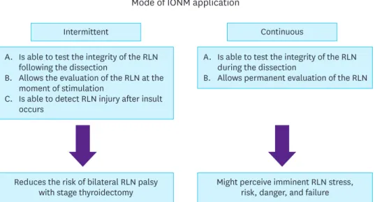
In the clinical practice, as a consequence, intermitted monitoring is able to test the integrity of RLN only following the thyroid dissection, not during the dissection (Fig. 1).
• The surgeon cannot dissect and stimulate at the same time.
• I-IONM cannot alert the surgeon to prevent injury.
• Intermitted technology records a LOS after the nerve damage has been done.
To reduce gap between intermitted stimulation and to monitor entire RLN continuously, a new mode of application of intraoperative neuromonitoring (IONM) was developed, i.e., the CIONM by VN stimulation (Fig. 1). Aim of CIONM is to recognize imminent RLN stress/
risk of injury, thus avoiding definitive nerve palsy and to overcome the main obstacle of intermitted IONM, i.e., the diagnosis of LOS when nerve lesion already has occurred.
Applying at the very beginning of surgery, an electrode on the VN and reducing the gap between 2 stimulations (increasing its frequency of stimulation, i.e., 1 per second),
monitoring of the functional integrity of the RLN all along its course and in real time for the duration of the intervention is achieved (7-9).
Mode of IONM application
A.
B.
C.
Is able to test the integrity of the RLN following the dissection
Allows the evaluation of the RLN at the moment of stimulation
Is able to detect RLN injury after insult occurs
A.
B.
Is able to test the integrity of the RLN during the dissection
Allows permanent evaluation of the RLN
Reduces the risk of bilateral RLN palsy
with stage thyroidectomy Might perceive imminent RLN stress, risk, danger, and failure
Intermittent Continuous
Fig. 1. Features of intermitted and continuous mode of IONM applications.
IONM = intraoperative neuromonitoring; RLN = recurrent laryngeal nerve.
FUNCTIONALITY
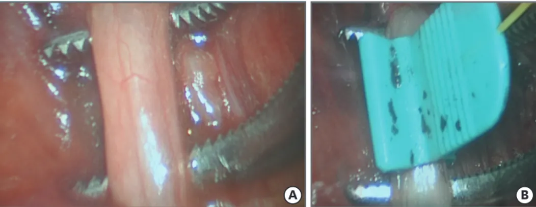
Monitoring is carried out by positioning of an electrode directly on VN in its intracervical course inside of the neck vascular-nerve sheet, that is, proximally to the RLN origin (Fig. 2).
A pulsatile stimulation of intensity and frequency reaches the VN through the electrode and it generates a potential for action that propagate through the RLN and stimulate the laryngeal muscle innervated by RLNs.
The electromyographic (EMG) signal is recorded from the electrodes of the endotracheal tube and reproduced both visually and acoustically both in amplitude and latency (Fig. 3).
The intensity of stimulation has proven optimal at the value of 1 mA, being superimposed (on fibers A and B), so it can guarantee the maximum amplitude of the EMG signal, but without causing any side effects due to recruitment of the C vagal fiber (10-13). The preferred frequency is usually 1 Hz allowing a sufficiently narrow gap, such as which monitoring can be termed “continuous.” The pulse is rectangular, negative, of the duration of 100/200 μs. The visual report consists of a curve typically biphasic or triphasic (Fig. 3). The amplitude and latency of the curve are characteristics for each case.
A B
Fig. 2. CIONM electrode position on the VN. In the specific case, this is the APS (Medtronic, Jacksonville, FL, USA).
Positioning of APS includes: (A) meticulous 360° dissection of the VN is required and (B) subsequent delicate positioning of the electrode.
CIONM = continuous intraoperative neuromonitoring; VN = vagus nerve; APS = automatic periodic stimulation.
Fig. 3. CIONM monitor provides real-time, permanent information about RLN latency and amplitude.
CIONM = continuous intraoperative neuromonitoring; RLN = recurrent laryngeal nerve.
These variations are determined by various factors, most commonly due to imperfection of contact of the surface electrodes of the endotracheal tube and the vocal cords (Table 1). Fore example, the use of paralyzing anesthetic agents will significantly reduce, if not completely eliminate, EMG responses to direct or passive neural stimulation.
While the amplitude is very much random variable (up to 100–2,000 μV), latency is usually between 5 and 7 ms per VN left and between 3 and 5 ms for the VN right, due to the different nerve length on both sides (14-18).
TYPE OF CIONM ELECTRODE
The stimulating CIONM electrodes must be configured in a manner that is protecting against dislocation and changes in their distance from the nerve during surgical manipulation within the surgical site (19). The ideal vagal electrode should meet many CIONM requirements: 1) geometry- design, 2) applicability, 3) easy removal, 4) low stimulation current, and 5) signal stability (20).
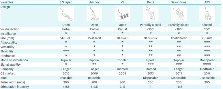
The evolution of continuous stimulation of RLN has seen the introduction of various types of electrodes different in shape, size, flexibility, mode application, and polarity (Table 2).
Table 1. Variation several variables unrelated to RLN injury
• Type of induction or maintenance of anesthesia
• Endotracheal tube malposition
• Small ID tube
• Manipulation of trachea
• Stimulation probe-nerve contact
• RLN preparation (i.e., degree of fluid in the surgical field, nerve ensheathed in fascia)
• Type of stimulation probe (i.e., monopolar vs. bipolar)
• Temperature
• Different metabolism of the patients for the drugs used for induction or maintenance of anesthesia RLN = recurrent laryngeal nerve; ID = internal diameter.
Table 2. CIONM electrodes for RLN monitoring availability
Variables S Shaped Anchor V3 Delta Saxophone APS
Design
Open Open Open Partially closed Partially closed Closed
VN dissection Partial Partial Partial <360° <360° 360°
Installation
* * * * * *
Size (mm) 54×8×0.8 25×0.8×18 20.8×4.8 18.05×5×7 17×different 2–3 mm
Adaptability
* * * ** ** ***
Versatility
* * * ** ** ***
Flexibility
*** * * ** ** ***
Hindrance
* * * ** ** ***
Mode of stimulation Tripolar Bipolar Tripolar Bipolar Tripolar Monopolar
Signal stability
* ** * *** *** ****
Company Langer Langer Inomed Inomed Langer Medtronic
CE market 2010 2009 2008 2013 2013 2011
Kind Reusable Reusable - Disposable Disposable Disposable
Pulse width (mcs) 200 200 200 200 200 200
Stimulation intensity 1–2.5 1–2.5 3–5 1 1–2.5 1
Probes have different proprieties according to geometry (closed vs. open design) VN dissection required, installation, size, adaptability, hindrance, mode of stimulation, and signal stability.
CIONM = continuous intraoperative neuromonitoring; RLN = recurrent laryngeal nerve; VN = vagus nerve; APS = automatic periodic stimulation; CE = combined event.
CIONM electrodes mainly distinguish by geometry and design into (a) open conformation, (b) closed, and (c) half-closed (Table 2). Open design may require that the electrode it is simply leaning on the surface of the nerve or that it is completely surrounded.
This determines the type of dissection of the VN which is required for the application: in the first case just open the vascular-nervous sheet, identify the nerve surface and position the CIONM electrode; in the second case, a 360° VN dissection is required (Fig. 2). These types of electrodes are maintained on site from adherence to the common carotid artery (CCA) and the internal jugular vein (IJV).
Their stability (condition necessary for its dislocation not to occur during operational maneuvers) is therefore given by the integrity of the vascular-nervous sheet, and by the VN dissection and it comes down in case of its wide dissection, for example in the case of lymphadenectomy of the lateral compartment or resection vascular due to a neoplastic infiltration (21).
Stability is considerably better for electrodes closed and half-closed. Both need a meticulous dissection of the entire VN circumference and they close on it completely surrounding it or
“croaking” it atraumaticly. The fair compromise between good stability and atraumaticity in case of accidental removal is given from the flexibility of the CIONM structure. More fine variations in ergonomics of design effect on ease of the application maneuver, reducing the operating time and improving learning curves.
As for the stimulation mode, CIONM electrodes differ in monopolar, bipolar and triplex (Table 2). To perform a VN pacing, is need a pulse of current, which then requires a closed circuit. Conventionally when the anode and cathode are placed at distance and only one of them is in contact with the excitable fabric is by definition a monopolar stimulation.
Generally, an EMG stimulation monopolar is done with a polarity negative on the probe (cathode, −). The anode is placed on the cutaneous (adhesive) or subcutaneous surface (needle) (22).
In bipolar stimulation, both poles are direct contact with the nerve, as well as in stimulation
“tripolar,” where the negative pole is placed at center of 2 positive poles. Such solutions have advantages and disadvantages. Monopolar stimulation is less focused, because of the current dispersion that can excite muscular or nervous structures adjacent. In contrast, bipolar stimulation is more focused, but may be subject to shunt, causing sub-maximal stimulation.
Furthermore, it is sensitive to orientation: positioning of the anode distal to the cathode can cause a block rather than a pacing. An electrode with triple configuration (+ − +) is even more focused, further reduces artifacts and is not sensitive to orientation.
Regarding dimensions is to be considered the size of the CIONM electrode, which it has to guarantee an adequate distance between the poles is greater in the bipolar type and further increased if triple. It should be noted that the shunt effect of bipolar and tripolar system is amplified by one wet surface even by modest amounts of liquids (washing water or blood), compromising the proper functioning of the electrode. This disadvantage is very small with the electrode monopolar, which is able to maintain a pacing stable even if the field is not completely dry. However, the presence of a larger liquid volume can cause the failure reaching the nerve by the stimulus, which tends to propagate through the material with a minor resistance (16,17, 22).
In conclusion, we can affirm that signal stability as a key requirement for CIONM. Most new CIONM electrodes design and geometry are in favor of closed design one because of 1) reduced displacement rate, 2) lower stimulation currents, 3) higher EMG amplitudes, and 4) EMG signal stable (Table 3).
PLACEMENT OF THE ELECTRODE
Contrary to intermittent RLN stimulation, the continuous stimulation of RLN requires the opening of the carotid vascular sheet and identification, visualization, isolation of the VN on which it is to be applied the CIONM electrode (Fig. 2). Proper handling, insertion and placement of the probes is critical for safe and accurate EMG monitoring. Access to the vascular-nervous sheet is possible through various types of surgical approaches (Figs. 4 and 5).
The VN is identified in the carotid sheath (between the middle and lower thyroid pole) at the early phase of the surgical intervention bilaterally. Typically for thyroid and parathyroid surgery, the favorite access is that medially to the strap muscle and laterally to the thyroid gland (Fig. 4).
Table 3. Reasons in favor of CIONM electrodes with a closed design and geometry
• Reduced displacement rate
• Lower stimulation currents
• Lower stimulation currents
• EMG signal stable
CIONM = continuous intraoperative neuromonitoring; EMG = electromyographic.
Fig. 4. Access to the VN, medially to the strap muscle and laterally to the thyroid gland.
VN = vagus nerve.
Fig. 5. Modified lateral access to VN. Access to the VN, laterally to the strap muscle and medially to spernocleidomuscle.
VN = vagus nerve.
However, this type of access is difficult in the case of massive goiters, re-do surgery because of difficult access to the carotid sheet and easier dislocation of the CIONM electrode during surgical maneuvers, as the cable runs inside the space operative (24).
The alternative is a modified lateral access, which provides for the margin between sterno- ioide muscle and sternocleidomastoidosis, spreading between the 2 muscle bundles to reach directly the vascular-nervous sheet. This type of procedure can keep isolated the CIONM wire cable from the operative field (Fig. 5).
This last modified side access is suggested in case of lymphadenectomy of the central
compartment of the neck (i.e., Dralle K1a + b and Robbins Livel 6). In detail, access to the carotid sheath is achieved in 2 different ways. The anterior (or median) approach is found between the space of the thyroid lobe medially and the sternothyroid muscle, but, in some cases, it depends on the extent of the goiter (Fig. 4). Positive identification of the carotid sheath is favored by gentle retraction and elevation of the thyroid gland in a medial direction and by using lateral countertraction on the sternothyroid muscle. The modified anterior (or intermedian) approach is the space between the sternothyroid muscle and the sternocleidomastoid muscle (Fig. 5). After finding the carotid sheath, surrounding muscle fibers are freed as these could interfere with the stimulation. By sharp dissection, the carotid sheath is opened and it is important to sweep away the fatty tissue in the carotid sheath. In order to allow safe stimulation of the VN, we dissected a 15 to 20 mm pouch in the carotid sheath, thus not all the sheath is routinely inspected (25).
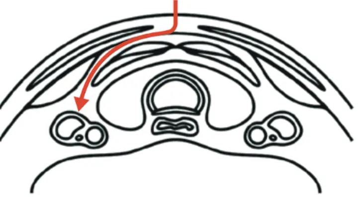
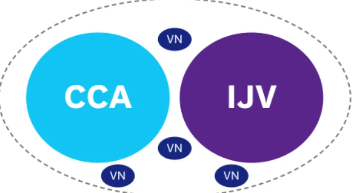
Furthermore, for a safer and standardized surgical approach, it is mandatory to have an accurate knowledge of the topographic relationships of the VN, the jugular and carotid vessels, and understand the neurophysiology of the CIONM system (Fig. 6). The topographic relationship of the VN with the carotid and jugular vessels have been previously studied in detail (11). In this study, we will provide an anatomic scheme for the identification of the VN within its course in the carotid sheath. In particular, the location of the VN in relation to the CCA and the IJV is determined and classified as anterior (A), posterior (P), posterior to internal jugular vein (Pj), and posterior to the common carotid artery (Pc). Literature suggests that most VN lay in the posterior region of the carotid sheath in the grove between the 2 vessels.
The P location of the VN is the most common configuration observed bilaterally in the neck followed by the Pc (15%) and the Pj (8%) locations. Less than 5% of cases of the A location are observed overall. Such classification is useful in the intraoperative setting. VN identification may be expedited with an increase in stimulation intensity of 2 to 3 mA. This is advantageous if the surgeon cannot initially identify the VN, or is not confident with carotid sheath dissection,
CCA IJV
VN
VN
VN VN
Fig. 6. Variability of VN localization in the carotid sheet.
VN = vagus nerve; CCA = common carotid artery; IJV = internal jugular vein.
especially in the case of redo surgery, or if the patient has a large goiter, a hostile neck (gross lateral metastatic lymph nodes that may displace the VN, tortuosity, previous neck procedures, kinking and coiling of the CCA, carotid sheath stuffed with fatty tissue, rotation of the patient's head, and variations of the IJV and CCA position), or during endoscopic thyroidectomy (Fig. 6).
Once identified the nervous structure, it is necessary to mobilize a portion for the positioning electrode. Mobilization a 360° is required when using an electrode closed, otherwise it is sufficient to expose the surface of the nerve. In both cases it is crucial importance not to preclude the vascularization of the VN during isolation and mobilization (26,27).
Most common CIONM electrode use is the automatic periodic stimulation (APS; Medtronic Inc., Jacksonville, FL, USA) device, which is a closed design probe. Thus, a circumferential dissection of the VN is required. Therefore, for optimal placement, the VN is dissected 3,608 at the carotid sheath for approximately 10 mm along the length of the nerve. VN dissection is carried out meticulously with fine-tip anatomic instruments, such as Shannessy brand.
Subsequently, the CIONM probe is gently positioned (Table 4).
Efforts were made to avoid dissecting arterial and venous blood supply of the VN while exposing it. The APS electrode is held at a 45° angle to the VN. A gentle sliding motion in a cranial-caudal direction is made to allow correct contact of the nerve between the jaws of the electrode without pinching the nerve and then the forceps are released.
It is important to consider the possibility of neuroanatomic variations that could prevent desired innervations of the APS. As it concerns for the right VN, it is advisable to proceed to the isolation of the most proximal section given the possibility that there is a non-RLN, the incidence of which is described between 0.3% and 1.6%.
Options for dissection and controlling of hemostasis during this stage excluded any energy- based devices to prevent thermal injury to the VN (therefore to the RLNs). Once identified, the VN is routinely stimulated with a current/intensity of 1-mA in the surgical field by the application of a stimulator probe. If the VN is not initially identified, probe stimulation amplitude is increased to 2 to 3 mA to expedite VN identification. The positive identification of the VN is heard from an audible signal and also is seen as a corresponding biphasic waveform EMG signal on the IONM monitor, which reflects an intact RLN.
Finally, attempts were made to keep the surgical field dry with a swab to avoid any artifacts and/or signal shunting. APS is available in 2 sizes: 2 and 3 mm. Initial baseline (V1) responses should be greater than 500 mcV according to guidelines (23).
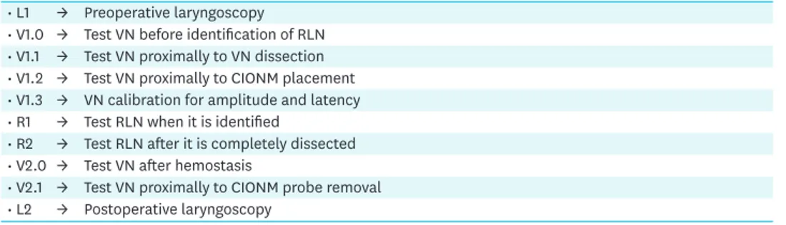
Table 4. Steps for a standardized procedure of CIONM technique
• L1 → Preoperative laryngoscopy
• V1.0 → Test VN before identification of RLN
• V1.1 → Test VN proximally to VN dissection
• V1.2 → Test VN proximally to CIONM placement
• V1.3 → VN calibration for amplitude and latency
• R1 → Test RLN when it is identified
• R2 → Test RLN after it is completely dissected
• V2.0 → Test VN after hemostasis
• V2.1 → Test VN proximally to CIONM probe removal
• L2 → Postoperative laryngoscopy
CIONM = continuous intraoperative neuromonitoring; VN = vagus nerve; RLN = recurrent laryngeal nerve.
Electrode integrity should be checked (by pressing electrodes check on the IONM system) after electrode insertion and before electrode removal to give additional assurance that electrode continuity was maintained throughout the entire procedure. If electrode impedance is very high, discontinue use and replace. Reuse of single use electrodes and probes increase the risk of infection and may cause degraded or ineffective monitoring (Table 4).
PROBE UNFORESEEABLE DISPLACEMENT
In detail, the rate of displacement is greater if the access is medial to the strap muscles. This may be due to interference of the cable in the operative field as opposed to when the access is lateral to the strap muscle (Tables 4 and 5). Displacement rate is between 0%–40% of cases (Table 5). Displacement requires subsequent need of re-baseline, thus, prolongation of surgery.
As a consequence, we recommend accurate movements by both the first operator and assistant, being careful not to stretch the cable with retractors when an anterior approach is decided upon.
Moreover, it is important to consider the possibility of neuroanatomic variation of the VN or the Table 5. Current published papers on CIONM: review on time effort, adverse systemic effects and local complications due to CIONM placement and displacement rate Author / Journal Year CIONM
probe VN VN stimulation
intensity (mA) Monitoring
adverse effects Adverse
effects Local
complications Time effort
(min) Displacement (%)
Terris / WJS 2015 12 1 ? 1 bradycardiahy
potension 1 VN injury 7.9+/−6.1
(6–26) 41
Schneider / LAS 2009 78 1.5 (0.8–2) Anesthesia monitor 0 0 1.45
(0.56–5.25) <5
Mangano / H&N 2015 400 1 Anesthesia monitor 0 0 15→6 11→6
Schneider / BJS 2015 2,581 1 Anesthesia monitor 0 0 - -
Phelan / Laryng. 2014 200 1 Anesthesia monitor 0 0 2.5 -
Schneider / H&N 2013 52 1 Anesthesia monitor 0 0 3.7 <5
Jonas / STI 2014 1,184 1–3 Anesthesia monitor 0 0 5.7 0
Ulmer / Friedrich 2012 79 1 HRVA 0 0 6.8 0
Van Slycke / LAS 2013 180 1 Anesthesia monitor 0 0 3.7 0
CIONM = continuous intraoperative neuromonitoring; VN = vagus nerve; HRVA = heart rate variability analysis.
presence of a non-RLN. We noticed that the A VN position requires less effort for identification, easier anatomic dissection, and simple CIONM probe installation and removal, but probe displacements are more likely. The P position is associated with more effort in VN identification, stiff dissection, and rougher installation. It is, however, less likely for displacement but needs more cautious removal. Therefore, we suggest making a smaller pocket for dissection in cases in which the VN is anterior so that the anterior fascia surrounding the carotid sheath will maintain the CIONM electrode within its position. However, if the nerve is in the rear posterior we suggest a larger pocket for a safer dissection. Finally, displacement was frequent when a 3-mm APS electrode size was used in a VN diameter <2 mm. The authors agree that the ideal CIONM probe may have different sizes or may adapt to different situations and distributions of the VN. If the operating surgeon has different sizes and adaptable CIONM electrodes available in the operating room, then the most appropriate one for the patient can be selected at the time of implantation.
According to the present study, time effort >15 minutes for the APS position was related to: VN P location, BMI >35 (class II obesity), redo surgery, and gland volume >75 mL (P<0.05). Time effort was not related to the size of the CIONM electrode used or the VN diameter.
TYPES OF INJURY
In general, nerve damage can be distinguished according to the traumatizing agent into mechanical, thermal and chemical. Chemical insult cannot be included for RLN damage as no lesive substances are used during thyroid surgery. As far as mechanical damage is concerned, these are subdivided into traction injuries (direct or indirect), by compression (clamping, stapling, bonding, buckling) or by cutting (shrinkage). Thermal insults are caused by the heat dispersion that occurs with use of haemostatic devices (ultrasonic or radiofrequency instruments, monopolar or bipolar scissors) or irrigation of the operating field with liquid at temperatures too high or too low (15).
The International Nerve Monitoring Study Group (INMSG) subdivides 2 types of injuries:
1) segmental lesions and 2) diffused/global injuries (23). Type 1 are mostly due to a direct damage to the nerve and the segment affected can be identified with the intermitted monitoring probe. Type 2 global injuries, whose etiopathogenesis is still unclear, perhaps seem to originate from indirect forces at the RLN laryngeal entry point (1-7).
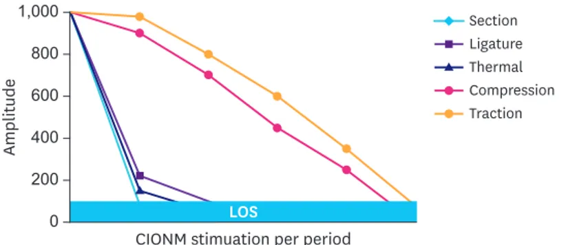
CIONM is able to prevent only some types of injuries (Fig. 7). In particular CIONM may avoid most of direct or indirect trauma that gradually action, showing a progressive, reversible, non acute, modification of the EMG signal in terms of increase latency and decrease amplitude, then the surgeon can interruption the maneuver. That is gradual nerve lesions,
600 400 200 0
Amplitude
800 1,000
CIONM stimuation per period LOS
Section Ligature Thermal Compression Traction
Fig. 7. Amplitude profile in different type of nerve injury.
slow lenses, that allow a slow modification of the amplitude and latency of the EMG signal, these allow the surgeon to notice the EMG modification and modify his surgical technique (syncronization of the surgical maneuver).
According to studies, 80% of the RLN trauma is due to a mechanism traction, in particular when applied to the trachea, ligament of Berry, at the lower thyroid pole intersection of RLN with inferior thyroid artery (7).
The interruption of the risk traction maneuver proved to be effective in the recovery of the initial EMG values above all if performed before a LOS (7).
INTERPRETATION
For CIONM signal to be effective it is necessary to ensure good sensitivity in identifying the variations of the EMG curve. High quality EMG signal in CIONM is fundamental to make any surgical deliberations, for safety, to reduce false positive results and limits of intermitted IONM.
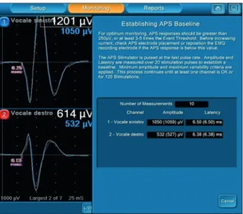
CIONM requires exact calibration of VN stimulation amplitude and latency at the beginning of surgery (Fig. 8). It is required, first, to achieve the calibration/baseline value of the amplitude and latency. This issue is reference on which is calculated the percentage of increase latency or decrease amplitude (2,7).
CIONM requires exact calibration of VN stimulation amplitude and latency at the beginning of surgery. To achieve net benefit, it is important to recruit an experienced surgeon who adheres to the strict standardization guidelines and a cooperate anesthesiologist. Additionally, if the surgeon has competence in troubleshooting algorithms and knowledge of monitoring limits, if they can implement training and experience and continuous audit, this would be beneficial in the future review of CIONM (1,13). Obtaining a baseline amplitude >500 mcV is the prerequisite for the diagnosis, interpretation, and verification of a functional intact RLN, an LOS, to monitor significant modification of EMG profile, and signal reentry or recovery. The observed increased
Fig. 8. Baseline achievement.
APS = automatic periodic stimulation.
EMG amplitude seen between V1 and V2 determinations in uneventful cases may reflect several phenomena as: 1) increasing VN diameter (i.e., better probe-VN contact), 2) increased excitability of ionic channels in neurons, and 3) vocal cord edema. This may be decreased in future studies by implementing more precise contact of the EMG tube electrodes to the vocal cords (8).
The INMSG established that the amplitude for reference must be 500 μV (23). In otherwise, the collaboration with the anesthesiologist is imperative who will re-position the EMG endotracheal tube by optimizing the contact of the surface electrode to the mucosa of the vocal cords until it reaches satisfactory EMG values. The 500 μV value is the prerequisite for the diagnosis, interpretation, verification of 1) functional intact RLN, 2) LOS, 3) definition of
“significant” modification of EMG signal, and 4) EMG signal re-entry/recovery.
Once the reference value has been acquired of the curve (baseline) the CIONM analyzes the changes of the same by displaying on a timeline the amplitude and latency values (Fig. 3). The CIONM software sets an audible and visual warning signal overcoming threshold values to assist the surgeon to early identify surgical risk maneuvers.
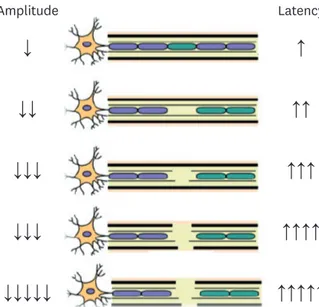
Vocal cord paralysis (VCP) is the outcome of RLN injury, which can be identified by CIONM as intraoperative progression of increasingly severe, adverse evoked EMG-events. The most typical sequence of adverse EMG events leading to VCP is multiple combined events (CEs) (increased latency + decrease amplitude) with evolution to complete LOS (Fig. 9). Again, CEs (generally reversible) and LOS (generally irreversible) track robustly with VCP and are identifiable intraoperatively with CIONM. Nine of 13 patients (70%) who developed adverse EMG changes based on these criteria, modification of the respective surgical maneuver resulted in EMG recovery (7,8).
If significant adverse EMG changes temporally associated with a given surgical maneuver occurs, then that maneuver should be immediately aborted. Subsequently, EMG changes should be closely followed-up to determine recovery. Once satisfactory EMG recovery is observed surgery can be continued. Otherwise, decision should be made with respect to the possible staged thyroidectomy.
Amplitude
↓
↓↓
↓↓↓
↓↓↓
↓↓↓↓↓
Latency
↑
↑↑
↑↑↑
↑↑↑↑
↑↑↑↑↑
Fig. 9. Severity of nerve injuries.
CIONM provides real-time RLN evaluation during surgical maneuvers. CEs and LOS, both easily identifiable intraoperatively, are related to the development of VCP. A CE represents a largely reversible electrophysiologic change when the associated surgical maneuver is aborted. If allowed to continue, it can advance to LOS (which typically is significantly less reversible) and to postoperative VCP. CIONM has utility in identifying real-time adverse concordant amplitude and latency changes (CEs), which can prompt modification of the associated surgical maneuver and may prevent RLN paralysis during thyroidectomy (10-20).
The definition of an intraoperative EMG signal loss (LOS) presupposes that the initial signal response was correct. If the intraoperative signal fails or the EMG drops below 100 μV with a primary intact signal and adequate stimulation with 1–2 mA, a LOS must be assumed.
Intraoperative signal loss has a predictive value positive (postoperative cordial paralysis) that varies between 92% and 100% of cases (23,26).
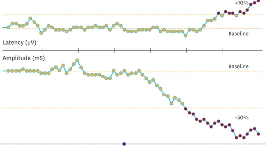
CIONM helps to prevent the occurrence of a LOS (Fig. 9). The risk of LOS is related to the presence of combined multiple events, where a CE is defined such as a 50% associated with a decrease in amplitude and an increase of 10% of latency relative to the calibration/baseline value (Fig. 10). These values are set to the CIONM signal of alert (Fig. 3) (7).
It is important to gain experience in distinguishing the curve changes due to artifacts from those that really indicate the nerve is under stress. Modern CIONM softwares analog filters can be effective in filtering respiratory interferences, artifacts, amplifiers, shunts, and other artifacts and especially those given by dissecting or hemostasis devices (7).
SAFETY OF CIONM PROBE POSITIONING
A detail schema for the CIONM standardized safety procedure is suggested (Table 4). This protocol is intended to control any adverse event in the dissection of VN and subsequent CIONM electrode positioning. We suggests to test the functionality of VN using the probe for
Latency (µV) Amplitude (mS)
Baseline
Baseline
−50%
+10%
Fig. 10. EMG reference values for the warning signal during CIONM
EMG = electromyographic; CIONM = continuous intraoperative neuromonitoring.
stimulation intermittent before proceeding to the positioning of the CIONM electrode and at it removal, proximally.
Mangano et al. have evaluated specifically the safety of CIONM electrode positioning (28). Four hundred VN dissections were analyzed considering VN diameter, mean time effort for CIONM probe positioning, and electrode dislocation rate. A significant superior dislocation rate in case of: 1) when a 3-mm APS electrode size was used in a VN diameter <2 mm, 2) anterior access, and 3) VN A subtype in relation (P<0.050). No related additional local or systemic morbidity was registered in this series. There was a statistically significant positive relationship between increased diameter of VN and increased EMG amplitude. There was also a significant increase of amplitude between initial and final VN stimulation in uneventful cases.
SIDE EFFECTS OF STIMULATION VAGAL
Table 5 reviews the literature reports about CIONM safety. Publications with the most number of procedures performed did not detect local or systemic complications.
Furthermore, VN stimulation counts numerous other applications in addition to the RLN monitoring. Implant electrodes are commonly used for permanent VN stimulation for the treatment of epilepsy, depression, tinnitus, anxiety, Alzheimer's disease, migraine, fibromyalgia, and obesity.
The spread of CIONM technology in thyroid surgery has contributed to promoting numerous animal model studies with the purpose of better understanding the neurophysiology, neurophysiological and safety of vagal stimulation. These studies are especially dedicated in the research part of recruitment of efferent nerve fibers autonomy and their short-term effect (during operative time) in determining side effects (10-13). It has come to the conclusion that the currents used for the CIONM technique do not activate the thin demyelinated fibers C responsible for the autonomous effects. The 1 mA stimulus is in fact super-mass only for the efferent fibers motor and fibers B myelinated autonomous.
Further studies must be performed to deepen the meaning of such experiences (10-13).
APRAISAL
CIONM is potentially superior to intermitted IONM because it enhances standardization by permanent VN stimulation, it provides entire and continuous RLN function monitoring, comprehensive EMG documentation, detects and potentially prevents imminent RLN failure by synchronization of surgical maneuvers, and clarifies RLN stress mechanism (Table 6).
The CIONM technique is no doubt a new great adjuvant for the surgeon in thyroid surgery, providing a useful tool to prevent improper surgical maneuvers. At the same time CIONM is still complementary to intermitted monitoring (Table 7).
Despite surgeon's experience, CIONM is useful in both preoperatively unpredictable RLN risk situations and preoperative predictable risk conditions: 1) preoperatively unpredictable RLN risk situations are represented by atypical RLN pattern, RLN anterior to the thyroid
gland, RLN fixed, splayed or entrapped, RLN posterior to Berry ligament, anteriorly located RLN to the Tuberculum-Zuckerkandl (posterior nodule), branched RLN, antevascular RLN, thin RLN, invaded RLN, non-RLN, 2) preoperative predicable high risk conditioning are recurrences with tenacious adherence, infiltrating tumors, massive goiter, tracheal deviation, graves' disease, lymph node dissection, short neck, obesity.
CIONM has utility in identifying real-time adverse concordant amplitude and latency changes (CEs), which can prompt modification of the associated surgical maneuver and prevent RLN paralysis during thyroidectomy (5-7). Authors recently reported electrophysiological parameters as waveform, amplitude decreased, and latency increased combined changes, relating these parameters to intraoperative surgical maneuvers that delineate nascent adverse but reversible electrophysiological parameters to prevent nerve injury (Table 8) (5-7). Surgical maneuvers were modified when adverse electrophysiological findings were noted. Both intraoperative CEs and LOS were significantly associated with development of VCP. If allowed to continue, it can advance to LOS, which typically is significantly less reversible, and to postoperative VCP (5-7).
The CIONM provides continuous feedback of the RLN during surgical maneuvers such as the mobilization of the thyroid lobe in the cranial direction or lateral to medial direction, Table 6. CIONM appears to be superior to intermitted RLN stimulation
• Enhance IONM standardization
• Entire and permanent RLN function monitoring
• Comprehensive EMG documentation
• Detects and prevent imminent RLN failure
• Elucidate RLN stress mechanism
• Syncronization of surgical maneuver
• EMG tube malposition identification
CIONM = continuous intraoperative neuromonitoring; RLN = recurrent laryngeal nerve; IONM = intraoperative neuromonitoring; ENG = electromyographic.
Table 7. CIONM and I-IONM are still 2 complementary devices
Proprieties CIONM I-IONM
Prevent transient RLNP − −
Prevent permanent RLNP ++ −
Syncronization of surgical manouvers +++ −
Awarness of nerve stress +++ +
Appreciation of nerve function recovery +++ +
Mechanism of nerve injury ++ +
RLN identification, mapping + ++
Site of nerve injury + ++
Nerve variability, branching + ++
Nerve dissection − ++
EBSLN monitoring − ++
EMG tube malpositioning ++ −
Documentation +++ +
CIONM = continuous intraoperative neuromonitoring; I-IONM = intermitted intraoperative neural monitoring;
RLNP = recurrent laryngeal nerve palsy; RLN = recurrent laryngeal nerve; EBSLN = external branch of the superior laryngeal nerve; EMG = electromyographic.
Table 8. CIONM events definitions
• mCE: amplitude decrease of >50% to 70% with a concordant latency increase of 5% to 10%
• sCE: Amplitude decrease of >70% with a concordant latency increase of >10%
• LOS: Complete loss of recognizable RLN EMG signal (amplitude <100 µV) intraoperatively as defined by the INMSG CIONM = continuous intraoperative neuromonitoring; mCE = mild combined event; sCE = severe combined event;
LOS = loss of signal; RLN = recurrent laryngeal nerve; EMG = electromyographic; INMSG = International Nerve Monitoring Study Group.
the capsular dissection or the digital extraction of a retrosternal portion of the goiter. Under these circumstances it is not always possible identify early the RLN, thus once positioned the CIONM electrode first, this continually warns the surgical team of security of every step of dissection in real time.
CIONM presents limitations. A continuous critical evaluation of neuromonitoring devices available to date is essential for the development and introduction of CIONM application method. Although various vagal electrodes are available for CIONM, there is still uncertainty regarding the electrophysiologic thresholds indicative of imminent RLN injury. Futher clinical research, especially in neurophysiology and neuropathology, is necessary in order to make a qualitative and quantitative analysis and interpretation of the change of signal obtained with CIONM before widely being used on Patients. These data are essential for surgeon's decision making.
CIONM ideal prognostic criterium must incorporate existing monitoring standards as outlined by the INMSG guidelines: of preoperative and postoperative laryngeal examination, routine vagal stimulation, recognition of standard normative recording parameters, and the application of a standard equipment trouble shooting algorithm. It must differentiate artifactual EMG changes (such as those occurring with intraoperative endotracheal tube displacement) from EMG changes truly reflective of impending neuropraxic injury EMG changes must track with impending neuropraxic injury and must be clearly recognizable intraoperatively. Of importance, these changes must be nascent and completely reversible once the associated surgical maneuver is aborted.
It is clear that further work is required to optimize a CIONM definition of meaningful warning threshold values. The negative predictive value (NPV) and positive predictive value (PPV) of various adverse EMG parameters are strongly related to the underlying prevalence of VCP in the study population. Before adverse EMG parameters of single CEs and LOS are applied to different surgical populations their prognostic accuracy should be tested by independent observers in varying surgical environment. The development and temporal course of the adverse EMG changes as a result of surgical trauma will likely vary with the nature of the given surgical maneuver and its force and duration.
REFERENCES
1. Lamadé W, Meyding-Lamadé U, Hund E, Senninger N, Herfarth C. Transtracheal monitoring of the recurrent laryngeal nerve. Prototype of a new tube. Chirurg 1997;68:193-5.
PUBMED | CROSSREF
2. Lamadé W, Brandner R, Brauer M, Hund E, Klar E, Herfarth C. Continuous monitoring of the recurrent laryngeal nerve. Langenbecks Arch Chir Suppl Kongressbd 1998;115:1055-7.
PUBMED
3. Lamadé W, Meyding-Lamadé U, Buchhold C, Brauer M, Brandner R, Uttenweiler V, et al. First continuous nerve monitoring in thyroid gland surgery. Chirurg 2000;71:551-7.
PUBMED
4. Lamadé W, Ulmer C, Seimer A, Molnar V, Meyding-Lamadé U, Thon KP, et al. A new system for continuous recurrent laryngeal nerve monitoring. Minim Invasive Ther Allied Technol 2007;16:149-54.
PUBMED | CROSSREF
5. Ulmer C, Koch KP, Seimer A, Molnar V, Meyding-Lamadé U, Thon KP, et al. Real-time monitoring of the recurrent laryngeal nerve: an observational clinical trial. Surgery 2008;143:359-65.
PUBMED | CROSSREF
6. Dralle H, Sekulla C, Lorenz K, Brauckhoff M, Machens AGerman IONM Study Group. Intraoperative monitoring of the recurrent laryngeal nerve in thyroid surgery. World J Surg 2008;32:1358-66.
PUBMED | CROSSREF
7. Schneider R, Randolph GW, Sekulla C, Phelan E, Thanh PN, Bucher M, et al. Continuous intraoperative vagus nerve stimulation for identification of imminent recurrent laryngeal nerve injury. Head Neck 2013;35:1591-8.
PUBMED | CROSSREF
8. Schneider R, Bures C, Lorenz K, Dralle H, Freissmuth M, Hermann M. Evolution of nerve injury with unexpected EMG signal recovery in thyroid surgery using continuous intraoperative neuromonitoring.
World J Surg 2013;37:364-8.
PUBMED | CROSSREF
9. Monticelli G. L'elettroneurostimolazione: neurofisiologia ed applicazione dei moderni stimolatori.
Milano: Università degli Studi di Milano; 2003.
10. Castoro MA, Yoo PB, Hincapie JG, Hamann JJ, Ruble SB, Wolf PD, et al. Excitation properties of the right cervical vagus nerve in adult dogs. Exp Neurol 2011;227:62-8.
PUBMED | CROSSREF
11. Groves DA, Brown VJ. Vagal nerve stimulation: a review of its applications and potential mechanisms that mediate its clinical effects. Neurosci Biobehav Rev 2005;29:493-500.
PUBMED | CROSSREF
12. Ulmer C, Friedrich C, Kohler A, Rieber F, Basar T, Deuschle M, et al. Impact of continuous intraoperative neuromonitoring on autonomic nervous system during thyroid surgery. Head Neck 2011;33:976-84.
PUBMED | CROSSREF
13. Yoo PB, Lubock NB, Hincapie JG, Ruble SB, Hamann JJ, Grill WM. High-resolution measurement of electricallyevoked vagus nerve activity in the anesthetized dog. J Neural Eng 2013;10:026003.
PUBMED | CROSSREF
14. Lorenz K, Sekulla C, Schelle J, Schmeiss B, Brauckhoff M, Dralle H, et al. What are normal quantitative parameters of intraoperative neuromonitoring (IONM) in thyroid surgery? Langenbecks Arch Surg 2010;395:901-9.
PUBMED | CROSSREF
15. Schneider R, Przybyl J, Pliquett U, Hermann M, Wehner M, Pietsch UC, et al. A new vagal anchor electrode for real-time monitoring of the recurrent laryngeal nerve. Am J Surg 2010;199:507-14.
PUBMED | CROSSREF
16. López JR. Cranial nerve monitoring. In: Galloway GM, Nuwer MR, Lopez J, Zamel K, editors.
Intraoperative Neurophysiologic Monitoring. Cambridge: Cambridge University Press; 2010. p. 109-41.
17. Tsui BC. Electrical nerve stimulation. In: Tsui BC, editor. Atlas of Ultrasound and Nerve Stimulation- guided Regional Anesthesia. New York (NY): Springer; 2007. p. 9-18.
18. Wu CW, Dionigi G, Chen HC, Chen HY, Lee KW, Lu IC, et al. Vagal nerve stimulation without dissecting the carotid sheath during intraoperative neuromonitoring of the recurrent laryngeal nerve in thyroid surgery. Head Neck 2013;35:1443-7.
PUBMED | CROSSREF
19. Dionigi G, Chiang FY, Rausei S, Wu CW, Boni L, Lee KW, et al. Surgical anatomy and neurophysiology of the vagus nerve (VN) for standardised intraoperative neuromonitoring (IONM) of the inferior laryngeal nerve (ILN) during thyroidectomy. Langenbecks Arch Surg 2010;395:893-9.
PUBMED | CROSSREF
20. Toniato A, Mazzarotto R, Piotto A, Bernante P, Pagetta C, Pelizzo MR. Identification of the nonrecurrent laryngeal nerve during thyroid surgery: 20-year experience. World J Surg 2004;28:659-61.
PUBMED | CROSSREF
21. Mahmodlou R, Aghasi MR, Sepehrvand N. Identifying the non-recurrent laryngeal nerve: preventing a major risk of morbidity during thyroidectomy. Int J Prev Med 2013;4:237-40.
PUBMED
22. Henry JF, Audiffret J, Denizot A, Plan M. The nonrecurrent inferior laryngeal nerve: review of 33 cases, including two on the left side. Surgery 1988;104:977-84.
PUBMED
23. Randolph GW, Dralle H, International Intraoperative Monitoring Study GroupAbdullah H, Barczynski M, Bellantone R, et al. Electrophysiologic recurrent laryngeal nerve monitoring during thyroid and parathyroid surgery: international standards guideline statement. Laryngoscope 2011;121 Suppl 1:S1-16.
PUBMED | CROSSREF
24. Asgharpour E, Maranillo E, Sañudo J, Pascual-Font A, Rodriguez-Niedenführ M, Valderrama FJ, et al.
Recurrent laryngeal nerve landmarks revisited. Head Neck 2012;34:1240-6.
PUBMED | CROSSREF
25. Kaisha W, Wobenjo A, Saidi H. Topography of the recurrent laryngeal nerve in relation to the thyroid artery, Zuckerkandl tubercle, and Berry ligament in Kenyans. Clin Anat 2011;24:853-7.
PUBMED | CROSSREF
26. Dralle H, Sekulla C, Lorenz K, Nguyen Thanh P, Schneider R, Machens A. Loss of the nerve monitoring signal during bilateral thyroid surgery. Br J Surg 2012;99:1089-95.
PUBMED | CROSSREF
27. Wu CW, Dionigi G, Sun H, Liu X, Kim HY, Hsiao PJ, et al. Intraoperative neuromonitoring for the early detection and prevention of RLN traction injury in thyroid surgery: a porcine model. Surgery 2014;155:329-39.
PUBMED | CROSSREF
28. Mangano A, Kim HY, Wu CW, Rausei S, Hui S, Xiaoli L, Chiang FY, Roukos DH, Lianos GD, Volpi E, Dionigi G Continuous intraoperative neuromonitoring in thyroid surgery: safety analysis of 400 consecutive electrode probe placements with standardized procedures. Head Neck 2016;38 Suppl 1:E1568-74.
PUBMED | CROSSREF