J. Exp. Biomed. Sci. 14 (2008) 147–155
Taxifolin Inhibited the Nitric Oxide Production and Expression of Pro-inflammatory Cytokine mRNA in Lipopolysaccharide-stimulated
RAW264.7 Cells
Man Hee Rhee
1,†, Mehari Endale
1, SM Kamruzzaman
1, Whi Min Lee
1, Hwa-Jin Park
2, Myung-Jo Yoo
3and Jae Youl Cho
41
Laboratory of Physiology & Signaling, College of Veterinary Medicine, Kyungpook National University, Daegu 702-701, Korea.
2Department of Biomedical Laboratory Science, College of Biomedical Science and
Engineering, Inje University, Gimhae 621-749, Korea.
3
College of Veterinary Medicine, Chonbuk National University, Jeonju 561-756, Korea
4
School of Biotechonology and Bioengineering, Kangwon National University, Chuncheon 200-701, Korea
In previous works, we found that solvent extract of Opuntia humifusa Raf., a member of the lactaceae family, displayed potent anti-oxidative and anti-inflammatory activities. Thus, all solvent fractions, except for the water layer, showed potent scavenging effects. According to activity-guided fractionation, one of active radical scavenging principles in the ethyl acetate fraction was found to be taxifolin. In this study, we investigated whether taxifolin showed anti-oxidative activity. In addition, taxifolin modulated nitric oxide (NO) release and the expression of pro-inflammatory cytokine mRNA such as interleukin-1β (IL-1β), IL-6, granulocyte-macrophage colony-stimulating factor (GM-CSF), and TNF-α.
Taxifolin showed potent anti-oxidant activity with the IC
50of 8.5±1.4 and 9.3±1.0 μM using xanthine/xanthine oxidase (XO) assay and 2,2-Diphenyl-lpicrylhydrazyl radical (DPPH) assay, respectively. We next determined the role of taxifolin on the immunomodulating activity using murine macrophage cell line RAW264.7 cells. Taxifolin dose- dependently inhibited NO production in lipopolysaccharide (LPS)-activated RAW264.7. It also significantly blocked the expression of inducible NO synthase (iNOS) mRNA in the LPS-stimulated RAW264.7 cells. In addition, taxifolin potently suppressed the expression of IL-1β, IL-6 and GM-CSF mRNA in LPS-activated RAW264.7 cells, but not that of TNF-α. Moreover, taxifolin significantly inhibited the transcriptional activity of nuclear factor-κB (NF-κB) and activator protein -1 (AP-1). These results suggest that taxifolin may downregulate inflammatory iNOS, IL-1β, IL-6 and GM-CSF gene expressions through inhibition of NF-K and AP-1 activation in LPS-stimulated RAW264.7 cells.
Key Words: Taxifolin; Opuntia humifusa Raf; Anti-oxidative activity; Anti-inflammatory activity, Nitric oxide;
Pro-inflammatory cytokines
INTRODUCTION
The use of natural products as antioxidant and anti- inflammatory agents has a long history that began with folk medicine. Focusing attention on plants medically used by indigenous people is the most efficient way to identify
herbal plants that may contain bioactive substances. The prickly pear (O. humifusa Raf.) is a member of the Cactaceae family, and is widely distributed in semiarid countries throughout the world, especially in the Mediterranean area and Central America (Goldstein & Nobel, 1994; Acuna et al., 2002; Lee et al., 2002). In Korea, O. humifusa Raf. was also cultivated for a long time (Cho et al., 2006). Concerning the pharmacological profile of Opuntia spp., reports indi- cated that the total phenols and some flavonoids in solvent extract from South Korean O. humifusa Raf. are respon- sible for the radical scavenging activity, and inhibitory activities of NO production and pro-inflammatory cytokine
*Received: July 16, 2008
Accepted after revision: September 16, 2008
†Corresponding author: Man Hee Rhee, Laboratory of Veterinary Physiology & Signaling, College of Veterinary Medicine, Kyungpook National University, Daegu 702-701, Korea.
Tel: +82-53-950-5967, Fax: +82-53-950-5955 e-mail: [email protected]
expression (Cho et al., 2006). In addition, Kim et al (2005) also reported that aqueous extract of O. humifusa Raf. has a protective effect on carbon tetrachloride-induced liver injury in rats (Kim et al., 2005). In addition, Opuntia humi- fusa extract suppressed DNCB-induced allergic contact dermatitis in BALB/c mice (Kim et al., 2007). Moreover, administration of leaf extracts of O. megacantha not only reduced blood glucose levels, but also resulted in a weight decrease in diabetic animals (Bwititi et al., 2000).
Chronic and acute inflammation are multiple processes that are mediated by activated inflammatory or immune cells (Guzik et al., 2003). Macrophages play a central role in managing many different immunopathological phenomena such as the over-production of pro-inflammatory cytokines and inflammatory mediators (i.e. ROS, NO and prosta- glandin E
2[PGE
2]) (Lundberg, 2003; Walsh, 2003). In the case of oxidative stress, NO and ROS affect virtually every step of the development of inflammation. Macrophages mediate the inflammatory process through the release of chemokines (e.g. GM-CSF) and cytokines (e.g. TNF-α, IL-1β and IL-6). Indeed, a number of inflammatory stimuli such as lipopolysaccharide (LPS) and pro-inflammatory cytokines activate immune cells to up-regulate such inflam- matory states (Gallucci et al., 1998) and therefore, they are important in exploring molecular mechanisms of action for the subsequent development of new potential anti- inflammatory drugs.
Therefore, in this study, first we determined whether taxifolin displayed potent antioxidant activity. We next investigated whether taxifolin inhibited LPS-induced NO production in RAW264.7 cells, and studied the mechanism of NO inhibitory activity.
MATERIALS AND METHODS 1. Reagents
Ascorbate, dimethylsulfoxide (DMSO), 2,2-Diphenyl- lpicrylhydrazyl radical (DPPH), LPS, Griess's reagent, and xanthine oxidase were obtained from the Sigma Co (St.
Louis, MO). Xanthine was from the Merck Co. (Milwaukee, WI). All other reagents were of the first grade.
2. Isolation of taxifolin
Solvent extraction and isolation of taxifolin were de- scribed in previous report (Cho et al., 2006). Briefly, O.
humifusa Raf. was collected in October from the province of Asan (Korea). Taxifolin was structurally identified by ESI-LC-MS/MS [Finnigan LCQ Advantage MAX ion trap mass spectrometer (San Jose, CA, USA)] and
1H-NMR (CD3OD, 300 MHz), and
13C-NMR (CD
3OD, 75 MHz) analysis.
3. Cell culture
RAW264.7 cells were maintained in DMEM supple- mented with 100 U/ml of penicillin and 100 μg/ml of streptomycin, and 10 % FBS. Cells were grown at 37℃
and 5% CO
2in humidified air.
4. DPPH radical scavenging activity
The DPPH assay measures hydrogen atom (or one electron) donating activity and hence provides an evaluation of antioxidant activity due to free radical scavenging. The DPPH, a purple-colored stable free radical, is reduced into the yellow-colored diphenylpicryl hydrazine. The Blios method was used with slight modifications in this experi- ment (Blois, 1958). The fresh batch of a radical stock solution was prepared daily. EDA described the difference of absorbance between the mixture and the control solution, as in a percentage: EDA (%) = (the absorbance of the control - the absorbance of the mixture) / the absorbance of the control × 100.
5. Xanthine oxidase activity assay
The activity of xanthine oxidase with xanthine, as a
substrate, was measured spectrophotometrically according
to the procedures of Noro et al. (Noro et al., 1983), with
the following modifications. Xanthine and xanthine oxidase
were mixed in cuvette with either compounds tested or
vehicles. The difference of absorbance was measured at
295 nm for 3 min and the enzyme activity was calculated
with references: (the activity of control - the activity of the
mixture) / (the activity of control) × 100. The final
concentration of xanthine oxidase was 250 μU/ml in a 0.1
mM phosphate buffer (pH 7.4).
6. Measurement of nitrite
In order to estimate the concentration of nitric oxide, nitrite (NO
2-) was measured using the Griess reagent (1%
sulfanilamide, 0.1% naphthylethylenediamine dihydrochlo- ride, and 2% phosphoric acid), as described previously (Hong et al., 2003). After preincubation of the RAW264.7 cells (1×10
6cells/ml) for 18 h, the cells were incubated with taxifolin with LPS (0.1 μg/ml) for 24 h. One-hundred μl of supernatant from each well of the cell culture plates was transferred into 96-well microplates, and the super- natant was then mixed with equal volume of Griess reagent at room temperature. The absorbance at 550 nm was determined in a Spectramax 250 microplate reader. The concentrations of nitrite were calculated from regression analysis using serial dilutions of sodium nitrite as a standard.
The percentage inhibition was calculated based on the ability of extracts to inhibit nitric oxide formation by cells compared with the control (cells in media without extracts containing triggering agents and DMSO), which was con- sidered as 0% inhibition.
7. MTT assay for the measurement of cell prolif- eration
A cell proliferation assay was also performed to exclude the possibility that the observed nitric oxide inhibition was false positive due to the cytotoxic effects. Cell proliferation was measured by 3-(4,5-dimethylthiazol-2-yl)-2,5-diphenyl tetrazolium bromide (MTT) assay as described previously (Hong et al., 2003). All experiments for the measurement of nitric oxide inhibition were conducted three times, each time with three independent observations and the results were averaged.
8. Extraction of total RNA
Total RNA from the LPS treated-RAW264.7 cells was prepared by using Easy Blue kit (iNtRON Biotechnology Co., Korea), according to the manufacturer's protocol. The total RNA extract was stored at -70℃ until use.
9. Semiquantitative RT-PCR amplification
Semiquantitative RT reactions were carried out using RT premix (Bioneer Co., Korea). Briefly, total RNAs (2 μg) were incubated with oligo-dT
18at 70℃ for 5 minutes and cooled on ice for 3 minutes. It was then incubated with RT premix for 90 min at 42.5℃. The reactions were terminated at 95℃ for 5 minutes for the inactivation of reverse tran- scriptase. The PCR reaction was further conducted using the PCR premix (Bioneer Co., Korea) with the appropriate sense and antisense primers for Glyceraldehyde-3-phosphate dehydrogenase (GAPDH, sense, 5'-CAC TCA CGG CAA ATT CAA CGG C-3'; antisense, 5'-CCT TGG CAG CAC CAG TGG ATG CAG G-3'), iNOS (sense, 5'-CCC TTC CGA AGT TTC TGG CAG CAG C-3'; antisense, 5'-GGC TGT CAG AGC CTC GTG GCT TTG G-3'), TNF-α (sense, 5'-TTG ACC TCA GCG CTG AGT TG-3'; antisense, 5'-CCT GTA GCC CAC GTC GTA GC-3'), IL-1β (sense, 5'-CAG GAT GAG GAC ATG AGC ACC-3'; antisense, 5'-CTC TGC AGA CTC AAA CTC CAC-3'), IL-6 (sense, 5'-GTA CTC CAG AAG ACC AGA GG-3'; antisense, 5'-TGC TGG TGA CAA CCA CGG CC-3'), and GM-CSF (sense, 5'-AGG ATG TGG CTG CAG AAT TTA CTT TTC-3'; antisense, 5'-TCA TTT TTG GAC TGG TTT TTT GCA TTC-3'). Incubation was made under conditions of a 5 min denaturation time at 94℃ followed by cycles of 95℃ for 45 seconds, an annealing time of 45 seconds at 55 to 60℃ and an elongation time of 45 seconds at 72℃.
Final extension was made for 10 min at 72℃ after the end of the cycles. The PCR products were separated in 1%
agarose using electrophoresis of BioRad Co. The relative intensities were calculated using Eagle eyes image analysis software (Stratagene Co., La Jolla). The resulting densities of the iNOS, TNF-α, IL-1β, IL-6, and GM-CSF bands were expressed relative to the corresponding densities of the GAPDH bands from the same RNA sample. GAPDH, a housekeeping gene, was used as RNA internal standard.
10. Luciferase reporter gene activity assay
HEK293 cells (1×10
6cells/ml) were transfected with 1
μg of plasmids with nuclear factor-kappa B dependent
reporter (NF-κB-), activator protein-1 dependent reporter
(AP-1-), and cAMP response element dependent reporter (CRE-Luc) as well as thymidine kinase promoter-Renilla luciferase (TK Renilla) reporter plasmid by using the calcium phosphate method according to the manufacturer's protocol. Luciferase assays were performed using the Luci- ferase Assay System (Promega) (Jung et al., 2006). Briefly, the transfected cells treated with taxifolin in the presence of PMA (0.1 μM), were lysed in the culture dishes with reporter lysis buffer. Lysates were centrifuged at maximum speed for 10 min in a microcentrifuge. Ten μl of the supernatant fraction were incubated with 10 μl of luciferase substrate, and the relative luciferase activity was determined with a GloMax (Promega, Madison, WI). Luciferase activity was normalized to TK Renilla luciferase activity.
11. Statistical analysis
One-way ANOVA with a post hoc Dunnett multiple comparison test of significance was used to determine the statistical significance of differences between values for the experimental and control groups. P values of 0.05 or less were considered as statistically significant. Data represent the means ± S.E.M. of three experiments conducted in triplicates.
RESULTS 1. Antioxidant activity of taxifolin
In order to compare the antioxidant capacity of active ingredients between taxifolin and quercetin, we adopted two well-known assay systems; xanthine/xanthine oxidase assay and DPPH radical scavenging assay. As shown in Table 1, taxifolin displayed potent antioxidant activity with
IC
50of 8.5±1.4 and 9.3±1.0 μM in xanthine/xanthine oxidase (XO) assay and 2,2-Diphenyl-lpicrylhydrazyl radical (DPPH) assay respectively. The antioxidant capacity of quercetin showed different profiles with methods used in the study. The antioxidant activity of quercetin using XO assay system is more potent than that of using DPPH radical scavenging system (i.e., IC
50of 97.5±5.1 nM vs 23.5±8.1 μM).
2. The inhibitory effect of taxifolin on NO production We next determined whether taxifolin, the potent anti- oxidant, ihhibited NO production in LPS-activated RAW- 264.7 cells. As shown in Fig. 1A, taxifolin significantly inhibited NO production in LPS (0.1 μg/ml) stimulated RAW264.7 cell in a dose-dependent manner. Since NO production in LPS-activated RAW264.7 cells is regulated by iNOS expression, we explored whether taxifolin modu- lated the expression level of iNOS mRNA with the concen- tration used for NO production's regulation. Fig. 2A and 2B displayed taxifolin's inhibition of iNOS mRNA in LPS- activated RAW264.7 cells in a dose-dependent manner. In order to exclude the confounding effect of taxifolin in the inhibitory activity on NO production, we performed it's cytotoxicity test in RAW264.7 cells using MTT assay.
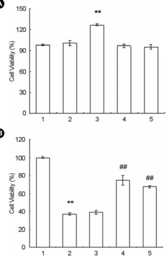
Taxifolin did not show any cytotoxicity effect with the concentration used in the present study (Fig. 3A). It is interesting that taxifolin (50 μM) significantly increased the cell viability. Moreover, LPS decreased the cell viability, which was reversed with the addition of taxifolin (Fig. 3B).
Table 1. Antioxidant activities of quercetin and taxifolin in xanthine oxidase assay and DPPH assay
IC50
XO assay DPPH assay
Quercetin 97.5±9.1 nM 23.5±8.1 μM
Taxifolin 8.5±1.4 μM 9.3±1.0 μM
Either phosphate buffer (0.1 mM, pH 7.4, for xanthine oxidase assay) or acetate buffer (10 mM, pH 5.5, for DPPH assay) and quercetin or taxifolin were mixed, and assay was carried out as described in 'Materials and Methods'. Each value is the mean ± SEM of three determinations, performed in duplicate
Fig. 1. Chemical structure of taxifolin used in the present study.
3. The effect of taxifolin on pro-inflammatory cytokine gene expression
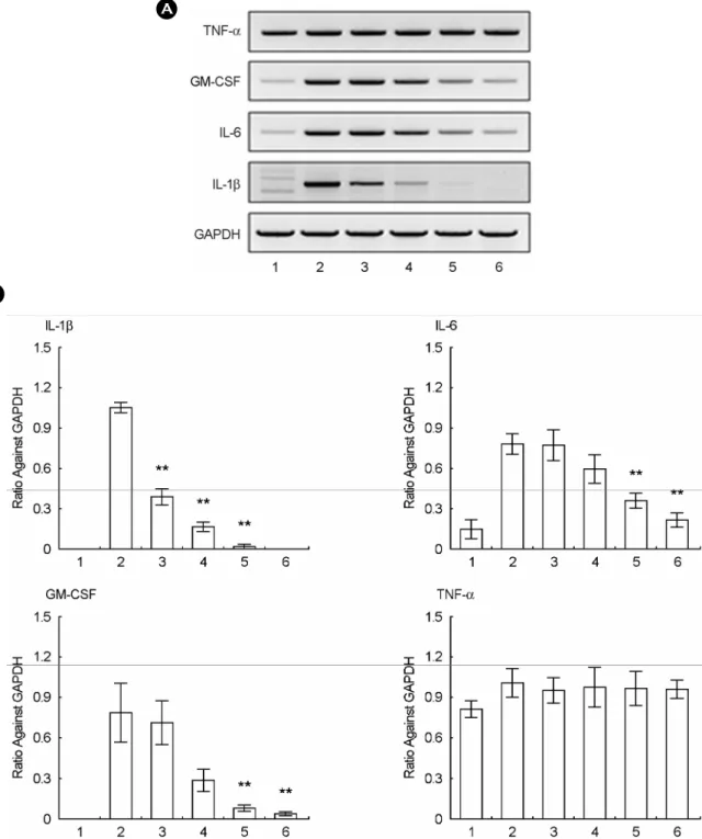
LPS (0.1 μg/ml) potently increased the expression of
IL-1β, IL-6 and GM-CSF in comparison to the basal level, but did slightly induce that of TNF-α (Fig. 4A). Taxifolin significantly inhibited the LPS (0.1 μg/ml)-induced expres- sions of IL-1β, IL-6 and GM-CSF in RAW264.7 cells.
However, taxifolin did not modulate the expression of TNF-α mRNA (Fig. 4A, B).
4. The effect of taxifolin on the transcription activity of NF-κB, AP-1 and CRE
Since transfection efficiency of murine macrophage RAW264.7 cell is very low, we transfected with cDNAs
Fig. 2. Effect of taxifolin on the NO production (A) and iNOSexpression (B, C) of RAW264.7 cells (1×10⁶ cells/ml). RAW264.7 cells (1×10⁶ cells/ml) were incubated with various concentration of taxifolin and LPS (0.1 μg/ml) for 18 hrs. A, NO production was determined with Griess's reagents. Each value is the means ± SEM of three independent experiments performed in triplicate.
*P<0.05 vs LPS-activated. B, C, After incubation with either taxifolin (25, 50, 100, 200 μM) and LPS (0.1 μg/ml) or LPS alone, the total RNA was prepared and RT-PCR were performed, as described in Materials and Methods, The gel picture was shown as representative one of the three independent experiments. Each value in bar graph is the means ± SEM of three independent experi- ments. *P<0.01 vs LPS-activated. Lane no. 1: basal, 2: LPS (0.1 μg/ml), 3: LPS + Taxifolin (25 μM), 4: LPS + Taxifolin (50 μM), 5: LPS + Taxifolin (100 μM), 6: LPS + Taxifolin (200 μM).
B
C A
Fig. 3. Effect of taxifolin on the cytotoxicity of RAW264.7 cells (1×10⁶ cells/ml). A, RAW264.7 cells (1×10⁶ cells/ml) were incubated with various concentration of taxifolin. Lane no. 1: basal, 2: Taxifolin (25 μM), 3: Taxifolin (50 μM), 4: Taxifolin (100 μM), 5: Taxifolin (200 μM). B, RAW264.7 cells (1×10⁶ cells/ml) were incubated with either taxifolin and LPS, or LPS (0.1 μg/ml) for 24 hrs. The determination of cell viability was performed using MTT assay, as described in Materials and Methods. Each value is the means ± SEM of three independent experiments performed in triplicate. **P<0.01 vs basal, ##P<0.01 vs LPS-activated. Lane no. 1: basal, 2: LPS (0.1 μg/ml), 3: LPS + Taxifolin (12.5 μM), 4:
LPS + Taxifolin (25 μM), 5: LPS + Taxifolin (50 μM).
B A
of NF-κB, AP-1 and CRE plasmids into HEK293 cells.
Taxifolin dose-dependently suppressed the transcriptional activity of NF-κB, which was stimulated by PMA (0.1 μM).
We next explored whether taxifolin modulated the AP-1 transcriptional activity, because it is reported that pro-
inflammatory cytokines were transcriptionally regulated by AP-1. As shown in Fig. 5B, taxifolin significantly inhibited the PMA (0.1 μM) stimulated AP-1 transcriptional activity.
However, taxifolin did not modulate the CRE luciferase activity, which was stimulated by PMA.
Fig. 4. Effects of taxifolin on the mRNA expression of pro-inflammatory cytokines in LPS-activated RAW264.7 cells. The mRNA levels of IL-1β, GM-CSF, IL-6, and TNF-α from the RAW264.7 cells were determined by semi-quantitative RT-PCR as described in Materials
& Methods. The figures present the representative results from three separate experiments, which give similar results. Lane no. 1: basal, 2:
LPS (0.1 μg/ml), 3: LPS + Taxifolin (25 μM), 4: LPS + Taxifolin (50 μM), 5: LPS + Taxifolin (100 μM), 6: LPS + Taxifolin (200 μM).
B
A
DISCUSSION
In our previous study (Cho et al., 2006), we found that taxifolin was contained in the chloroform extract of the cacti with a minor quantity using LC-ESI-MS analysis.
Furthermore, we examined the total phenolic contents of each fraction: that of ethyl acetate fraction was about 4 times higher than that of chloroform fraction. Indeed, quercetin and taxifolin showed potent anti-oxidant capability in our assay systems, xanthine/xanthine oxidase assay and DPPH assay. This suggests that taxifolin and its derivatives take a major role in the antioxidative effects of the cacti. In an agreement with this, it has been previously reported that some flavonoids quercetin, (+)-dihydroquercetin, quercetin 3-methyl ether, rutin, and isorhamnetin were found in the ethyl acetate fraction from the prickly pears of O. ficus- indica var. saboten (Butera et al., 2002; Dok-Go et al., 2003; Galati et al., 2003). Taxifolin also significantly inhi-
bited LPS-stimulated NO production (Fig. 2A). Taxifolin potently suppressed the expression of iNOS mRNA, which was stimulated by LPS (Fig. 2B and 2C). In order to exclude the possibility that the inhibitory effect of taxifolin in NO production is due to the it's cytotoxic effect in itself, we carried out cell viability test using the MTT assay. As shown in Fig. 3A, taxifolin did not show cytotoxicity even at a concentration of 200 μM and did increase cell viability at a concentration of 50 μM. On the other hand, LPS induced cell toxicity to macrophages via the NO generation and similarly the NO inhibitor, L-NAME, strongly blocked LPS-induced cytoxicity (data not shown). It seems that co-exposure to taxifolin and LPS block the LPS-induced cytotoxicity. We reported that the ethyl acetate fraction of O. humifusa Raf. may contain cell-protective components.
In this regard, it is well known that the NO rapidly and spontaneously reacts with a superoxide anion (O
2-) in order to form a peroxynitrite anion (ONOO
-), which is more toxic to biological systems than O
2-or NO, by causing a
Fig. 5. Effects of taxifolin on the transcriptional activity of NF-κB, AP-1 and CRE. The cDNA of NF-κB, AP-1 and CRE and TK Renilla were cotransfected. One day before assay, the cells transferred into 24 sell plate and further incubated for 24 hr.Luciferase activity was determined using Promega kit as described in Materials and Methods. Each value is the means ± SEM of three independent experiments performed in triplicate. RLU stands for relative luciferase unit. *P<0.05 vs PMA-activated, **P<0.01 vs PMA-activated. Lane no. 1: basal, 2: PMA (0.1 μM), 3: LPS + Taxifolin (50 μM), 4: LPS + Taxifolin (100 μM), 5: LPS + Taxi- folin (200 μM).
modification of proteins or DNA damage (Kim et al., 2000). We assume that some components, having radical scavenging activity, play an important role in protecting the cell from NO-induced cell toxicity by directly scavenging the peroxynitrite anion (ONOO
-). In addition, flavonoid (e.g., quercetin and rutin) and nonflavonoid (e.g., caffeic and its derivatives), which are distributed in many medicinal plants (e.g., Opuntia ficus-indica), were known to inhibit the expression of iNOS through modulation of NFκB signaling (Shen et al., 2002; Song et al., 2002; Dok-Go et al., 2003; Shin et al., 2004), and probably have a role in cytoprotective effect against LPS-induced cytotoxicity. This suggested that taxifolin play an important role in the development of protection against reactive species-induced cell damage, resulting from the radical scavenging activity.
In an agreement with this, it was reported that antioxidative flavonoids, such as taxifolin and quercetin, showed pro- tective effects against oxidative neuronal injuries induced in primary cultured rat cortical cells (Dok-Go et al., 2003).
In the current study, the potential for taxifolin to inhibit the LPS-induced expression of cytokines, TNF-α, IL-1β, IL-6 and GM-CSF, was tested because of their significance in inflammation conditions (Franzen et al., 2004; Blanco et al., 2008; Chen & O'Shea, 2008). IL-1β is important cyto- kine in chronic inflammatory diseases such as rheumatoid arthritis and host defense (Dayer, 2004; Blanco et al., 2008).
The IL-6 family, IL-6, IL-11, and oncostatin M, display pro-inflammatory effect as well as anti-inflammatory effect.
Moreover, the IL-6 family play an important role in innate and adaptive immune responses (Blanco et al., 2008). GM- CSF is known to be a pro-inflammatory hemopoietic cyto- kine widely used in hematological disorders to stimulate proliferation and differentiation of neutrophilic, eosinophilic and monocytic lineages (Franzen et al., 2004). As shown in Fig. 4A, B, taxifolin potently suppressed the expressions of IL-1β, IL-6, and GM-CSF mRNA, but did not modulate that of TNF-α. This implies that anti-inflammatory activity of taxifolin is cytokine specific pattern. NO, an inflam- matory mediator, and inflammatory cytokines, such as IL-1β, IL-6, and GM-CSF, are known to be under the control of transcriptional factor such as NF-κB and AP-1 (Castranova, 2004; Karin & Greten, 2005; Karin, 2006). In
this study, taxifolin significantly inhibited the activities of NF-κB and AP-1. However, CRE transcriptional activity is not affected by taxifolin. This suggested that the anti- inflammatory effects, including modulation of NO pro- duction and pro-inflammatory cytokine gene expression are mediated via suppression of transcriptional activity of NF-κB and AP-1.
In this study, among the solvent extracts of Opuntia humifusa Raf., the activity-guided fractionation of ethyl acetate extract showed that taxifolin has strong antioxi- dative activity. Furthermore, taxifolin exhibited potent anti- inflammatory activity. It inhibited LPS-induced NO pro- duction and inducible NO synthase expression. In addition, taxifolin suppressed pro-inflammatory (IL-1β, IL-6 and GM-CSF) cytokine expressions. These results suggest that taxifolin may modulate LPS-induced inflammation through the inhibition of the transcription factors, NF-κB and AP-1.
REFERNES
Acuna UM, Atha DE, Ma J, Nee MH, Kennelly EJ. Antioxidant Capacities of Ten Edible North American Plants. Phytother Res. 2002. 16: 63-65.
Blanco P, Palucka AK, Pascual V, Banchereau J. Dendritic Cells and Cytokines in Human Inflammatory and Autoimmune Diseases. Cytokine Growth Factor Rev. 2008. 19: 41-52.
Blois MS. Antioxidant Determinations by the Use of a Stable Free Radical. Nature 1958. 181: 1199-1200.
Butera D, Tesoriere L, Di Gaudio F, Bongiorno A, Allegra M, Pintaudi AM, Kohen R, Livrea MA. Antioxidant Activities of Sicilian Prickly Pear (Opuntia Ficus Indica) Fruit Extracts and Reducing Properties of Its Betalains: Betanin and Indica- xanthin. J Agric Food Chem. 2002. 50: 6895-6901.
Bwititi P, Musabayane CT, Nhachi CF. Effects of Opuntia Megacantha on Blood Glucose and Kidney Function in Streptozotocin Diabetic Rats. J Ethnopharmacol. 2000. 69:
247-252.
Castranova V. Signaling Pathways Controlling the Production of Inflammatory Mediators in Response to Crystalline Silica Exposure: Role of Reactive Oxygen/Nitrogen Species. Free Rad Biol Med. 2004. 37: 916-925.
Chen Z, O'Shea JJ. Regulation of Il-17 Production in Human Lymphocytes. Cytokine 2008. 41: 71-78.
Cho JY, Park SC, Kim TW, Kim KS, Song JC, Kim SK, Lee HM, Sung HJ, Park HJ, Song YB, Yoo ES, Lee CH, Rhee MH.
Radical Scavenging and Anti-Inflammatory Activity of Extracts from Opuntia Humifusa Raf. J Pharm Pharmacol.
2006. 58: 113-119.
Dayer JM. The Process of Identifying and Understanding Cyto- kines: From Basic Studies to Treating Rheumatic Diseases.
Best Pract Res Clin Rheumatol. 2004. 18: 31-45.
Dok-Go H, Lee KH, Kim HJ, Lee EH, Lee J, Song YS, Lee YH, Jin C, Lee YS, Cho J. Neuroprotective Effects of Antioxidative Flavonoids, Quercetin, (+)-Dihydroquercetin and Quercetin 3-Methyl Ether, Isolated from Opuntia Ficus-Indica Var.
Saboten. Brain Res. 2003. 965: 130-136.
Franzen R, Bouhy D, Schoenen J. Nervous System Injury: Focus on the Inflammatory Cytokine 'Granulocyte-Macrophage Colony Stimulating Factor'. Neurosci Lett. 2004. 361: 76-78.
Galati EM, Mondello MR, Giuffrida D, Dugo G, Miceli N, Pergolizzi S, Taviano MF. Chemical Characterization and Biological Effects of Sicilian Opuntia Ficus Indica (L.) Mill.
Fruit Juice: Antioxidant and Antiulcerogenic Activity. J Agric Food Chem. 2003. 51: 4903-4908.
Gallucci S, Provenzano C, Mazzarelli P, Scuderi F, Bartoccioni E.
Myoblasts Produce Il-6 in Response to Inflammatory Stimuli.
Int Immunol. 1998. 10: 267-273.
Goldstein G, Nobel PS. Water Relations and Low-Temperature Acclimation for Cactus Species Varying in Freezing Tolerance.
Plant Physiol. 1994. 104: 675-681.
Guzik TJ, Korbut R, Adamek-Guzik T. Nitric Oxide and Super- oxide in Inflammation and Immune Regulation. J Physiol Pharmacol. 2003. 54: 469-487.
Hong S, Kim SH, Rhee MH, Kim AR, Jung JH, Chun T, Yoo ES, Cho JY. In Vitro Anti-Inflammatory and Pro-Aggregative Effects of a Lipid Compound, Petrocortyne a, from Marine Sponges. Naunyn-Schmiedeberg's archives pharmacol. 2003.
368: 448-456.
Jung KK, Lee HS, Cho JY, Shin WC, Rhee MH, Kim TG, Kang JH, Kim SH, Hong S, Kang SY. Inhibitory Effect of Curcumin on Nitric Oxide Production from Lipopolysaccharide- Activated Primary Microglia. Life Sci. 2006. 79: 2022-2031.
Karin M. Nuclear Factor-Kappab in Cancer Development and Progression. Nature 2006. 441: 431-436.
Karin M, Greten FR. Nf-Kappab: Linking Inflammation and Immunity to Cancer Development and Progression. Nat Rev Immunol. 2005. 5: 749-759.
Kim KS, Ahn MR, Kim KS, Park SC, Rhee MH, In JG, Kim BH, Nah YL, Kim HH, Han SH. Effects of Opuntia Humifusa Extract on Dncb-Induced Allergic Contact Dermatitis in Balb/C Mice. Lab Anim Res. 2007. 23: 169-173.
Kim OK, Murakami A, Nakamura Y, Takeda N, Yoshizumi H, Ohigashi H. Novel Nitric Oxide and Superoxide Generation Inhibitors, Persenone a and B, from Avocado Fruit. J Agric Food Chem. 2000. 48: 1557-1563.
Kim YH, Park SC, Son HY, Rhee MH, Kim TW, Han SH, Kim BH, Kim KS. Hepatoprotective Effects of Opuntia Humifusa against Carbon Tetrachloride-Induced Acute Liver Injury in Rats. Lab Anim Res. 2005. 21: 263-266.
Lee JC, Kim HR, Kim J, Jang YS. Antioxidant Property of an Ethanol Extract of the Stem of Opuntia Ficus-Indica Var.
Saboten. J Agric Food Chem. 2002. 50: 6490-6496.
Lundberg IE. Clinical Symptoms in Patients with Myositis-an Acquired Metabolic Myopathy? Idiopathic Inflammatory Myopathies: Why Do the Muscles Become Weak? Curr Opin Rheumatol. 2003. 15: 675-678.
Noro T, Oda Y, Miyase T, Ueno A, Fukushima S. Inhibitors of Xanthine Oxidase from the Flowers and Buds of Daphne Genkwa. Chem Pharm Bull. (Tokyo) 1983. 31: 3984-3987.
Shen SC, Lee WR, Lin HY, Huang HC, Ko CH, Yang LL, Chen YC. In Vitro and in Vivo Inhibitory Activities of Rutin, Wogonin, and Quercetin on Lipopolysaccharide-Induced Nitric Oxide and Prostaglandin E(2) Production. Eur J Pharmacol.
2002. 446: 187-194.
Shin KM, Kim IT, Park YM, Ha J, Choi JW, Park HJ, Lee YS, Lee KT. Anti-Inflammatory Effect of Caffeic Acid Methyl Ester and Its Mode of Action through the Inhibition of Pro- staglandin E2, Nitric Oxide and Tumor Necrosis Factor- Alpha Production. Biochem Pharmacol. 2004. 68: 2327-2336.
Song YS, Park EH, Hur GM, Ryu YS, Lee YS, Lee JY, Kim YM, Jin C. Caffeic Acid Phenethyl Ester Inhibits Nitric Oxide Synthase Gene Expression and Enzyme Activity. Cancer Lett. 2002. 175: 53-61.
Walsh LJ. Mast Cells and Oral Inflammation. Crit Rev Oral Biol Med. 2003. 14: 188-198.