Streptozotocin 유도 당뇨 흰쥐에서 복령약침의 β-cell 손상 방지 효과
서창완
*
ᆞ서병관*
ᆞ김종인**
ᆞ강성길*
*
경희대학교 한의과대학 침구학교실**
한국한의학연구원목적 : 정상 췌장조직 속에 존재하는 췌장 소도세포들을 파괴시켜 고혈당을 유발시키고 복령 물추출물로 약침을 시술하여 췌장 조직의 보호효과와 항당뇨 효과를 살펴보고자 실험을 진행하였다.
방법 : 5주령의 Sprague-Dawley rat을 통제된 실험실 환경에 적응시킨 후 1주일간 복령약침액 (125mg/kg 복령약침군 및 250mg/kg 복령약침군)을 좌우 신수(BL
23
)에 교대로 각각 피하에 약침하고 streptozotocin을 복강내 주사하여 3일 후 diabetes mellitus 유도 정도를 평가하고 2주일간 추가 치료를 진행 한 뒤, 혈액지표(plasma glucose, insulin, TG, TC, NEFA, sGOT, sGPT, ALP, BUN, CRE)와 췌장조직의 형태학적 분석 및 염증 관련 단백질의 발현을 평가하였다.결과 : 복령약침군(125mg/kg 복령약침군 및 250mg/kg 복령약침군)에서 insulin과 triglyceride, NEFA 수 치가 유의하게 감소하였으며, 간 기능 효소수치인 sGOT가 감소하는 경향을 나타내었으나, 신장기능지수는 유의한 감소를 나타내지 않았다. 특히 250mg/kg 복령약침군에서 streptozotocin 투여로 인한 pancreatic islet 의 형태학적 변성이 현저하게 개선되었다. Western blot 결과 JNK-2, P-JNK-2, P-JNK-1, ERK1/2 및 phosphorylated ERK1의 발현이 감소되었다.
결론 : 복령약침이 고인슐린혈증과 고지질질혈증을 개선시키고 streptozotocin에 의한 pancreatic islet의 파괴를 억제하며, 이는 inflammation-related transcription factor인 NF-kB와도 관련이 있는 것으로 판단된 다. 향후 복령약침의 항당뇨 효과와 그 기전에 관한 추가 연구가 필요할 것으로 사료된다.
핵심단어 : Poria cocos, streptozotocin, diabetes mellitus, hyperinsulinemia
1)
Poria cocos Herbal Acupuncture Prevents β-cell Damage on Streptozotocin-induced Diabetic Rat
Seo Chang-wan * , Seo Byung-kwan * , Kim Jong-in ** and Kang Sung-keel *
* Department of Acupuncture & Moxibustion, College of Oriental Medicine, Kyung Hee University
** Korea Institute of Oriental Medicine
․Acceptance : 2009. 9. 4. ․Adjustment : 2009. 9. 18. ․Adoption : 2009. 9. 20.
․Corresponding author : 강성길, 서울특별시 동대문구 회기동 1. 경희의료원 한방병원 침구과 Tel. 82-2-958-9193 E-mail : [email protected]
국문초록
Original Article
Ⅰ. Introduction
From recent studies, diabetets mellitus is thought of as a group of metabolic disease characterized by hyperglycemia resulting from defects in insulin secretion, insulin action, or both, associated with long-term damage, dysfunction, and failure of various organs, especially the eyes, kidneys, nerves, heart, and blood vessels
1)
. Type I diabetes results from a cellular-mediated autoimmune destruction of the β-cells of the pancreas, leading to absolute insulin deficiency2)
.In the progress of development of diabetes mellitus, destruction of insulin-producing pancreatic β-cells are mediated by infiltration of CD4+ and CD8+ T cells and macrophages
3)
, and local produc- tion of inflammatory cytokines such as interleukin (IL)-1β, tumor necrosis factor (TNF)-α, and inter- feron (IFN)-γ4)
. Particularly in spontaneous animal models such as the non-obese diabetic mouse and the Bio-breeding rat, macrophages and dendritic cells are the first cells that appear at the periphery of pancreatic ducts and islets of Langerhans5)
.Poria cocos
(P. cocos
), which is known as Fu Ling, is often used in Korean traditional medicine.Dried sclerotia of
P. cocos
is prescribed to cause diuresis for edema, oliguria, to invigorate the spleen and calm the mind for loose stools, and to treat diarrhea, restlessness, insomnia6)
. Recent studies reported thatP. cocos
exhibited anti-inflammatory7)
, anti-emetic8)
, renal protective effect from inflamma- tion9)
, anti-spastic, anti-ulcerative, and reducing gas- tric hyperacidity effect10)
, and inhibitory effect on tumor promotion11)
.In this study, analysis of the blood parameters and the inflammatory protein expression, and his- tological observation of the pancreas in the rodent streptozotocin-induced diabetic model were conducted to evaluate anti-diabetic, pancreas protective effect of
P. cocos
.Ⅱ. Materials and Methods
1. Materials
P. cocos
used in this study was purchased from Dukhyundang (South Korea), a reputable Korean herb vender and identified and authenticated with the assistance of Department of Herbology, College of Oriental Medicine, Kyung Hee university.P.
cocos
(600g) was extracted by boiling in distilled water (6,000ml) for 4 hours in a decocting machine, and then filtered through filter papers (Whatman, England). The crude water extract solution was concentrated with a vacuum rotary evaporator under low pressure, and then the residue was lyophilized in a freezing dryer.2. In vivo experiment 1) Animals
Male SD (Sprague-Dawley) rats were obtained at 5 weeks of age from Orient Company (Seoul, Korea) and acclimated for 1 week before being randomly assigned into the groups. The animals were housed in polypropylene cages with steel grid tops, and fed with water and standard rodent chow (Lab diets, Richmond, USA)
ad libitum
. The animal rooms were controlled at a 12 : 12-hours light-dark cycle (8 : 00 am to 8 : 00 pm), a temperature of 23±2℃, and a humidity of 50±10% throughout the acclimatization and experimental periods.
The animals were randomly divided into four groups; control group (citrate buffer only), STZ (Streptozotocin only), and herbal acupuncture groups (P125 and P250). Herbal acupuncture groups were injected subcutaneously at left and right
Sinsu
(BL23
) alternately on exactly the same time every day with either 125mg/kg or 250mg/kg of theP. cocos
(P125 and P250). After 1 week of administration ofP. cocos
herbal acupuncture, in order to induce type I diabetes, 65mg/kg of Streptozotocin (STZ) was dissolved in 0.1 M sodium citrate buffer (pH 4.5) and injected intraperitoneally. After 72 hoursfrom intraperitoneal injection of STZ, animals with serum glucose level above 300mg/dL were selected and continuously administrated with each herbal acupuncture for 2 weeks. The animals assigned as a control group were only injected intraperitoneally with citrate buffer. The experiments were conducted in accordance with internationally-accepted experi- mental protocols for laboratory animal use and care as found in the U.S. guidelines.
2) Blood Analysis
At the end of the study, the animals were sacrificed for blood analysis after 12 hours of fasting period. The blood samples withdrawn from heart were centrifuged at 5,000rpm for 15 minutes, and the plasma was stored at -70℃ until assay.
The plasma glucose concentration was determined using the glucose oxidase method (Trinda medhod), and the spectrophotometric analysis was determined using the UV spectrophotometer (U-3210, Hitachi
™, Japan). The plasma insulin concentration was measured by ELISA reader (Labsystems, Finland), using the Rat insulin ELISA kit (Shibayagi, Japan).
Plasma triglyceride (TG), total cholesterol (TC) were determined using commercially available kits (Asan Pharmaceutical Co, Korea), and the plasma nonest- erified fatty acid (NEFA) concentrations were assayed by an enzymatic colorimetric method (Shinyang Chem- ical Co, Korea). sGOT (serum Glutamic Oxal- oacetic Transaminase), sGPT (serum Glutamic Pyruvic Transaminase), ALP (alkaline phosphatase), BUN (Blood Urea Nitrogen), CRE (creatinine), indices of functional enzymes of liver and kidney, were an- alyzed using the International Federation of Clinical Chemistry (IFCC) method with a SMAR-TLAB (STANBIO Co, USA).
3) Histopathology
Wedges of pancreatic tissues were fixed in 10%
neutral buffered formalin, embedded in paraffin, cut into thin sections (5㎛), and then deparaffinized with xylene, hydrophilized by 100%, 95%, 90%, 80%, 70% of alcohol, Canada embalsamed and then stained with hematoxylin and eosin (H&E). The
slides were observed through light microscope (Olym- pus, Japan).
4) Protein analysis and western blot
The pancreatic tissues which were equivalently weighed and homogenized in PRO-PREP Solution, were centrifuged at 5,000rpm. The protein concen- tration was determined with Bio-Rad Dc protein assay method. For immunoblotting, the protein samples were subjected to 10% SDS-PAGE, elec- trotransferred to nitrocellulose paper, and blocked with 5% skim milk solution before incubating with primary antibody in PBS buffer solution for more than 12 hours. The samples were then washed with TPBS three times and subsequently incubated with secondary antibody attached with horseradish per- oxidase for 1 hour, and finally washed with TPBS three times. To detect proteins, a chemiluminescent signal was developed using the ECL kit.
3. In vitro experiments 1) Cell culture
Rat insulinoma (RINm5F) cell strain was purchased from ATCC (American Type Culture Collection). Stocks of RINm5F cells were main- tained in RPMI1640 medium containing 10% fetal bovine serum (FBS), penicillin (100U/ml), strepto- mycin (100U/ml), glutamine) (Gibco) supplemented with 2.5㎍/ml of amphotericin B in the CO
2
culti- vator at 37℃, 5% CO2
.2) Analysis of cellular toxicity
Cellular toxicity of aqueous extraction of
P.
cocos
was analyzed using 3-(4,5-dimethlthiazol-2- yl)-5-(3-carboxymethoxyphenyl)-2-(4-sulfophenyl)- 2H-tetrazolium inner salt (MTS) assay method at the concentration of 0, 0.0625, 0.125, 0.25, 0.5 and 1mg/ml. RINm5F stain cells (1 x 10⁵
cell/well) were grown in the 96 well plate and treated with the varied concentrations ofP. cocos
extract for 24 hours. After 30 minutes incubation at 37℃ with additional 20㎕ of MTS solution, cellular toxicity was analyzed with OD value spectrophotometricallyNC STZ P125 P250
Glucose (mg/dL) 163.9±9.40 378.0±11.8 364.3±26.7 386.6±35.2
Insulin (ng/ml) 2.18±0.17 0.65±0.03 0.94±0.10
**
1.11±0.12**
TG (mg/dL) 69.2±3.9 249.7±60.1 190.4±44.9
*
113.5±22.0*
TC (mg/dL) 64.5±2.8 78.6±5.8 67.9±5.3 71.5±5.8
NEFA (uEq/L) 469±24 806±85 564±67
*
549±59*
sGOT (U/L) 1 19.6±34.7 208.2±26.5 124.5±31.9
**
145.4±32.2**
sGPT (U/L) 42.5±7.3 86.6±18.0 86.0±7.7 103.5±14.2
ALP (U/L) 1 83.5±29.8 317.6±37.5 418.5±44.3 360.8±78.7
BUN (mg/dL) 14.4±1.4 35.8±6.9 42.4±8.6 33.9±7.5
CRE (mg/dL) 1.014±0.1 0.91±0.1 0.91±0.1 0.84±0.1
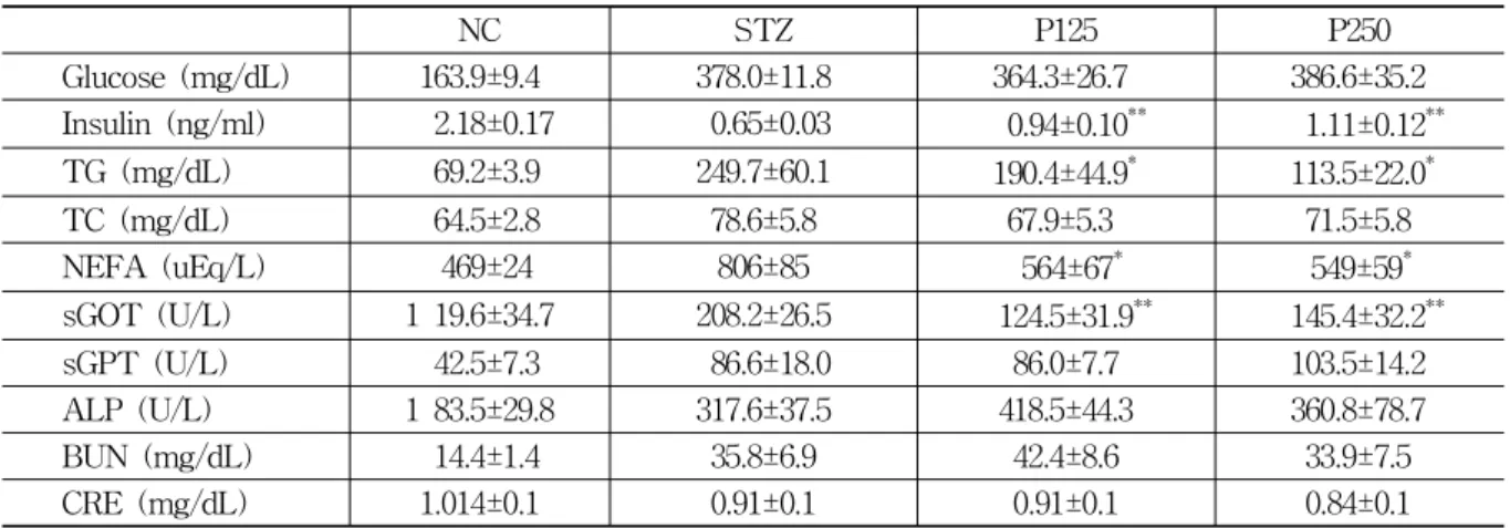
Values represent the mean±SE (n=6). Plasma parameters were analyzed in plasma samples obtained from blood of 12 hr fasted mice.
* :p<0.05. ** : p<0.01. *** : p<0.001 vs. STZ group.
Table 1. Effects of
P. cocos
Herbal Acupuncture on Plasma ParametersFig. 1. Protective effect of
P. cocos
herbal acu- puncture atSinsu
(BL23
) on pancreatic islets from STZ-induced destructionMicroscopic views on the sections of the pancreas obtained from NC, STZ, P125 and P250. H&E, magni- fication×200.
NC : citrate buffer.
STZ : streptozotocin intraperitoneally injected.
P125 : 125mg/kg of theP. cocus herbal acupuncture.
P250 : 250mg/kg of theP. cocus herbal acupuncture.
measured at 450nm.
3) Western blot
After 1 x 10
6
cells/well of RINm5F cells in 6 wells were acclimatized for 1 day, and then admin- istered with STZ (10mM) andP. cocos
aqueous extration (0.125mg/ml, 0.25mg/ml, 0.5mg/ml) for 24 hours, inflammation-related protein such as iNOS was measured.4. Statistical analysis
All data were expressed as a mean±S.E. Comparisons of data were done by Dunnett’s two-tailed
t
test.Mean values were considered significantly different when
p
<0.05.Ⅲ. Results
1. Fasting plasma glucose, and insulin resistance index
The fasting plasma glucose, insulin, TG, TC, NEFA, sGOT, sGPT, ALP, BUN and CRE level were compared between groups (Table 1). Fasting insulin, TG, and NEFA level in the P125, P250 herbal acupuncture treatment groups showed sig-
nificant differences when compared to STZ group.
There were no significant differences in the plasma glucose, TC levels between groups. sGOT level in the P125, P250 herbal acupuncture treatment groups showed a significant difference, but there were no significant differences in the BUN and CRE level between the groups(Table 1).
2. Morpholgy of pancreas
Morphology of pancreatic islets from the control
iNOS
NC STZ P125 P250
1 1.8 0.8 0.9
2 1.5 1 0.5 0
Fold
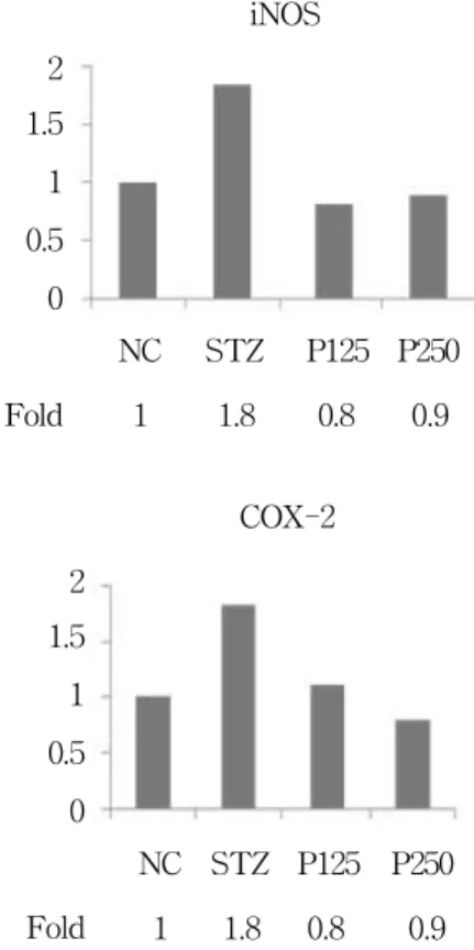
Fig. 3. Effects of
P. cocos
herbal acupuncture solution on cytotoxicity measured by MTS assayNC STZ P125 P250 1 1.8 0.8 0.9 2
1.5 1 0.5 0
Fold
COX-2 and the
P. cocos
herbal acupuncture in SD ratsafter 3 weeks of treatment was shown in Fig. 1.
Morphology of islets of the rats of P250 group was remarkably improved when compared to those of STZ group (Fig. 1).
3. Effect of P. cocos on the expression of inflammation-related proteins in pancreas
Changes of expression of inflammation-related factors (extracellular signal-regulated kinase (ERK), c-jun N terminal kinase (or, stress-activated protein kinase)) analyzed by Western blot in SD rats after 3 weeks of treatment was shown in Fig. 2. The expressions of JNK-2 and P-JNK-2 were signif- icantly decreased when compared to STZ group.
There were no significant differences in the ex- pression of JNK-1 between groups, however the expression of P-JNK-1 was significantly decreased when compared to STZ group. The expression of ERK1/2 was decreased when compared to STZ group.
There were no significant differences in the ex- pression of phosphorylated ERK2 between groups; how- ever, phosphorylated ERK1 was decreased (Fig. 2, 3).
NC STZ P125 P250
iNOS →
COX-2 → P-JNK-2 → P-JNK-1 → JNK-2 → JNK-1 → P-ERK-1 → P-ERK-2 → ERK-1 ERK-2 actin actin
Fig. 2. Effects of
P. cocos
herbal acupuncture atSinsu
(BL23
) on iNOS, COX-2, TNF-a and pho- sphorylation of ERK1/2 and JNK 1/2 in STZ- induced rat pancreasⅣ. Discussion
Type I diabetes mellitus is an autoimmune disease characterized by selective destruction of pancreatic β-cells and related with insulitis by infiltration of T cells and macrophages and local production of inflammatory cytokines such as IL-1 β, TNF-α, IFN-γ
1-4)
. In type I diabetes, β-cell mass is reduced by 70-80% at the time of diagnosis. Because of the variable degree of insulitis and absence of detectable β-cell necrosis, it was suggested that β-cell loss occurs slowly over years12)
. The rate of β-cell destruction is quite variable, being rapid in some individuals (mainly infants and children) and slow in others (mainly adults)13)
.Autoimmune destruction of β-cells has multiple genetic predispositions and is also related to envir- onmental factors that are still poorly defined.
Although patients are rarely obese when they present with this type of diabetes, the presence of obesity is not incompatible with the diagnosis.
These patients are also prone to other autoimmune disorders such as Grave’s disease, Hashimoto’s thyroiditis, Addison’s disease, vitiligo, and pernicious anemia
1)
. Markers of the immune destruction of the β-cell include islet cell autoantibodies (ICAs), autoantibodies to insulin (IAAs), autoantibodies to glutamic acid decarbo xylase (GAD65), autoantibodies to the tyrosin phosphatases IA-2 and IA-2β. One and usually more of these autoantibodies are present in 85-90% of the individuals when fasting hyperglycemia is initially detected14)
. β-cell death in the course of insulitis is probably caused by direct contact with activated macrophages and T-cells, and/or exposure to soluble mediators secreted by these cells, including cytokines, nitric oxide (NO), and oxygen free radicals15)
. In the insulitis lesion in type I diabetes, invading immune cells produce cytokines, such as IL-1, TNF-α, and IFN-γ, and induce β-cell apoptosis via the activation of β-cell gene networks under the control of the trans- cription factors NF-B and STAT-1. The execution of β-cell death occurs through activation of mitogen-activated protein kinases, via triggering of ER stress and by the release of mitochondrial death signals16)
.STZ (N-methyl-N-nitrosoureido D-glucosamine) derived originally from
Streptomyces achromogenes 17)
is a natural product that selectively kills pancreatic β-cells, and is widely used to generate insulin- dependent diabetes18)
in mouse models or treat pancreatic tumors19)
. Intraperitoneal administration of STZ is one of efficacious method to induce diabetes in rodents20)
, and assumed that it is mediated by two mechanisms; the chemical poison model and the O-GlcNAc-dependent model. The first is linked to the N-nitrosourea group act as a DNA alkylating agent and/or nitric oxide donor21)
. Several studies propose that STZ induces apoptosis by inhibiting O-GlcNAcase, an enzyme that, together with O-GlcNAc transferase, is important for dynamic intracellular protein O-glycosylation22)
. STZ actionin β-cells is accompanied by characteristic alteration in blood insulin and glucose concentrations. These changes reflect abnormalities in β-cell function (glucose oxidation, insulin biosynthesis and secretion, responsiveness to glucose)
23)
.Recently, studies for treatments of Type I diabetes mellitus have been done using various modalities such as acpuncture, moxibustion, electroacupunc- ture, herbal acupuncture and laser acupuncture
24)
.Of them, previous studies show the effect of
Cinnamomi Ramulus 25,26)
,Ginseng Radix 27) , Lycii Radicis Cortexa 28)
in reducing hyperglycemia. Also,Acanthopanacis Cortex 29) , Astragali Radix 30)
intra- venous herbal acupuncture have been proven to be effective in protecting intradermal cells by decreas- ing the filtration rate of proteinuria and intradermal cell count in plasma and urine, and thereby main- taining renal function. There have been few studies so far; however, about preventing β-cell damage mechanism of Type I diabetes mellitus usingP.
cocos
herbal acupuncture. In this study, we found thatP. cocos
herbal acupuncture on streptozotocin- induced diabetic rat can prevents β-cell damage and thus protect pancreas.Dried sclerotia of
P. cocos
,Fu ling
is prescribed to cause diuresis for edema, oliguria, to invigorate the spleen and calm the mind for loose stools, and to treat diarrhea, restlessness, insomnia6)
.P. cocos
consists of 90% β-glucan and 10% various terpenes by dry weight. Three major compounds were isolated and identified as pachymic acid, 3β- hydroxylanosta-7,9(11),24-trien-21-oic acid, and dehy- droeburicoic acid31)
. Recent studies reported thatP.
cocos
exhibited anti-inflammatory7)
, antiemetic8)
, renal protective effect from inflammation9)
, anti-spastic, anti-ulcerative, and reducing gastric hyperacidity effect10)
, and inhibitory effect on tumor promotion11)
. Satoet al
. demonstrated that the triterpene acid compound dehydrotrametenolic acid isolated from dried sclerotia ofP. cocos
promotes adipocyte differ- entiationin vitro
and acts as an insulin sensitizerin vivo
. Dehydrotrametenolic acid reduced hyper- glycemia in obese hyperglycemicdb/db
mice and acted as an insulin sensitizer as indicated by theresults of the glucose tolerance test
32)
. However, Sonet al
. reported that aqueous extract ofP. cocos
had some decreasing effect on the level of blood glucose in the fructose rich diet fed hyperglycemic rats and cholesterol rich diet fed hyperlipidemic rats but had no diminishable effects on the glucose tolerance, so the anti-diabetic effect was not dependent on insulin secretions33)
.Several points are in discord on the anti-diabetic effect of
P. cocos
and its mechanisms between reports because there is some differences of its experimental protocols. Satoet al
. for example, useddb/db
C57BLKS/J mice (genotype +/+) orally administered dehydrotrametenolic acid isolated fromP. cocos
for 14 days to estimate the anti-diabetic effect by measuring the levels of blood glucose, serum insulin, and the glucose tolerance test, but Son et al. measured blood glucose level in the fructose and cholesterol - rich diet fed hyper- glycemic SD rats with orally administering crude extract ofP. cocos
for 3 weeks. And the insulin sensitizing effect was estimated in the SD rats intravenously injected with STZ by the glucose tolerance test, without measuring the serum insulin level.In this study, with decreased plasma insulin level, lipid profile (TG, NEFA) of the P125, P250 herbal acupuncture treatment group was also reduced.
P. cocos
herbal acupuncture also improved liver profile (sGOT). These results suggest thatP.
cocos
herbal aqueous extract inhibits hyperinsuline mia induced by STZ-induced diabetes.With 3 weeks of treatment,
P. cocos
herbal acupuncture prevented the morphological deter- ioration induced by STZ. These results suggest thatP. cocos
aqueous extract has preventative effect from STZ-induced pancreatic islet destruc- tion.Furthermore, in this study, the significant decrease of expression of inflammation-related factors (JNK- 2, P-JNK-2, and P-KNK-1) were observed, and also the decrease of expression ERK1/2 and phos- phorylated ERK1 were also detected. These results suggest that
P. cocos
aqueous extract protects theβ-cells from STZ-induced pancreatic islet destruction and the protective effect of
P. cocos
aqueous extract is related with NF-kB, the inflammation- related transcription factor.Further study on the anti-diabetic effect of
P.
cocos
and its mechanisms is needed.Ⅴ. Conclusion
In this study,
P. cocos
aqueous extract was sub- cutaneously injected atSinsu
(BL23
) for 3 weeks to STZ-induced diabetic rodent model, and the plasma glucose, insulin, TG, TC, NEFA, sGOT, sGPT, ALP, BUN, CRE level were examined. Also, histological examination and quantitative analysis of expression of inflammation related proteins were conducted. As shown in the results,P. cocos
herbal acupuncture improved hyperinsulinemia and hyperlipidemia, and protected pancreatic destruction induced by STZ.And the protective effect of
P. cocos
aqueous extract is related to NF-kB, the inflammation- related transcription factor.Ⅵ. References
1. The expert committee on the diagnosis and classification of diabetes mellitus. Report of the expert committee on the diagnosis and classi- fication of diabetes mellitus. Diabetes Care.
2003 ; 26 : s5-s20.
2. Atkinson MA, Maclaren NK. The pathogenesis of insulin dependent diabetes. N Engl J Med.
1994 ; 331 : 1428-36.
3. Foulis AK, McGill M, Farquaharson MA. Insulin in type I (insulin-dependent) diabetes mellitus in man-macrophages, lymphocytes, and interferongamma containing cells. J Pathol. 1991 ; 165 : 97-103.
4. HE Hohmeier, VV Tran, G Chen, R Gasa, CB Newgard. Inflammatory mechanisms in diabetes : lessons from the β-cell. International Journal of
Obesity. 2003 ; 27 : s12-s16.
5. Francoise Homo-Delarche, Hemmo AD Rexhage.
Immune cells, pancreas development, regeneration and type 1 diabetes. Trends in immunology.
2004 ; 25(5) : 222-9.
6. Department of herbology. Herbology. 4th edition.
Seoul : Younglimsa. 1998 : 302-4.
7. Han Duckyong. Modern Pharmacognosy. Seoul : Hakchangsa. 1989 : 214.
8. Otsuka H, Fujioka S, Kimiya T, Mizuta E and TAkamoto M. Studies on anti-inflammatory agents.
VI, Anti-inflammatory constituents of Cinna- momum sieboldii Meissn. Yakugaku-zasshi. 1982 : 102, 162.
9. Han DS. Pharmacognosy. Seoul : Dongmyungsa.
1988 : 110, 116, 156, 159, 229, 174, 283, 356.
10. Wei Chen, Konstantin V Salojin, Qing-sheng Mi, Marsha Grattan, T Craig Meagher, Peter Zucker and Terry L Delovitch. Insulin-like gro- wth factor (IGF)-I/IGF-binding protein-3 complex : therapeutic efficacy and mechanism of pro- tection against type1 diabetes. Endo crinology.
2003.
11. Kaminaga T, Yasukawa K, Kanno H, Tai T, Nunoura Y, Takido M. Inhibitory effects of lanostane-type triterpene acids, the compo nents of
Poria cocos
, on tumor promotion by 12-O- tetradecanoylphorbol-13-acetate in two stage carcinogenesis in mouse skin. Oncology. 1996 ; 53 : 382-5.12. Kloppel G, Lohr M, Habich K, Oberholzer M, Heitz PU. Islet pathology and the pathogenesis of type 1 and type 2 diabetes mellitus revisited.
Surv Synth Pathol Res. 1985 ; 4 : 110-25.
13. Zimmet PZ, Tuomi T, Mackay R, Rowley MJ, Knowles W, Cohen M, Lang DA. Latent aut- oimmune diabetes mellitus in adults(LADA) : the role of antibodies to glutamic acid decar- boxylase in diagnosis and prediction of insulin dependency. Diabet Med. 1994 ; 11 : 299-303.
14. Cantor AB, Krischer JP, Cuthbertson DD, Schatz DA, Riley WJ, Malone J, Schwartz S, Quattrin T, Maclaren NK. Age and family relationship accentuate the risk of IDDM in relatives of
patients with insulin dependent diabetes. J Clin Endocrinol Metab. 1995 ; 80 : 3739-43.
15. Eizirik DL, Mandrup-Poulsen T. A choice of death: the signal-transduction of immune-mediated- cell apoptosis. Diabetologia. 2001 ; 44 : 2115-33.
16. Miriam Cnop, Nils Welsh, Jean-Christophe Jonas, Anne Jorns, Sigurd Lenzen, Decio L Eizirik.
Mechanisms of pancreatic β-cell death in type 1 and type 2 diabetes. Diatetes. 2005 ; 54 : s97- s107.
17. Vavra JJ, Deboer C, Dietz A, Hanka LJ, Sokolski WT. Streptozotocin, a new antibacterial antibiotic.
Antibiot Annu. 1959 ; 7 : 230-5.
18. Mansford KR, Opie L. Comparison of metabolic abnormalities in diabetes mellitus induced by streptozotocin or by alloxan. Lancet. 1968 ; 1 : 670-1.
19. Brentjens R, Saltz L. Islet cell tumors of the pancreas : the medical oncologist’s perspective.
Surg Clin North Am. 2001 ; 81 : 527-42.
20. Katsumata K, Katsumata K Jr, Katsumata Y.
Protective effect of diltiazem hydro chloride on the occurrence of alloxan-or streptozotocin- induced diabetes in rats. Horm MetabRes. 1992 ; 24 : 508-10.
21. Turk J, Corbett JA, Ramanadham S, Bohrer A, Mcdaniel ML. Biochemical evidence for nitric oxide formation from streptozotocin in isolated pancreatic islets. Biochem Biophys Res Commun.
1993 ; 197 : 1458-64.
22. Shalini Pathak, Helge C Dorfmueller, Vladimir S Borodkin and Daan MF van Aalten. Chemical Dissection of the Link between Streptozotocin, OGlcNAc, and Pancreatic Cell Death. Chem Biol.
2008 ; 15(8) : 799-807.
23. T Szkudelski. The Mechanism of alloxan and streptozotocin action in b cells of the rat pan- creas. Physiol. Res. 2001 ; 50 : 536-46.
24. Cao Shao Ming. Comparison of acupuncture, moxibustion, warming acupuncture therapy on diabetes mellitus. Zhongguo Zhenjiu. 1997 ; 17 (10) : 586.
25. Mang B, Wolters M, Schmitt B, Kelb K, Lich- tinghagen R, Stichtenoth DO, Hahn A. Effects
of a cinnamon extract on plasma glucose, HbA1c, and serum lipid in diabetes mellitus type 2. Eur J Clin Invest. 2006 ; 36 : 340-4.
26. Khan A, Safdar M, Ali Khan MM, Khattak KN, Anderson RA. Cinnamon improves glucose and lipids of people with type 2 diabetes. Diabetes Care. 2003 ; 26(12) : 3215-8.
27. Hye Jung Lee, Yong Tae Choi. (A)Study of ginseng radix aqua-acupuncture on the allo xan- induced diabetic rabbits and retrograde transport of horseradish peroxidase. Journal of college of Oriental medicine, Kyunghee university. 1987 ; 10 : 169-87.
28. Jang Jae Lee. The effect of Lycii Radicis Cortex Hexane herbal acupuncture on analgesia and blood glucose. Journal of college of Daejeon uni- versity. 1997.
29. Luo Su Sheng et al. The effect of Acanth- opanacis Cortex intravenous herbal acupuncture on intradermal cell count in plasma and urine in diabetes mellitus. Journal of Integrated Tradi-
tional and Western Medicine. 2001 ; 21(2) : 105- 7.
30. Liu Zhi Oiang et al. The effect of Astragali Radix intravenous herbal acupuncture on platelet in diabetes mellitus. Journal of Integrated Tradi- tional and Western Medicine. 2001 ; 21(4) : 274-6.
31. Lam Hoang, Soon-ho Kwon, Kyung-ah Kim, Jong-moon Hur, Young-hwa Kang, Kyung-sik Song. Chemical standardization of
Poria cocos
. Kor J Pharmacogn. 2005 ; 36(3) : 177-85.32. Sato M, Tai T, Nunoura Y, Yajima Y, Kawa- shima S, Tanaka K. Dehydrotramete nolic acid induces preadipocyte differentiation and sensitizes animal models of non insulindependent diabetes mellitus to insulin. Biol Pharm Bull. 2002 Jan ; 25(1) : 81-6.
33. Son youngjong, Lee youngjong. The effects of Sinomenii caulis and Hoelen on the levels of blood glucose in rats. Kor J Herbology. 2003 ; 18(1) : 65-71.