Introduction
Range of motion (ROM) is generally applied as clinical criteria for purposes of diagnosis and to as- sess outcome and effectiveness of shoulder joint treatments (Muir et al, 2010). Limitations in shoulder
ROM, specifically glenohumeral internal rotation defi- cit (GIRD), are associated with shoulder pathology (Downar and Sauers, 2005; Tyler et al, 2000; Wilk et al, 2009a). Furthermore, soft tissue tightness in the posterior glenohumeral joint has been confirmed as a cause of restricted glenohumeral internal rotation Corresponding author: Oh-yun Kwon [email protected]
Reliability Study of Measuring Range of Motion Glenohumeral Joint Internal Rotation With Pressure Biofeedback Stabilization
Ui-jae Hwang1,2, BPT, PT, Sung-min Ha3, PhD, PT, In-chul Jeon1,2, MSc, PT, Sung-hoon Jung1,2, BPT, PT, Kyu-hwan Choi4, PhD, PT, Su-jung Kim5, PhD, PT,
Oh-yun Kwon1,6,7, PhD, PT
1Kinetic Ergocise Based on Movement Analysis Laboratory
2Dept. of Physical Therapy, The Graduate School, Yonsei University
3Dept. of physical Therapy, College of Health Science, Sangji University
4Dept. of Physical Therapy, Ansan University
5Dept. of Physical Therapy, College of Health Sciences, Youngsan University
6Dept. of Physical Therapy, College of Health Science, Yonsei University
7Dept. of Ergonomic Therapy, The Graduate School of Health and Environment, Yonsei University
Abstract
1)The aims of the current study were to assess reliability of range of motion (ROM) measurement of glenohumeral internal rotation (GIR) with a pressure biofeedback stabilization (PBS) method and to compare the reliability between manual stabilization (MS) and the PBS method. In measurement of pure glenohumeral joint motion, scapular stabilization is necessary. The MS method in GIR ROM measurement was used to restrict scapular motion by pressing the palm of the tester’s hand over the subject’s clavicle, coracoid process, and humeral head. The PBS method was devised to maintain consistent pressure for scapular stabilization during GIR ROM measurement by using a pressure biofeedback unit. GIR ROM was measured by 2 different stabilization methods in 32 subjects with GIR deficit using a smartphone clinometer application. Repeated measurements were performed in two test sessions by two testers to confirm inter- and intra-rater reliability. After tester A performed measurements in test session 1, tester B’s measurements were conducted one hour later on the same day to assess the inter-rater reliability and then tester A performed again measurements in test session 2 for confirming the intra-rater reliability.
Intra-class correlation coefficient (ICC) (2,1) was applied to assess the inter-rater reliability and ICC (3,1) was applied to determine the intra-rater reliability of the two methods. In the PBS method, the intra-rater reliability was excellent (ICC=.91) and the inter-rater reliability was good (ICC=.84). The inter-rater and intra-rater reliability of the PBS method was higher than in the MS method. The PBS method could regulate manual scapular stabilization pressure in inter- and intra-rater measuring GIR ROM. Results of the current study recommend that the PBS method can provide reliable measurement data on GIR ROM.
Key Words: Glenohumeral internal rotation; Manual stabilization; Pressure biofeedback stabilization; Reliability.
(GIR) (Gerber et al, 2003; Kelley et al, 2009;
Ludewig and Reynolds, 2009; Myers et al, 2007).
GIRD has been confirmed not only in throwing ath- letes, who are apt to generate limited ROM because of repetitive microtrauma at the posterior gleno- humeral capsule (Burkhart et al, 2000; Burkhart et al, 2003b), but also in populations diagnosed with im- pingement syndrome (Ticker et al, 2000; Tyler et al, 2000) and participating in recreational weight-training (Kolber et al, 2009; Kolber and Corrao, 2011).
Because the increased interest in detecting and treat- ing GIRD, reliable standard of measuring is required in measurement of GIR ROM.
As concern has increased for treating and detect- ing GIRD, various methods have been devised to measure GIR ROM. Several methods of GIR ROM measurement have been suggested that measured the vertebral level that could be reached behind the back (Bigliani et al, 1997) and the “sleeper stretch posi- tion” where the subject is in a side-lying position for scapular stabilization, with the shoulder flexed 90° so that GIR may be measured to confirm tight- ness of posterior glenohumeral soft tissues (Burkhart et al, 2003a; Laudner et al, 2008). Recently, a shoulder mobility test was evaluated for the Functional Movement Screen, in which the subject rotates one hand internally behind the back and the other hand externally rotates from the head side above, attempting to bring the hands as close to- gether as possible in the back region to measure glenohumeral joint mobility (Sprague et al, 2014).
Although various methods have been developed for GIR ROM measurement, scapular stabilization was not applied to measure pure GIR ROM in the gleno- humeral joint. Thus, a method to apply scapular sta- bilization in a supine position at 90° of shoulder ab- duction and 90° elbow flexion was generally used for GIR ROM measurement.
Ellenbecker et al (1996) noted that scapulothoracic motion confounded shoulder rotation ROM measure- ments in standard methods in which the scapula was relatively free to move at 90° of shoulder abduction
with a supine position. They experimented with man- ually restricting scapular motion by applying scapular stabilization pressure in the posterior direction to the coracoid process and clavicle with a supine position and proposed that this method represented a more reliable measure of glenohumeral motion rather than non-scapular stabilization (Ellenbecker et al, 1996).
Numerous studies have underlined the importance of using scapulothoracic joint stabilization to restrict scapular motion (Boon and Smith, 2000; Burkhart et al, 2003a; Ellenbecker et al, 2002; Meister et al, 2005).
Unfortunately, in many instances, the scapular stabili- zation pressure applied to the coracoid process of subjects was not clearly stated by the investigators.
A pressure biofeedback unit (PBU) is made up of an inflatable cushion that is combined with a pres- sure gage displaying feedback on pressure for stabilization. A PBU has been used in various bio- feedback methods generally to monitor stabilization of the cervical and lumbar spine and pelvis during ex- ercise (Cairns et al, 2000; Chiu et al, 2005; Hudswell et al, 2005; Park et al, 2011). However, a PBU has not been used to stabilize the scapula during GIR ROM measurement, although scapular stabilization pressure as a pressing cushion could be easily confirmed by using a PBU. Thus, in the current study, a PBU was used to supply biofeedback for the investigator.
Although manual stabilization (MS) has been used for applying scapular stabilization, the pressure force was not consistent but subjective. The current study devised a new method to measure GIR using a PBU for applying consistent scapular stabilization pressure.
Measuring GIR by putting a PBU on acromion of a subject’s scapula, the investigator was provided bio- feedback that could confirm stabilization pressure as pressure. The purpose of the current study was to determine the inter- and intra-rater reliability of pressure biofeedback stabilization (PBS) used to ap- ply consistent manual pressure on the coracoid proc- ess of the subject’s scapula by using a PBU during shoulder internal rotation to measure GIR and to compare it to traditional MS methods.
Methods
Subjects
Sixty-three subjects at Yonsei University were measured to identify 32 subjects (27 males, 5 females;
mean age 23.0±1.8 years; mean height 172.1±6.5 ㎝;
mean weight 69.4±10.0 ㎏) with GIRD. Inclusion cri- teria included (1) no history of neurological disease, arthritis, connective tissue disorder, or shoulder/neck injury or surgery and (2) a difference in the passive GIR ROM of above 10° between the right and left side (between-side difference; mean±standard devia- tion, 14.6±5.5°) (Crockett et al, 2002; Myers et al, 2009; Thomas et al, 2011). Exclusion criteria consisted of reported shoulder pain at the time of data collec- tion, recent shoulder surgery for which the partic- ipant was still receiving care, or ongoing shoulder rehabilitation program for shoulder. Before the study, the principal tester explained the experimental protocol to the subjects in detail. All subjects signed an in- formed consent form, and this study was approved by the Yonsei University Wonju Institutional Review Board (approval number: 1041849-201510-BM-071-01).
Instrumentation
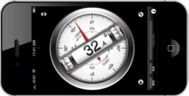
The Clinometer in iPhone application used to measure GIR ROM is Clinometer (Plaincode Software Solutions, Stephanskirchen, Germany), an application designed using the three inbuilt accelerometers (LIS302DL accelerometer) (Figure 1). The Clinometer application for measuring GIR ROM with the arm abducted 90°, including the iPhone is readily avail- able at a low cost for several smartphones including iPhone; the intra-class correlation coefficients (ICC) for GIR ROM measurement was .81 (95% confidence interval: .70∼.88) (Werner et al, 2014). Two testers downloaded the application to their smartphones and practiced the two methods of GIR ROM measure- ment using the Clinometer application before the test.
Testers
The two testers performed GIR ROM measurement
with two methods: MS and PBS. They trained in the two methods before the start of the study. They had 3 and 4 years of clinical orthopedic physical therapy ex- perience, and had experience in ROM measurement.
The order of the tester’s measurements was the same;
tester A always tested first and tester B last. An in- dependent observer, blinded from this study, read the GIR ROM data displayed by the smartphone Clinometer application. Data for each measurement were recorded by the independent observer on separate data sheets so that the testers were not able to view any measure- ments from encounters with previous subjects.
Procedures
Repeated GIR ROM measurements using the same protocol were performed to assess the intra-rater re- liability on two different days with an interval of 7 days between testing sessions (Figure 2). Each rater measured the GIR ROM of the subject’s side with GIRD once for each subject in a session. Subjects were asked not to perform excessive upper extremity activity between the first and second sessions. After tester A completed measurements in test session 1, tester B’s measurements were conducted one hour later on the same day to assess the inter-rater reli- ability (Figure 2). The order in which testers per- formed the two methods measuring GIR ROM was randomized using a table of random numbers created using the Randomization online software program at www.randomization.com. Testing was performed with the individuals positioned supine with the shoulder at 90° of abduction and the elbow at 90° of flexion.
Figure 1. Smartphone Clinometer application shown on an iPhone.
Testers maintained the subject’s shoulder at 90° of abduction during GIR ROM measurement in a paral- lel line with the subject’s humerus by attaching tape to the table. Testers hold the iPhone running Clinometer with one third distal part of the subject’s forearm. The end point for passive motion was de- termined by the positioning testers, both by subject discomfort and by capsular end-feel (Boon and Smith, 2000). The independent observer read the GIR ROM data displayed by iPhone Clinometer when the testers determined the end point of GIR ROM.
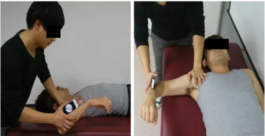
Measurements: Two methods of GIR ROM In the first method, MS was used to measure GIR
ROM by placing and pressing the palm of the test- er’s hand over the subject’s clavicle, coracoid proc- ess, and humeral head (Wilk et al, 2009b). The sub- ject was positioned supine with the shoulder at 90°
of abduction and the elbow at 90° of flexion. The testers performed MS with their left hand, and used their right hand to push on the subject’s right fore- arm toward internal rotation (Figure 3).
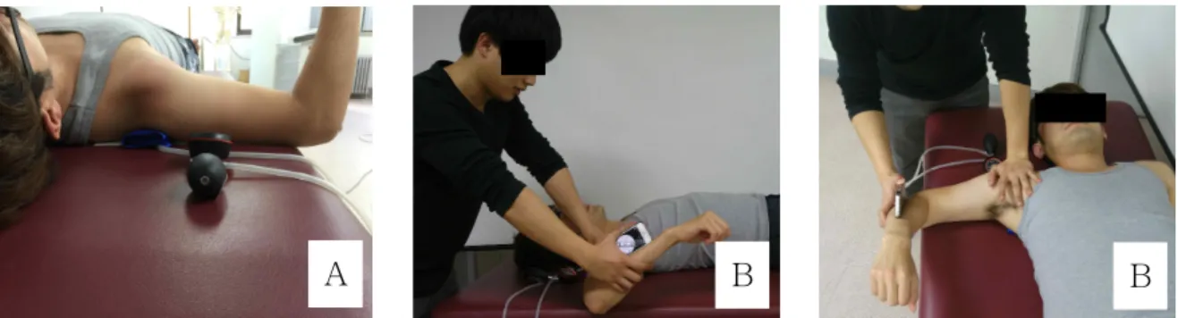
In the second method, PBS was applied to meas- ure GIR ROM using PBU by placing the palm of the tester’s hand over the subject’s clavicle, coracoid process, and humeral head. The testers folded the PBU up to one third of its size as a line between air-tube. The folded PBU was put under the sub- ject’s acromion, and then testers regulated initial pressure to 20 ㎜Hg. The testers applied 30 ㎜Hg of pressure to the subject’s humeral head before push- ing on the subject’s forearm. By receiving feedback about scapular motion from the PBU, the testers maintained constant pressure at 30 ㎜Hg during measurement. The testers conducted PBS in their left hand, and pushed with their right hand on the subject’s right forearm toward internal rotation, confirming that pressure of the PBU was held stable (Figure 4).
Figure 2. Flow chart of progression of measurements through the study.
Figure 3. The MS method for measurement of GIR ROM: the tester applies anteroposterior-directed pressure for stabilization against the subject’s coracoid process, blocking anterior tilting of the scapula.
Statistical analysis
All statistical analyses were conducted using SPSS ver. 18.0 software (SPSS Inc., Chicago, IL, USA).
ICC was used with a single measure for consistency among measurements for each movement. The ICC (3,1) model was used to estimate intra-rater reli- ability by calculating across test sessions and the ICC (2,1) model was used to test inter-rater reli- ability by calculating across raters in session 1.
An independent t-test was used to determine differ- ences between GIR ROM according to the two methods. Values were considered statistically sig- nificant at p<.05.
The standard error of measurement (SEM) is not affected by inter-subject variability (Weir, 2005) and is important for clinical utilization of a measurement procedure; therefore it was reported in conjunction
with the ICC using the formula: SEM=SD√(1-ICC) (SD: standard deviation) (Portney and Watkins, 2008).
The minimal detectable change (MDC) was calculated for the inter-rater measurements using the formula:
MDC95=1.96×SEM×√2 to determine the magnitude of change that would exceed the threshold of measure- ment error at the 95% confidence level (Haley and Fragala-Pinkham, 2006; Portney and Watkins, 2008).
Results
The means and standard deviations of GIR ROM from measurement of the two methods are shown in Table 1. A statistically significant difference was al- so found between methods (p<.001). Table 2 shows the intra-rater reliability with MS and PBS, along
Measurement method Mean±SDa p value
MSb 55.41±5.51
<.001
PBSc 56.75±6.41
amean±standard deviation, bmanual stabilization, cpressure biofeedback stabilization.
Table 1. Mean and standard deviation of GIR ROM according to methods of measurement
A B B
Figure 4. The PBS method for measurement of GIR ROM (A: The position of the PBU is under the center of the acromion. B: The tester applies an anteroposterior-directed stabilization pressure against the subject’s coracoid process, blocking anterior tilting of the scapula to confirm applied pressure through the PBU.).
Measurement
method ICCa SEMb (°) MDCc (°) CVd (%)
MSe .72 3.05 8.45 10.44
PBSf .91 1.82 5.05 10.67
aintra-class correlation coefficients, bstandard error of measurement, cminimal detectable change,dcoefficient of variation, emanual stabilization, fpressure biofeedback stabilization.
Table 2. Intra-rater reliability of GIR ROM measurement according to methods
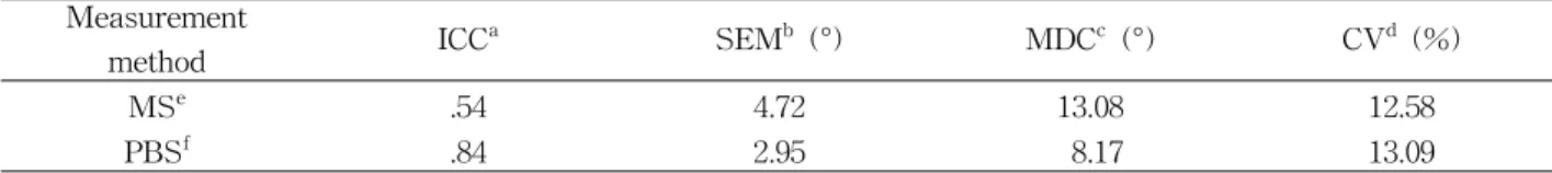
with coefficient of variation (CV) and the SEM and the ICC value including MDC. Table 3 shows the inter-rater reliability with MS and PBS, along with CV and the SEM and the ICC value including MDC.
Our interpretation of the ICC value was based on guidelines offered by Lunden et al (2010), in which ICC values were classified for reliability, using the following criteria: excellent (.90∼.99), good (.80∼.89), fair (.70∼.79), and poor (≤.69).
Discussion
This study investigated GIR ROM gauged by the Clinometer iPhone application and determined the in- tra- and inter-rater reliability of two methods of measurement, MS and PBS. Previous researches demonstrated that the MS method minimizes or pre- vents accessory scapulothoracic motion during GIR measurement (Boon and Smith, 2000; Ellenbecker et al, 1996). Although the scapular stabilization with MS method was considered as valid measurement method of GIR (Boon and Smith, 2000; Ellenbecker et al, 1996), there was no study to determine wheth- er applying pressure can be maintained consistently while measuring of GIR ROM with the MS method.
It is important to find applied pressure on scapula can be maintained consistently during MS using a PBU to measure pure GIR ROM for improving test reliability. The results of this study demonstrated that the intra-rater reliability was excellent (.91) and the inter-rater reliability was good (.84) for PBS method. And the inter-rater and intra-rater reliability of the PBS method was higher than the MS method.
This study confirmed that MS by using a PBU to
apply a consistent pressure force improved the reli- ability for GIR ROM measurement.
Previous studies suggested that a hand-held dyna- mometer (HDD) can be applied to control pressure force for improving reliability of measuring joint range of motion (Gajdosik and Bohannon, 1987;
Hayes et al, 2001; Lea and Gerhardt, 1995). Because HDD was relatively expensive, the current study used a PBU to regulate the amount of applied pres- sure for scapular stabilization. The MS method can restrict the normal arthrokinematics of the gleno- humeral joint. When stabilization or pressure is ap- plied to the anterior humeral head during passive GIR ROM measurement or activity, the normal ante- rior translation of the humeral head could be re- stricted (Howell et al, 1988). Comerford and Mottram (2012) suggested that various stabilization exercises using a PBU be used with a minimum applied pres- sure of 10 ㎜Hg to regulate uncontrolled movement.
Thus, PBS was conducted to exert a force of 10 ㎜ Hg for consistent MS in this study.
For the MS method, intra-rater reliability (ICC=.72) across the session was higher than inter-rater reli- ability (ICC=.54). Previous studies also suggested that intra-rater reliability was higher than inter-rater reli- ability in the MS method (Awan et al, 2002; Boon and Smith, 2000; Wilk et al, 2009b). Awan et al (2002) suggested that inter- and intra-rater reliability of the MS method was good, although inter-rater re- liability (ICC=.50) was lower than intra-rater reli- ability (ICC=.64). Boon and Smith (2000) confirmed that intra-rater reliability was good (ICC=.60) and inter-rater reliability was poor (ICC=.38) and suggested that MS be modified to measure GIR ROM for clinical decision making. The MS meth- Measurement
method ICCa SEMb (°) MDCc (°) CVd (%)
MSe .54 4.72 13.08 12.58
PBSf .84 2.95 8.17 13.09
aintra-class correlation coefficients, bstandard error of measurement, cminimal detectable change,dcoefficient of variation, emanual stabilization, fpressure biofeedback stabilization.
Table 3. Inter-rater reliability of GIR ROM measurement according to methods
od may generate tension on the glenohumeral joint capsule via direct contact with articulating surfaces, which may restrict normal glenohumeral motion (Wilk et al, 2009b). The amount of pres- sure applied on the humeral head significantly af- fects the amount of GIR; for instance, greater posteriorly directed pressure results in less GIR (Wilk et al, 2009b). Because GIR ROM in the MS method depends on applied scapular stabilization pressure, it is difficult to maintain consistent pressure in raters, thus it is hard to make a co- herent measurement of GIR ROM.
The GIR ROM angle in PBS method (56.75±6.41°) was significantly greater than the MS method (55.41±5.51°). The possible reason that the PBS method was significantly higher than the MS method (p<.001) could be the application of pressure above 10 ㎜Hg in the MS method, and that might restrict normal glenohumeral motion more than the PBS method. The current study calculated SEM and MDC to determine measurement error and the minimum threshold of measurement to find that differences be- tween methods. The SEM and MDC values of the PBS method were lower than the MS method in both inter- and intra-rater reliability. That might be because pressure was more subjectively applied in the MS method than the PBS method. The varia- bility of applied pressure in the MS method may raise the values of SEM and MDC higher than PBS.
The present study demonstrated that the PBS method is more reliable for measuring GIR ROM than the MS method. The high reliability of the PBS method was influenced by the consistent applied pressure force used to ensure uniformity of stabilization. The PBS method may make similar tension in the glenohumeral joint capsule and that may feel similar for the tester when compared to the MS method. The current study demonstrated that consistent applied pressure force for scapular stabili- zation made inter- and intra-rater reliability higher in measurement of GIR ROM. Thus, it is essential that consistent scapular stabilization pressure be ap-
plied for any study measuring GIR ROM in order to obtain consistent and reliable data.
The current study has several limitations. First, the generalizability of the results of our study is limited because our subjects were young and may have relatively few problems with GIRD compared with older patients. Thus, additional research is needed to examine the reliability of GIR ROM meas- urement by the PBS method in different age groups and in individuals with shoulder dysfunction. Second, the PBS method was applied only up to 10 ㎜Hg.
Further study is required to find the reliability of the PBS method applied in various force levels and to confirm the most reliable manual pressure force for measurement of GIR ROM.
Conclusion
The present study confirmed that intra- and in- ter-rater reliability were excellent and good, main- taining a consistent applied pressure force while the same and different testers measured GIR ROM.
Based on the results of the current study, we rec- ommend that the PBS method could be provided for reliable measurement of GIR ROM through regulating consistent applied scapular stabilization pressure in the clinical station. And inter- and intra-rater reli- ability of PBS method was higher than MS method in GIR ROM measurement. The results in this study indicated that the PBS method can regulate manual scapular stabilization pressure across multiple testers measuring GIR ROM.
References
Awan R, Smith J, Boon AJ. Measuring shoulder in- ternal rotation range of motion: A comparison of 3 techniques. Arch Phys Med Rehabil. 2002;
83(9):1229-1234.
Bigliani LU, Codd TP, Connor PM, et al. Shoulder
motion and laxity in the professional baseball player. Am J Sports Med. 1997;25(5):609-613.
Boon AJ, Smith J. Manual scapular stabilization: Its effect on shoulder rotational range of motion.
Arch Phys Med Rehabil. 2000;81(7):978-983.
Burkhart SS, Morgan CD, Kibler WB. Shoulder in- juries in overhead athletes. The “dead arm”
revisited. Clin Sports Med. 2000;19(1):125-158.
Burkhart SS, Morgan CD, Kibler WB. The disabled throwing shoulder: Spectrum of pathology. Part I: Pathoanatomy and biomechanics. Arthroscopy.
2003a;19(4):404-420.
Burkhart SS, Morgan CD, Kibler WB. The disabled throwing shoulder: Spectrum of pathology. Part III: The SICK scapula, scapular dyskinesis, the kinetic chain, and rehabilitation. Arthroscopy.
2003b;19(6):641-661.
Cairns MC, Harrison K, Wright C. Pressure biofeed- back: A useful tool in the quantification of ab- dominal muscular dysfunction? Physiotherapy.
2000;86(3):127-138.
Chiu TT, Law EY, Chiu TH. Performance of the craniocervical flexion test in subjects with and without chronic neck pain. J Orthop Sports Phys Ther. 2005;35(9):567-571.
Comerford M, Mottram S. Kinetic Control: The manage- ment of uncontrolled movement. 1st ed. Chatswood, Churchill Livingstone Australia, 2012:64-88.
Crockett HC, Gross LB, Wilk KE, et al. Osseous adaptation and range of motion at the gleno- humeral joint in professional baseball pitchers.
Am J Sports Med. 2002;30(1):20-26.
Downar JM, Sauers EL. Clinical measures of should- er mobility in the professional baseball player. J Athl Train. 2005;40(1):23-29.
Ellenbecker TS, Roetert EP, Bailie DS, et al.
Glenohumeral joint total rotation range of motion in elite tennis players and baseball pitchers.
Med Sci Sports Exerc. 2002;34(12):2052-2056.
Ellenbecker TS, Roetert EP, Piorkowski PA, et al.
Glenohumeral joint internal and external rotation range of motion in elite junior tennis players. J
Orthop Sports Phys Ther. 1996;24(6):336-341.
Gajdosik RL, Bohannon RW. Clinical measurement of range of motion. Review of goniometry empha- sizing reliability and validity. Phys Ther. 1987;
67(12):1867-1872.
Gerber C, Werner CM, Macy JC, et al. Effect of se- lective capsulorrhaphy on the passive range of motion of the glenohumeral joint. J Bone Joint Surg Am. 2003;85-A(1):48-55.
Haley SM, Fragala-Pinkham MA. Interpreting change scores of tests and measures used in physical therapy. Phys Ther. 2006;86(5):735-743.
Hayes K, Walton JR, Szomor ZR, et al. Reliability of five methods for assessing shoulder range of motion. Aust J Physiother. 2001;47(4):289-294.
Howell SM, Galinat BJ, Renzi AJ, et al. Normal and abnormal mechanics of the glenohumeral joint in the horizontal plane. J Bone Joint Surg Am.
1988;70(2):227-232.
Hudswell S, von Mengersen M, Lucas N. The cra- nio-cervical flexion test using pressure biofeed- back: A useful measure of cervical dysfunction in the clinical setting? Int J Osteopath Med.
2005;8(3):98-105.
Kelley MJ, McClure PW, Leggin BG. Frozen should- er: Evidence and a proposed model guiding rehabilitation. J Orthop Sports Phys Ther. 2009;
39(2):135-148. http://dx.doi.org/10.2519/jospt.2009.2916 Kolber MJ, Beekhuizen KS, Cheng MS, et al.
Shoulder joint and muscle characteristics in the recreational weight training population. J Strength Cond Res. 2009;23(1):148-157. http://dx.doi.org/
10.1519/JSC.0b013e31818eafb4
Kolber MJ, Corrao M. Shoulder joint and muscle characteristics among healthy female recreational weight training participants. J Strength Cond Res.
2011;25(1):231-241. http://dx.doi.org/10.1519/JSC.
0b013e3181fb3fab
Laudner KG, Sipes RC, Wilson JT. The acute effects of sleeper stretches on shoulder range of motion. J Athl Train. 2008;43(4):359-363.
Lea RD, Gerhardt JJ. Range-of-motion measurements.
J Bone Joint Surg Am. 1995;77(5):784-798.
Ludewig PM, Reynolds JF. The association of scap- ular kinematics and glenohumeral joint pathologies.
J Orthop Sports Phys Ther. 2009;39(2):90-104.
http://dx.doi.org/10.2519/jospt.2009.2808
Lunden JB, Muffenbier M, Giveans MR, et al.
Reliability of shoulder internal rotation passive range of motion measurements in the supine versus sidelying position. J Orthop Sports Phys Ther. 2010;40(9):589-594.
Meister K, Day T, Horodyski M, et al. Rotational motion changes in the glenohumeral joint of the adolescent/little league baseball player. Am J Sports Med. 2005;33(5):693-698.
Muir SW, Corea CL, Beaupre L. Evaluating change in clinical status: Reliability and measures of agreement for the assessment of glenohumeral range of motion. N Am J Sports Phys Ther.
2010;5(3):98-110.
Myers JB, Oyama S, Goerger BM, et al. Influence of humeral torsion on interpretation of posterior shoulder tightness measures in overhead athletes.
Clin J Sport Med. 2009;19(5):366-371. http://
dx.doi.org/10.1097/JSM.0b013e3181b544f6
Myers JB, Oyama S, Wassinger CA, et al. Reliability, precision, accuracy, and validity of posterior shoulder tightness assessment in overhead athletes.
Am J Sports Med. 2007;35(11):1922-1930.
Park KN, Cynn HS, Kwon OY, et al. Effects of the abdominal drawing-in maneuver on muscle ac- tivity, pelvic motions, and knee flexion during active prone knee flexion in patients with lumbar extension rotation syndrome. Arch Phys Med Rehabil. 2011;92(9):1477-1483. http://dx.doi.org/
10.1016/j.apmr.2011.03.020
Portney L, Watkins M. Foundations of Clinical Research: Applications to practice. 3rd ed. New Jersey, Pearson Prentice Hall, 2008:77-96.
Sprague PA, Monique Mokha G, Gatens DR, et al.
The relationship between glenohumeral joint to- tal rotational range of motion and the functional movement screen™ shoulder mobility test. Int J Sports Phys Ther. 2014;9(5):657-664.
Thomas SJ, Swanik CB, Higginson JS, et al. A bi- lateral comparison of posterior capsule thickness and its correlation with glenohumeral range of motion and scapular upward rotation in collegiate baseball players. J Shoulder Elbow Surg. 2011;
20(5):708-716. http://dx.doi.org/10.1016/j.jse.2010.08.031 Ticker JB, Beim GM, Warner JJ. Recognition and treat- ment of refractory posterior capsular contracture of the shoulder. Arthroscopy. 2000;16(1):27-34.
Tyler TF, Nicholas SJ, Roy T, et al. Quantification of posterior capsule tightness and motion loss in patients with shoulder impingement. Am J Sports Med. 2000;28(5):668-673.
Weir JP. Quantifying test-retest reliability using the intraclass correlation coefficient and the SEM. J Strength Cond Res. 2005;19(1):231-240.
Werner BC, Holzgrefe RE, Griffin JW, et al.
Validation of an innovative method of shoulder range-of-motion measurement using a smart- phone clinometer application. J Shoulder Elbow Surg. 2014;23(11):e275-e282.
Wilk KE, Obma P, Simpson CD, et al. Shoulder in- juries in the overhead athlete. J Orthop Sports Phys Ther. 2009a;39(2):38-54. http://dx.doi.org/
10.2519/jospt.2009.2929
Wilk KE, Reinold MM, Macrina LC, et al.
Glenohumeral internal rotation measurements dif- fer depending on stabilization techniques. Sports Health. 2009b;1(2):131-136.
This article was received October 5, 2015, was reviewed October 5, 2015, and was accepted November 7, 2015.