The primary objectives of imaging in congenital heart disease (CHD) are the precise delineation of the cardio- vascular anatomy and the quantitative assessment of function. The evaluation of CHD was one of the first ap- plications of cardiac magnetic resonance imaging (MRI) and the findings of this technique continue to be one of its important indications. In some cases, MRI can even replace echocardiography or cardiac catheterization, and the absence of ionizing radiation is a major advan- tage of MRI in cases involving children. MRI has proven itself to be useful for the diagnosis of lesions incomplete- ly evaluated by echocardiography, most notably the coarctation of the aorta, stenosis of the pulmonary arter- ies, as well as for the depiction of three-dimensional re- lationships of cardiovascular structures in complex cas- es of CHD.
Techniques
Morphologic information is obtained primarily with electrocardiogram (EGC)-gated multislice spin-echo im- ages through the heart, usually with a slice thickness of 3-7 mm and a gap between the slices of 3-5 mm, with the slices being performed at increased numbers-of-exci- tations through areas of interest. Spin-echo techniques are used to assess the morphology, while flowing blood is depicted as signal voids (dark signals) when using this imaging sequence. Gradient-recalled techniques can generate much faster images, in which cardiac blood flow is observed in the form of a high signal intensity on these gradient echo images (bright signal). The velocity- encoded cine MRI technique can be used for the mea- surement of blood flow. In most cases, the optimal scan plane, when using MRI for the diagnosis of congenital heart disease, is the axial plane, however other planes are needed for a detailed diagnosis to be obtained.
MR Imaging of Congenital Heart Disease
1Dong Hun Kim, M.D., Sang Wan Ryu, M.D.2, Yun Woo Chang, M.D., Ji Youn Jang, M.D., Jung Hwa Hwang, M.D.
1Department of Radiology, Soonchunhyang University Medical School
2Department of Thoracic Surgery, Chonnam National University Medical School
Received March 16, 2004 ; Accepted October 5, 2004
Address reprint requests to : Dong Hun Kim, M.D., Department of Radiology, Soonchunhyang University Medical School
657 Hannam-dong, Youngsan-gu, Seoul 140-743, Korea.
Tel. 82-2-790-9396 Fax. 82-2-795-3928 E-mail: [email protected]
MRI is useful for demonstrating the anatomy of various congenital cardiac lesions and plays an important role in the diagnosis of congenital cardiac lesions. Its large field of view and unlimited imaging planes enable the depiction of complex lesions, the complicated three-dimensional relations of the cardiac chambers and anomalies of the central pulmonary arteries, the systemic and pulmonary veins, and aorta. We describe the normal MR anatomy and MR imaging findings of a variety of congenital patholo- gies of the heart, in order to provide a better understanding and facilitate the interpre- tation of the MR features of various congenital heart diseases.
Index words :Heart, abnormalities Heart, MR
Normal Cardiac Anatomy
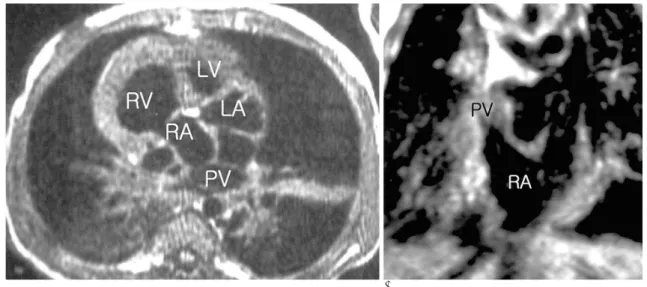
The most definitive distinguishing features of the right (RA) and left atria (LA) are the atrial appendages. On an axial MR image, the RA appendage appears as a triangu- lar structure with a broad-based opening into the RA.
The LA appendage is a long, narrow, finger-like projec- tion with a narrow orifice (Fig. 1A). The morphologic right ventricle (RV) has a muscular infundibulum and a prominent moderator band (Fig. 1B). The muscular in- fundibulum and well-defined outflow region separate the RV atrioventricular valve from the semilunar valve.
On the other hand, in the morphologic left ventricle (LV), the atrioventricular valve and semilunar valve are in fibrous continuity. The atrioventricular valve (tricus- pid valve) of the morphologic RV is slightly more apical in terms of its septal attachment than the mitral valve of the morphologic LV. As a result, this small septum, i.e.
the atrioventricular septum, divides the LV from the RA. The ventricular septum at the apex is heavily tra- beculated in the RV, but smooth in the LV. A moderator band (muscular band), connecting the free wall of the RV and the interventricular septum, is prominent on the axial images (Fig. 1B).
Congenital Heart Diseases
In the segmental approach to the diagnosis of congeni-
tal heart disease, the heart is divided into three main seg- ments: the atria, the ventricles, and the great vessels and the connections between them, namely the venous, atri- oventricular and ventriculoarterial connections (1). The atria and the venous connects form at the same time, so that abnormalities of atrial position are usually associat- ed with abnormalities in the situs of organs in the rest of the body. If both atria have the morphologic features of the RA, then each side structure of the visceral and tho- racic organs tends to have the characteristics of a right- sided structure. This situation is often associated with as- plenia syndrome. In left isomerism, both lungs have two lobes, and both the right and left pulmonary arteries pass over their respective bronchi (hyparterial bronchi). This condition is associated with polysplenia and interrupted inferior vena cava with azygos continuation.
Venous Connections
Systemic Venous Connection
The left-sided superior vena cava (SVC) is the most common anomalous systemic venous connection. Most commonly, the left SVC drains from the left brachio- cephalic vein and joins the coronary sinus to drain into the RA. In situs anomaly, however, the inferior vena ca- va (IVC) may drain into the LA. The interruption of the IVC with azygos continuation can be readily demon- strated by MRI, which allows the noninvasive diagnosis of this anomaly.
A B
Fig.1. Normal MR anatomy of the heart.
A. Axial T1-weighted spin-echo image shows normal atrial appendages. The right atrial appendage has a characteristic wide attach- ment to the right atrium (asterisk). The opening of the left atrial appendage is narrow (arrow).
B. Axial T1-weighted spin-echo image shows a prominent moderator band (asterisk), a characteristic feature of the normal right ventricle and normal fibrous continuity of the aortic valve and mitral valve (arrow).
Total Anomalous Pulmonary Venous Connection The pulmonary veins which grow from the lung buds form a confluence. If all of the pulmonary veins join the systemic route, this anomaly is called total anomalous pulmonary venous connection (TAPVC). TAPVC is clas- sified according to the location of the venous insertion;
TAPVC of the supracardiac type shows pulmonary ve- nous confluence draining into the SVC or vertical vein.
This is the most common form of TAPVC. In the cardiac type, there is anomalous venous drainage to the coro- nary sinus or to the right atrium (Figs. 2A, B).
Infracardiac type TAPVC involves pulmonary venous confluence draining into the IVC or portal venous sys- tem. Obstruction to flow is common, and occurs due to a combination of factors, namely the length of the com- mon venous channel, obstruction at the diaphragmatic hiatus, closure or restriction of the ductus venosus, and resistance to blood flow through the hepatic sinusoids (2).
Partial Anomalous Pulmonary Venous Connection Partial anomalous pulmonary venous connection (PA- PVC) is diagnosed when a part of the pulmonary veins drains into the systemic circulation. In such cases, it is important to identify the connections of all four pul- monary veins. The most common pattern involves the right upper pulmonary vein connecting to the SVC. PA- PVC is sometimes associated with a sinus venosus atrial septal defect.
Ventricles and Atrioventricular Connection
Congenitally Corrected Transposition of the Great Arteries
This condition is said to occur when the morphologic ventricles are on the wrong side of the heart (L-loop).
Corrected transposition can be understood as a ventricu- lar inversion. In almost all cases, the aorta is anterior to and to the left of the pulmonary artery (spatial relation- ship known as L-transposition). In MRI, the ventricular morphology and the positions of the great vessels can be determined directly (Figs. 3A, B).
In corrected transposition, if no other abnormalities are present, then the patients involved are asymptomatic.
However, the morphologic RV is not designed to pump against systemic pressures and, consequently, failure of the morphologic RV or arrhythmia generally occurs in the fifth or sixth decade of life. Other anomalies, such as Ebstein’s anomaly, ventricular septal defect (VSD) or pulmonary valve stenosis (PS) may also be present.
Tricuspid Atresia
In tricuspid atresia, communication between the RA and RV is lacking. To compensate for this, an atrial sep- tal defect (ASD) is obligatorily present, in order for blood to be shunted from the RA to the LA, and a VSD in order for blood to be shunted from the LV to the RV (3). If the ASD, VSD or pulmonary outflow tract is small, the RV may be small and rudimentary. In tricuspid atresia, MRI shows a fibrous band (imperforated tricuspid valve), fat,
A B
Fig. 2. Total anomalous pulmonary venous return to right atrium and double-outlet right ventricle with ventricular septal defect.
Axial (A) and coronal (B) T1-weighted spin-echo images show the pulmonary vein converge on the right atrium.
RA, right atrium; LA, left atrium; RV, right ventricle; LV, left ventricle; PV, pulmonary vein.
and muscle interposed between the atrium and ventri- cle, and allows the size of the VSD to be measured.
Postoperatively, MRI can be used to assess the Fontan shunt and any complications that may be present.
Hypoplastic Left Heart Syndrome
Hypoplastic left heart syndrome is diagnosed when there is underdevelopment of the LV. It is usually caused by aortic stenosis or atresia, mitral stenosis or atresia, or both. In such cases, there is little blood flow through the ascending aorta, and blood tends to flow from the pulmonary artery through the ductus arterio- sus into the aorta, and then in retrograde fashion to the aortic root to supply the coronary arteries. As a result, the main pulmonary artery is very large. MRI can readi- ly depict the enlargement of the chamber and the diam- eters of the great arteries. Another important role for MRI in hypoplastic left heart syndrome is the evaluation of the morphology and function of the various stages of the postoperative period (the Norwood procedure) (4).
Ebstein’’s Anomaly
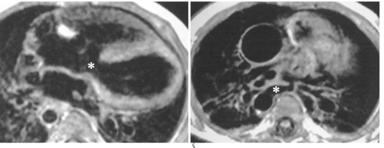
This anomaly is a primary abnormality of the tricus- pid valve, in which the septal and posterior leaflets of the valve adhere to the RV wall. The leaflets become free at a variable distance, at a location more apical than usual, so that the tricuspid valve orifice is displaced to- ward the apex (5). As a result, the portion of the RV that is basal to the valve orifice becomes “atrialized ventri- cle,”meaning that it functions as part of the atrium.
The atrialized portion of the RV becomes progressive- ly thin-walled and smooth-walled, and it may become markedly enlarged (Fig. 4). Blood within the atrialized ventricle is pressed back into the true right atrium. This to-and-fro motion causes functional obstruction to the emptying mechanism of the atrium. Ebstein anomaly is frequently associated with an ASD. MRI can be useful in the assessment of the degree of right atrial dilation, the size and function of the right ventricle, and the severity of tricuspid regurgitation.
A B
Fig. 3. Corrected transposition of the great vessels. A. Axial T1-weighted spin-echo image shows that the aorta (Ao) is anterior to and to the left of the pulmonary artery (PA). This spatial orientation is called L-transposition. B. Coronal T1-weighted spin-echo im- age shows that the aorta (Ao) arises from the rudimentary right ventricle (RV) and that the pulmonary artery (PA) arises from the left ventricle (LV).
Fig. 4. Ebstein’s anomaly. Axial T1-weighted spin-echo image shows that the tricuspid valve leaflets have been displaced api- cally away from the atrioventricular groove, so that the right ventricle is divided into a proximal, atrialized portion and a distal, small functional right ventricle. The markedly displaced attachment of the tricuspid valve leaflet results in the forma- tion of a large atrialized right ventricle (asterisk).
Endocardial Cushion Defect (Atrioventricular Septal Defect)
Endocardial cushion defects involve an abnormal sep- tation between the atria and the ventricles. These de- fects are situated in the inferior portion of the interatrial septum, the basal portion of the interventricular sep- tum, the septal leaflet of the tricuspid valve, and the sep- tal leaflet of the mitral valve (3). Defects in the endocar- dial cushion tissue cause a spectrum of cardiac anom- alies including primum ASD and complete atrioventric- ular septal defect (Fig. 5). The atrioventricular septum is created by the normal apical displacement of the tricus-
pid valve relative to the mitral valve. However, in endo- cardial cushion defect, the tricuspid and mitral valves originate at the same level, and the atrioventricular sep- tum is absent. In severe cases (complete atrioventricular cancal), both the atrial and ventricular portions of the septum in the region around the valve origins are ab- sent. This creates a common atrioventricular valve ori- fice with continuous, common atrioventricular valve leaflets. Axial MRI shows this abnormal relationship.
Ventriculoarterial connection
Complete Transposition of the Great Arteries
In complete transposition of the great arteries, the pul- monary artery arises from the LV and the aorta arises from the RV. The pulmonary and systemic circulations are in separate loops. The aorta is usually anterior to and to the right of the pulmonary artery, and the pulmonary and systemic circulations exist in parallel (Figs. 6A, B).
Communications between these two circulations include a patent foramen ovale, ASD, VSD, PDA, and systemic collateral arteries. Complete transposition of the great ar- teries is usually associated with the existence of a VSD, which is present for the purpose of allowing oxygenated blood to reach the systemic circulation. The preferred surgery for this anomaly is the Jatene (arterial switch), in which the roots of the great arteries are transected and
A B
Fig. 6. Transposition of the great vessels. A. Axial T1-weighted spin-echo image shows the right atrium and left atrium in their nor- mal positions. The RA connects to the morphologic right ventricle (asterisk within moderator band), which is on the right side of the heart. The LA connects to the morphologic left ventricle, which is on the left side of the heart. B. Sagittal T1-weighted spin-echo image shows that the aorta and pulmonary artery are transposed. The ascending aorta (Ao) arises from the right ventricle (RV).
Fig. 5. Endocardial cushion defect. Axial T1-weighted spin- echo images show a shortened ventricular septum and a pri- mum atrial septal defect (arrow). The secundum portion of the atrial septum is intact.
reattached to the opposite ventricle. MRI can depict pathologic anatomy and assess the size and thickness of the LV prior to performing the Jatene operation (4, 6).
Tetralogy of Fallot (TOF)
Tetralogy of Fallot consists of pulmonary stenosis, an overriding aorta, a perimembranous VSD, and resultant RV hypertrophy. The key feature of tetralogy is malalignment of the infundibular septum. This anterior- ly displaced infundibular septum encroaches on the RV outflow, causing the RV outflow tract to be abnormally small and the aorta to override the VSD. The pulmonary valve is usually bicuspid or unicuspid, and is stenotic in approximately two thirds of cases. Central pulmonary artery stenosis occurs commonly in subjects with TOF.
MRI plays an important role in the assessment of steno- sis of the main or branch arteries, the size of the RV out- flow tract, and the size of the RV.
Pulmonary Atresia
Pulmonary atresia with VSD is an extreme form of TOF in which a direct connection from the RV to the pul- monary arteries is lacking (7). This absence of connection between the RV and the pulmonary artery confluence can be followed on sequential axial MR images. The atre- sia can be focal, limited to the valve level, or more exten- sive. In such cases, a markedly enlarged aorta is observed to override a perimembranous VSD, and blood is usually delivered to the lungs via systemic-to-pulmonary collater- al channels, which can be seen as abnormal vessels origi- nating from the descending aorta and traveling toward
the lungs or connecting with the pulmonary arteries.
MRI is excellent for defining the main pulmonary arter- ies and the collateral arteries (Figs. 7A, B).
Double-Outlet Right Ventricle
Various definitions exist for double-outlet right ventri- cle (DORV). In DORV, more than 50% of the orifices of both great vessels are situated over the RV or the aortic valve and the mitral valves lack their normal fibrous continuity. DORV is always associated with a VSD, which is an important factor in determining the type of surgical repair to be performed. Axial MRI can be used to determine the location of the VSD, by identifying the great vessels on the images and then inspecting the im- ages immediately inferior to them, in order to determine which arterial outflow tract is confluent with the VSD.
In DORV, the great arteries usually have a side-by-side relationship at the base of the heart, but in some cases, the aorta is anterior to the pulmonary artery and slightly to the left or right of the pulmonary artery (Figs. 8A, B).
Associated anomalies include valvular, subvalvular ob- struction, and coarctation of the aorta.
Truncus Arteriosus
This anomaly is due to the failure of the division of the primitive truncus arteriosus into the aorta and pul- monary artery (8), as a result of which a single arterial trunk forms over both ventricles. A VSD is always pre- sent in such cases. Collet and Edwards established a sys- tem of classification of truncus arteriosus according to the origin of the pulmonary artery arising from the com-
A B
Fig. 7. Pulmonary atresia with VSD.
A. Axial T1-weighted spin-echo image shows a VSD (asterisk).
B. Prominent aortopulmonary collaterals originate from the descending thoracic aorta (asterisk).
mon arterial trunk. In type I truncus arteriosus, a short, separate pulmonary trunk is present, while in type II, the right and left pulmonary arteries are close to each other, but arise separately from the pulmonary trunk (Fig. 9). In type III, the right and left pulmonary arteries arise further laterally, while in type IV, no pulmonary vessels arise from the aorta, but branches from the de- scending thoracic aorta supply the pulmonary vascula- ture. MRI can be used to distinguish truncus arteriosus from pulmonary atresia and to measure the relative sizes and confluence of the pulmonary artery.
Intracardiac and Extracardiac Shunts (Left-to-Right Shunts)
Ventricular Septal Defect
Perimembranous Ventricular Septal Defect
The membranous part of the ventricular septum is a small area, just inferior to the root of the aorta, between the right coronary cusp and the noncoronary cusp (Fig.
10). Most VSDs in this region extend beyond the anatomic membranous ventricular septum and so are often called perimembranous. This portion of the inter- ventricular septum is the last part to form, so it is the most frequent location for a VSD to occur.
Supracristal Ventricular Septal Defect
The crista supraventricularis is a muscular ridge that defines the outlet part of the RV. The supracristal part of the ventricular septum is the part of the ventricular sep- tum that is inferior to the right and left coronary cusps
(9) . A supracristal VSD is one that is located just below the pulmonary valve and the aortic valve (Fig. 11). One of the complications of a supracristal VSD is prolapse of the right coronary cusp into the septal defect, which re- sults in aortic insufficiency.
Muscular Ventricular Septal Defect
The muscular septum is the largest part of the septum.
A B
Fig. 8. Double-outlet right ventricle with subaortic VSD.
A. Axial T1-weighted spin-echo image shows the aorta (Ao) and pulmonary artery (PA), and that both have a complete muscular in- fundibulum.
B. More inferiorly located is a VSD (asterisk) connecting the aorta (Ao). The arrow indicates the infundibular septum.
Fig. 9. Truncus arteriosus. Sagittal T1-weighted spin-echo im- age shows that the pulmonary artery (PA) arises from posterior part of the truncus (TR).
This is located more apically than the membranous part.
Multiple VSDs can be present in this area, giving the septum the appearance of a “Swiss cheese”. Because of the trabeculation on the right side of the heart, these de- fects are often difficult to visualize at surgery and must be clearly delineated by imaging.
Atrial Septal Defect
Ostium Primum Atrial Septal Defect
Ostium primum atrial septal defect is defined as the persistence of the ostium primum, with a large defect being present in the interatrial septum at the junction of the atrial and ventricular septa (3). This defect is known to include a spectrum of endocardial cushion defects
(atrioventricular septal defects).
Ostium Secundum Atrial Septal Defect
Normally, after the septum primum forms, a second primitive septum develops, namely the septum secun- dum, which eventually forms the posterior and inferior part of the interatrial septum, including the foramen ovale.
Atrial tissue separates the edge of the defect from the atri- oventricular valves. Deficiencies of this part of the septum constitute the most common type of ASD (Fig. 12).
Sinus Venosus Atrial Septal Defect
In this case, deficient incorporation of the sinus veno- sus creates an ASD near the junction of the RA with the superior vena cava. This portion of the interatrial sep- tum also normally creates part of the separation of the superior right pulmonary vein from the LA. When a si- nus venosus ASD is present, the right superior pul- monary vein drains into the RA.
Patent Ductus Arteriosus (PDA)
PDA is defined as continued patency of the ductus ar- teriosus beyond three months after birth. It should be distinguished from the delayed closure of the ductus, which is found, for example, in premature and low birth weight infants. PDA is usually observed in the top of the left pulmonary artery, close to the pulmonary artery bi- furcation (Fig. 13). The origin and the shape of the duc- tus arteriosus in TOF and pulmonary atresia with VSD are quite different from those of the uncomplicated duc- tus, in that it arises from the undersurface of the aortic arch and requires a longer distance to insert itself into Fig. 10. Membranous ventricular septal defect. Axial T1-
weighted spin-echo image shows the presence of a defect in the membranous septum (arrow), which is situated between and inferior to the right and noncoronary sinuses of Valsalva.
Fig. 11. Supracristal ventricular septal defect. Axial T1-weight- ed spin-echo image shows a ventricular septal defect (arrow).
The right coronary cusp is covering the defect.
Fig. 12. Secundum atrial septal defect. Axial T1-weighted spin- echo image shows a defect in the posterior, inferior atrial sep- tum, which is the secundum portion of the atrial septum. The residual septum primum is clearly seen (arrow). Note the dila- tion of the right atrium caused by the left-to-right shunt.
the proximal segment of the left pulmonary artery.
Thoracic Aortic Anomalies
Coarctation of the Aorta and Interruption of Aortic Arch (IAA)
The coarctation of the aorta is a congenital narrowing of the aorta. It is a focal narrowing just inferior to the origin
of the left subclavian artery. This abnormality includes both localized and diffuse types. The localized type (re- ferred to as the postductal, adult or juxtaductal type) in- volves those cases in which the narrowing is short and
Fig. 14. Coarctation of the aorta in an infant. Sagittal T1- weighted spin-echo image shows a discrete narrowing of the aorta, distal to the origin of the left subclavian artery.
A B
Fig. 15. Double aortic arch.
A. Axial T1-weighted spin-echo image demonstrates that both the right and left limbs of the double arch encircle the trachea and esophagus (vascular ring).
B. Coronal T1-weighted spin-echo image shows that the right arch (R) is larger and higher than the left arch (L).
Fig. 13. Patent ductus arteriosus. Axial T1-weighted spin-echo image shows patent ductus arteriosus (asterisk) that extends from the origin of the left pulmonary artery to the descending thoracic aorta, just distal to the origin of the left subclavian artery.
대한영상의학회지 2004;51:563-572
선천성 심질환의 자기공명영상1
1순천향대학교 의과대학 방사선과학교실
2전남대학교 의과대학 흉부외과학교실
김동훈・류상완2・장윤우・장지연・황정화
MRI는 선천성 심질환의 해부학적 구조를 보여주고 진단과 치료에 있어서 중요한 역할을 한다. MRI는 다양한 방 향의 영상을 통해 복잡한 심방구조의 3차원적인 관계를 보여주며 대동맥뿐만 아니라 대정맥, 폐정맥 및 폐동맥의 구조를 잘 나타낸다. 저자들은 본 화보에서 정상 심장과 선천성 심질환의 MR영상을 통해 질병의 이해를 돕고자 하 였다.
discrete, and is located just beyond the left subclavian artery. The diffuse type (known as the preductal, infantile or tubular hypoplastic type) involves those cases in which there is a long segment of aortic narrowing and the blood flow to the lower extremities depends on the patent duc- tus arteriosus (3, 10). MRI is effective for both the preop- erative assessment of coarctation and its postoperative monitoring (Fig. 14). It is important to measure the diame- ter of the distal arch, because it can influence the type of surgery that needs to be performed. In aortic coarctation, a collateral supply to the descending thoracic aorta devel- ops from the internal mammary artery and other branch- es of the subclavian arteries and intercostal arteries. The interruption of the aortic arch (IAA) is due to a disconti- nuity of the aortic arch which results from the complete absence of an arch segment. The site of interruption may be at the isthmus, between the left common carotid artery and the left subclavian artery or between the in- nominate artery and the left common carotid artery.
Abnormal Branching of the Aorta and Vascular Rings Anomalies of the aortic arch result from either the per- sistent patency of a vascular segment that should nor- mally regress, or the abnormal regression of a segment that should normally remain patent. There are numer- ous variations of this anomaly, some of which create vascular rings and cause symptoms of tracheal and esophageal compression. MRI can be used to identify arch anomalies, including right aortic arch with retroe- sophageal left subclavian artery and double aortic arch.
In the case where a vascular ring is present, the trachea and esophagus are surrounded by vascular structures (Figs. 15A, B). In the case of a right aortic arch, the vas-
cular ring is formed by the combination of the aortic arch, an aberrant vessel passing posterior to the trachea, and the left ligamentum arteriosum. MRI has the advan- tage of showing both the vascular structures and the air- ways and esophagus.
References
1. Freedom RM, Yoo SJ, Benson LN. The segmental and sequential ap- proach to congenital heart disease. In: Freedom RMM, Yoo SJ, Benson LN, eds. Congenital heart disease: a textbook of angiocardiog- raphy, vol 1. Armonk, NY: Futura Publishing, 1997:95-120 2. Julsrud PR. Anomalous pulmonary venous connection. In: Baum S, ed.
Abrams’angiography, vol 1. Boston: Little, Brown and Company, 1997:868-890
3. Strife JL, Bisset III GS, Burrows PE, Cardiovascular system. In:
Kirks DR, ed. Practical pediatric imaging: Diagnostic radiology of in- fants and children. 3rd ed. Philadelphia: Lippincott-Raven Publishers, 1998:511-618
4. Higgins CB, de Roos A. Congenital heart disease: morphology and function. In: Higgins CB, de Roos A, eds. Cardiovascular MRI &
MRA. Philadelphia: Lippincott Williams & Wilkins, 2003:307-338 5. Giuliani ER, Fuster V, Brandenburg RO, Mair DD. Ebstein’s
anomaly: the clinical features and natural history of Ebstein’s anomaly of the tricuspid valve. Mayo Clin Proc 1979;54:163-173 6. Amplatz KM. Complete transposition of the great vessels. In: Amplatz
KM, ed. Radiology of congenital heart disease. St. Louis: Mosby-Year Book, 1993:675-707
7. Gomes AS, Lois JF, Williams RG. Pulmonary atresia: MR imaging in patients with congenital obstruction of the right ventricular out- flow tract. Radiology 1990;174:51-57
8. Donnelly LF, Higgins CB. MR imaging of conotruncal abnormali- ties. AJR Am J Roentgenol 1996;166:925-928
9. Bremerich J, Reddy GP, Higgins CB. MRI of supracristal ventricu- lar septal defects. J Comput Assist Tomogr 1999;23:13-15
10. Rees S, Somerville J, Ward C, Martinez J, Mohiaddin RH, Underwood R, et al. Coarctation of the aorta: MR imaging in late postoperative assessment. Radiology 1989;173:499-502