Copyrights © 2015 The Korean Society of Radiology
168
Case Report
pISSN 1738-2637 / eISSN 2288-2928 J Korean Soc Radiol 2015;73(3):168-171 http://dx.doi.org/10.3348/jksr.2015.73.3.168
INTRODUCTION
Spontaneous breast infarction is a very rare episode, repre- senting about 0.1% of biopsied (1) and 0.3–1.5% of fibroadeno- ma cases including the reported cases of pregnant women (2-4).
Although several cases of breast infarction have been reported in the literature, we found only two reports that described the mammographic findings of an infarcted breast tumor involving five patients (1, 5). In the present study, we describe a case of spontaneous breast infarction in a postpartum woman and dis- cuss the mammography and ultrasonography imaging findings.
CASE REPORT
A 33-year-old woman with a palpable mass on her right breast visited our hospital four weeks postpartum. She initially discov- ered the palpable breast mass during her 3rd trimester of preg- nancy and reported no change in its size after delivery. She did not experience nipple discharge or pain and had no history of
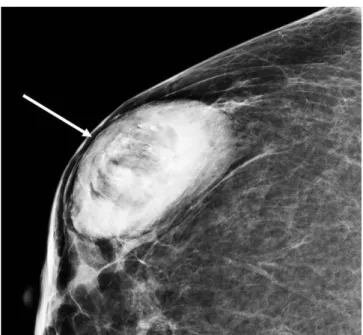
trauma or use of anticoagulant drugs. The physical examination revealed a hard and non-tender mass in the right breast, with- out nipple retraction and skin thickening. The patient had no axillary or supraclavicular lymphadenopathy. Mammography revealed a large and oval shaped fat-containing mass with a cir- cumscribed margin and microcalcifications (Fig. 1). On ultra- sonography, the mass was seen as an oval shaped structure with a circumscribed margin and heterogeneous echogenicity along with a combined pattern of posterior features (Fig. 2A). A few echogenic dots were observed within the mass. Minimal vascu- larity was noted at the periphery of the mass on a color Doppler image (Fig. 2B). Considering these radiologic findings as prob- ably benign, the mass was classified as a category 3 lesion. How- ever, the patient wanted to perform biopsy for the fear of malig- nancy. The mass was pathologically verified as a breast infarction.
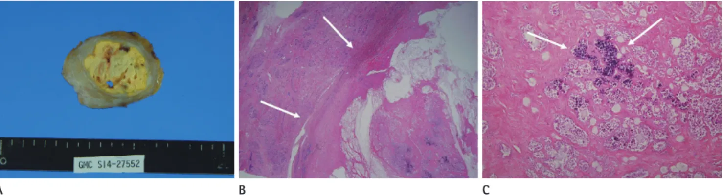
Three months later, there was no change in size of the mass and the patient underwent excisional biopsy. The specimen was re- ported as a circumscribed solid mass measuring 4.8 × 3.6 × 2.3 cm with necrosis on the cut surface (Fig. 3A). There was exten-
Spontaneous Infarction of Hyperplastic Breast Tissue: A Case Report
과성장 유방조직의 자발적 괴사: 증례 보고Eun Young Yoo, MD, Sang Yu Nam, MD*, Hye-Young Choi, MD, Min Ji Hong, MD
Department of Radiology, Gil Medical Center, Gachon University School of Medicine and Science, Incheon, Korea
Spontaneous breast infarction is a very rare complication of fibroadenoma of the breast. We present an interesting case of a 33-year-old woman with spontaneous infarction of hyperplastic breast tissue related to pregnancy and lactation. Mam- mography showed an oval, circumscribed, fat-containing mass with microcalcifica- tions. Ultrasonography revealed an oval, circumscribed mass with echogenic dots.
Color Doppler imaging revealed presence of minimal vascularity at the periphery of the mass.
Index terms Breast
Spontaneous Infarction Mammography Ultrasonography Microcalcification
Received May 11, 2015 Revised June 30, 2015 Accepted July 18, 2015
*Corresponding author: Sang Yu Nam, MD Department of Radiology, Gil Medical Center, Gachon University School of Medicine and Science, 21 Namdong-daero 774beon-gil, Namdong-gu, Incheon 21565, Korea.
Tel. 82-32-460-3682 Fax. 82-32-460-3065 E-mail: [email protected]
This is an Open Access article distributed under the terms of the Creative Commons Attribution Non-Commercial License (http://creativecommons.org/licenses/by-nc/3.0) which permits unrestricted non-commercial use, distri- bution, and reproduction in any medium, provided the original work is properly cited.
169
Eun Young Yoo, et al
jksronline.org J Korean Soc Radiol 2015;73(3):168-171 sive necrosis with adjacent fibrosis and residual epithelium, as well as lactation hyperplasia (Fig. 3B). Microcalcifications and cystic changes were also seen within the mass (Fig. 3C). There was no evidence of malignancy and the histologic examination confirmed the features of infarcted hyperplastic breast tissue.
DISCUSSION
Spontaneous breast infarction is a very rare complication of
fibroadenoma of the breast. According to a recent study by Oh et al. (1), 0.1% of all biopsied lesions (9/8310) were spontaneous infarctions. Infarctions in benign lesions may occur in various conditions such as fibroadenoma, intraductal papilloma, and hyperplasia associated with pregnancy and lactation, as well as during anticoagulant therapy and with oral contraceptive use (1-3, 6-8). The malignant etiologies of breast infarction include invasive carcinoma, ductal carcinoma in situ, as well as medul- lary carcinoma, and papillary carcinoma (1, 9). Spontaneous breast infarction may occur as an uncommon complication of fine needle aspiration biopsy (10). The vascular trauma during the needling procedure may induce thrombosis and infarction.
The presenting symptom is most frequently a rapidly growing palpable mass that is sometimes painful and hard. Occasionally, bloody discharge secondary to tissue necrosis can be observed (7). In such patients, the clinical diagnosis of an inflammatory or malignant breast disease may be possible. The cytopatholog- ic features of infarction include necrosis and worrisome nuclear features, which are often confused with inflammation or malig- nancy (8).
Spontaneous infarction has been mostly described during the third trimester of pregnancy and lactation. In these cases, the proposed pathogenesis includes a relative vascular ischemia during phases of increased metabolic requirements of the breast tissue (3). The possibility of a probable torsion due to the in- creased mobility of these tumors has been suggested in cases of spontaneous infarction occurring in the absence of pregnancy Fig. 1. A 33-year-old woman with spontaneous breast infarction of
hyperplastic tissue. Craniocaudal mammogram of her right breast shows an oval shaped large fat-containing mass with a circumscribed margin and microcalcifications (arrow).
Fig. 2. Ultrasonography findings of infarcted hyperplastic breast tissue.
A. On ultrasonography, the mass appears as an oval shaped structure with a circumscribed margin, and heterogeneous echogenicity with a com- bined pattern of posterior features. A few echogenic dots can be seen within the mass (arrows).
B. Color Doppler sonogram shows only minimal vascularity at the periphery of the mass.
A B
170
Spontaneous Infarction of Hyperplastic Breast Tissue
jksronline.org
J Korean Soc Radiol 2015;73(3):168-171 and lactation.
Until date, we were able to locate only two reports that de- scribed the mammographic findings of an infarcted breast tu- mor involving five patients. In one study, an infarcted lactating adenoma presented as a circumscribed mass without additional findings on mammography (5). Two infarcted fibroadenomas and one medullary carcinoma similarly exhibited oval shaped, indistinct, and highly dense masses (1). An infarcted intraduct- al papilloma presented as a round, circumscribed, equal density mass on mammography. As indicated by these reports, it is evi- dent that no characteristic mammographic findings lead to dif- ferentiation between malignant and benign tumor infarctions of the breast.
There are a few reports on the ultrasonographic findings of spontaneous breast infarction. On ultrasonography, the most common reported features include an oval shaped, parallel-ori- ented, indistinct, heterogeneously hypoechoic mass with or without posterior acoustic enhancement. In addition, these le- sions are sometimes irregular shaped and angularly margined mass that can be shown for malignancy.
In conclusion, we report a recent case of infarcted hyperplas- tic breast tissue in a young postpartum woman. The spontane- ous breast infarction presented as a circumscribed mass with microcalcifications on mammography, and a heterogeneous echoic mass with echogenic dots on ultrasonography. There was minimal vascularity observed at periphery of the mass on color Doppler imaging. It is proposed that even though the incidence of a spontaneous breast infarction is very low and it has no specif-
ic radiologic features, it should be included in the differential di- agnoses of women with a recent history of pregnancy or lactation.
REFERENCES
1. Oh HJ, Kim SH, Kang BJ, Lee AW, Song BJ, Kim HS, et al. Ul- trasonographic features of spontaneous breast tumor in- farction. Breast Cancer 2014 Mar 15 [Epub]. http://dx.doi.
org/10.1007/s12282-014-0525-3
2. Lucey JJ. Spontaneous infarction of the breast. J Clin Pathol 1975;28:937-943
3. Raju GC, Naraynsingh V. Infarction of fibroadenoma of the breast. J R Coll Surg Edinb 1985;30:162-163
4. Skenderi F, Krakonja F, Vranic S. Infarcted fibroadenoma of the breast: report of two new cases with review of the liter- ature. Diagn Pathol 2013;8:38
5. Behrndt VS, Barbakoff D, Askin FB, Brem RF. Infarcted lac- tating adenoma presenting as a rapidly enlarging breast mass. AJR Am J Roentgenol 1999;173:933-935
6. Jimenez JF, Ryals RO, Cohen C. Spontaneous breast infarc- tion associated with pregnancy presenting as a palpable mass. J Surg Oncol 1986;32:174-178
7. Fowler CL. Spontaneous infarction of fibroadenoma in an adolescent girl. Pediatr Radiol 2004;34:988-990
8. Deshpande KM, Deshpande AH, Raut WK, Lele VR, Bobhate SK. Diagnostic difficulties in spontaneous infarction of a fibroadenoma in an adolescent: case report. Diagn Cyto- pathol 2002;26:26-28
Fig. 3. Histopathology of infarcted hyperplastic breast tissue.
A. Gross examination of the specimen shows a 4.8 × 3.6 × 2.3 cm, well circumscribed yellow colored mass with areas of hemorrhage, which is distinct from the surrounding breast parenchyma.
B. Low-power microscopic view of the lesion shows extensive necrosis of hyperplastic breast tissue (arrows) (H&E, × 10).
C. Higher power photomicrograph shows microcalcifications within the mass (arrows) (H&E, × 100).
H&E = hematoxylin and eosin
A B C
171
Eun Young Yoo, et al
jksronline.org J Korean Soc Radiol 2015;73(3):168-171 9. Greenberg ML, Middleton PD, Bilous AM. Infarcted intra-
duct papilloma diagnosed by fine-needle biopsy: a cytolog- ic, clinical, and mammographic pitfall. Diagn Cytopathol 1994;11:188-191; discussion 191-194
10. Pinto RG, Couto F, Mandreker S. Infarction after fine needle aspiration. A report of four cases. Acta Cytol 1996;40:739- 741
과성장 유방조직의 자발적 괴사: 증례 보고
유은영 · 남상유* · 최혜영 · 홍민지
자발적 유방 괴사는 매우 드문 질환이다. 저자들은 임신과 수유와 관련하여 유방조직이 과성장(hyperplastic tissue)한 33 세 여성에서 자발적 괴사(spontaneous infarction)로 진단된 1예의 영상 소견을 보고하고자 한다. 유방촬영술에서 난원형 의 경계가 좋은 지방을 함유한 종괴로 보였고 석회화를 동반하였다. 초음파에서는 고에코성 점들을 동반한 난원형의 경 계가 좋은 종괴로 보였으며, 색도플러에서 종괴의 주변부를 따라 혈관이 보였다.
가천대학교 길병원 영상의학과