178
Copyrights © 2014 The Korean Society of RadiologyINTRODUCTION
The serratus anterior is a flattened sheet of muscle originating from the first through the ninth ribs and inserting along the me- dial border of the costal surface of scapula. The function of ser- ratus anterior is to protract and rotate the scapula, thus provid- ing a full range of motion at glenohumeral joint. It also keeps the scapula closely opposed to the thoracic wall (1, 2).
Only a few cases of isolated aplasia of the serratus anterior muscle have been reported (2, 3). Usually, it presents as a part of the combination of shoulder girdle muscle anomalies (4-6).
Here, we report a case of a 30-year-old male with unilateral complete aplasia of the right serratus anterior muscle and wing- ing of the right scapula, which was incidentally found through
chest radiography and CT scan. This unusual case emphasizes the importance of paying careful attention to chest wall struc- tures while interpreting a plain radiography.
CASE REPORT
A 30-year-old Korean male presented with left flank pain. He complained of tenderness on his left ribs and left scapula areas, and showed decreased range of motion in the left shoulder. He described having more pain when he took left-side down lateral decubitus. There was tenderness, rated 6 to 7 on the numerical rating scale, of the area. He had a 1 month history of strenuous physical activities.
His medical history was not significant. He had never been
Case Report
pISSN 1738-2637 / eISSN 2288-2928 J Korean Soc Radiol 2014;71(4):178-181 http://dx.doi.org/10.3348/jksr.2014.71.4.178
Received April 21, 2014; Accepted July 15, 2014 Corresponding author: Jeong Min Ko, MD
Department of Radiology, St. Vincent’s Hospital, College of Medicine, The Catholic University of Korea, 93 Jungbu-daero, Paldal-gu, Suwon 442-723, Korea.
Tel. 82-31-249-7482 Fax. 82-31-249-5713 E-mail: [email protected]
This is an Open Access article distributed under the terms of the Creative Commons Attribution Non-Commercial License (http://creativecommons.org/licenses/by-nc/3.0) which permits unrestricted non-commercial use, distri- bution, and reproduction in any medium, provided the original work is properly cited.
The isolated aplasia of the serratus anterior muscle with winging of scapula is very rare, and only a few cases are reported. Here, we present a case of a 30-year-old Korean male who initially presented with a left flank pain. His physical exam did not show any significant finding in his right shoulder. However, his chest radiograph showed absence of right serratus anterior muscle and slightly elevated and medially rotated right scapula. Subsequent CT scan showed the right serratus anterior mus- cle aplasia and medial winging of the right scapula. This case is unique in two as- pects. First, the combination of abnormalities is different from the typical congeni- tal abnormalities involving shoulder girdle, such as Sprengel deformity or Poland syndrome. Secondly, this was incidentally diagnosed with chest radiograph, without clinical impression. Careful reading of chest radiograph can help the radiologists to detect such clinically silent abnormalities.
Index terms Congenital Serratus Anterior Aplasia
Winged Scapula Chest Radiograph
Incidental Finding of Unilateral Isolated Aplasia of Serratus Anterior Muscle and Winged Scapula on Chest Radiograph: A Case Report
흉부 X-선 사진에서 우연히 발견된 일측성 앞톱니근 단일 무형성과 날개 견갑골의 증례 보고Joonsung Choi, MD, Hyun Jin Park, MD, Jeong Min Ko, MD
Department of Radiology, St. Vincent’s Hospital, College of Medicine, The Catholic University of Korea, Suwon, Korea
Joonsung Choi, et al
179
jksronline.org J Korean Soc Radiol 2014;71(4):178-181
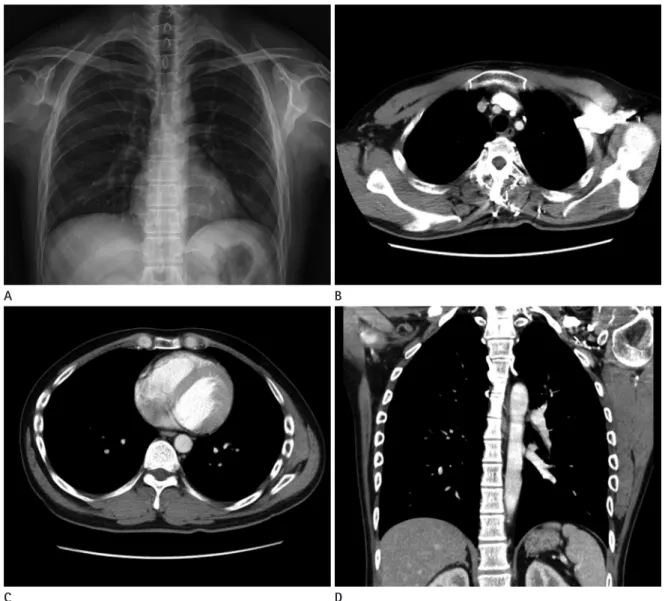
or pole medially rotated (Fig. 1A). A subsequent chest CT scan showed an increased distance between the medial border of right scapula and the rib cage compared to the opposite side, which is compatible with medial winging of right scapula (Fig.
1B). Although the other muscles contributing to the shoulder girdle were normal, the CT scan showed an absence of the right serratus anterior muscle (Fig. 1C, D). There was no remarkable difference in the size and shape of the two scapulae.
After conservative treatment, the left flank pain was relieved.
Because the patient didn’t complain of any symptoms on his right shoulder, no treatment about his was performed on his hospitalized or operated on before. His family history was non-
specific and there was no known congenital disorder. He had smoked for fifteen pack-years and socially drunk alcohol. The patient’s review of systems was unremarkable.
A chest radiograph showed noted absence of right serratus anterior muscle. The lateral chest wall shadow normally is com- posed of serratus anterior muscle, intermuscular fat plane, latis- simusdorsi muscle, and subcutaneous fat from medial to lateral.
But the patient’s chest radiograph didn’t show shadows of the right serratus anterior muscle or the intermuscular fat plane, and it showed the right scapula elevated by 0.9 cm and its inferi-
Fig. 1. Chest radiograph (A) shows absence of right serratus muscle shadow compared to the other side showing normal chest wall with the shadows of serratus anterior, intermuscular fat plane, latissimus dorsi, and subcutaneous fat from medial to later- al. The right scapula is elevated by 0.9 cm compared to the opposite side, and the inferior pole of the right scapula is medially rotated. Axial chest CT scan shows increased distance between the medial border of right scapula and right rib cage, compatible with medial winging of right scapula (B). Axial (C) and coronal (D) chest CT scans show the absence of right serratus anterior muscle.
A
C
B
D
Incidental Finding of Unilateral Isolated Aplasia of Serratus Anterior Muscle and Winged Scapula on Chest Radiograph
180
J Korean Soc Radiol 2014;71(4):178-181 jksronline.orgcompatible with the new disease category that Chernev suggest- ed; but this case is different in the following aspects. First, the degree of high scapula was subtle. This case shows only 0.9 cm elevation of the scapula, contrary to 2−7 cm elevation in the usual cases (8). Secondly, there was no remarkable scapula hy- poplasia. Therefore, we can reasonably assume that this case could be a new type of deformity or a mild variant of the defor- mity that Chernev suggested.
This case is also unique in the course of diagnosis. The previ- ous cases of isolated aplasia of the serratus anterior muscles were diagnosed by either physical exam alone or by physical exam and magnetic resonance imaging. But this case is the first where it was diagnosed incidentally, while examining the chest radiog- raphy. The role of the radiologist was important in diagnosing this anomaly. When the shadow of the serratus anterior muscle and the medial rotation of the inferior pole of scapula are not present on the chest radiograph, it can be reasonably assumed that there is aplasia or hypoplasia of the serratus anterior and winging of scapula. While interpreting chest radiograph, the awareness of the characteristic finding of this rare deformity can lead to correct diagnosis of this clinically vague deformity. It can also help make an early diagnosis of the cause of winged scapula and reduce unnecessary examinations. This case shows us the importance of thorough reading with high index of suspicion for abnormalities, when interpreting chest radiographs in every- day practice.
REFERENCES
1. Martin RM, Fish DE. Scapular winging: anatomical review, diagnosis, and treatments. Curr Rev Musculoskelet Med 2008;1:1-11
2. Chernev I, Pessina MA. Unilateral complete congenital ser- ratus anterior muscle aplasia: a case report. PM R 2009;1:
587-589
3. Levin SE, Trummer MJ. Agenesis of the serratus anterior muscle: a cause of winged scapula. JAMA 1973;225:748 4. David TJ, Winter RM. Familial absence of the pectoralis
major, serratus anterior, and latissimus dorsi muscles. J Med Genet 1985;22:390-392
5. Van Heest AE. Congenital disorders of the hand and upper extremity. Pediatr Clin North Am 1996;43:1113-1133 right shoulder.
DISCUSSION
Scapular winging is a rare debilitating condition that limits the function of the upper extremity (1), and it can be subdivided into medial and lateral according to the direction of the rotation of the scapula.
Medial winging of the scapula is caused by an abnormality of the serratus anterior muscle. The serratus anterior is solely in- nervated by a long thoracic nerve, originating from the anterior rami of the fifth through seventh nerves (1). Most of the cases of medial winging of the scapula have been associated with neuro- logic causes from the injury of the long thoracic nerve (1). The medial scapular winging caused by isolated aplasia of the serra- tus anterior muscle is extremely rare (2, 3).
The most common congenital abnormality involving shoul- der girdle is Sprengel deformity (2), which consists of congenital high scapula and limited motion of the shoulder, often includes the regional muscle hypoplasia (7, 8). The most commonly af- fected muscles are the trapezius, the pectoralis major, and the sternocleidomastoid muscles. Other muscles such as the serra- tus anterior, the levator scapulae, the infraspinatus, latissimus- dorsi, the teres major, the pectoralis minor, and the rhomboids are less affected (2). Sprengel deformity is almost always associ- ated with other malformations of cervicothoracic vertebrae or thoracic rib cage (9). In Sprengel deformity, congenital elevation of the scapula is caused by an interrupted caudal migration of the scapula and is associated with arrested growths of bone and muscle (2, 8, 9).
Poland syndrome is another congenital abnormality involving shoulder girdle. It is characterized by partial or complete aplasia of the pectoralis major muscle (6). Poland syndrome is often accom- panied by serratus anterior aplasia and scapular hypoplasia (2).
Chernev and Pessina (2) suggested a new category of defor- mity composed of serratus anterior muscle aplasia and winged, high-riding, and hypoplastic scapula. They thought this defor- mity may become a new variant of known congenital syn- dromes affecting the scapula, such as Sprengel deformity or Po- land syndrome.
This case shows isolated absence of serratus anterior and winging and medial rotation of the scapula. These features are
Joonsung Choi, et al
181
jksronline.org J Korean Soc Radiol 2014;71(4):178-181
8. Gonen E, Simsek U, Solak S, Bektaser B, Ates Y, Aydin E.
Long-Term Results of Modified Green Method in Spren- gel’s Deformity. J Child Orthop 2010;4:309-314
9. Cavendish ME. Congenital elevation of the scapula. J Bone Joint Surg Br 1972;54:395-408
6. Hegde HR, Shokeir MH. Posterior shoulder girdle abnor- malities with absence of pectoralis major muscle. Am J Med Genet 1982;13:285-293
7. Collins JD. Congenital and acquired atrophy of the shoul- der girdle muscles in a patient with Sprengel’s deformity. J Natl Med Assoc 2011;103:635-643
흉부 X-선 사진에서 우연히 발견된 일측성 앞톱니근 단일 무형성과 날개 견갑골의 증례 보고
최준성 · 박현진 · 고정민
앞톱니근의 단일 무형성으로 인한 날개 견갑골은 매우 드물며, 단지 몇 개의 증례만 보고되었다. 본 저자들은 처음에 왼쪽 옆구리 통증을 주소로 내원한 30세 한국 남자의 증례를 보고하고자 한다. 이 환자는 신체검사에서 오른쪽 어깨에 특이 소 견을 보이지 않았던 환자이다. 그러나 흉부 X-선 사진에서 오른쪽 앞톱니근의 무형성과 오른쪽 날개 견갑골이 관찰되었 고 이어 시행한 전산화단층촬영에서 확인된 증례이다. 이 증례는 두 가지 측면에서 특이한 증례이다. 첫째로 본 증례에서 관찰된 이상소견이 Sprengel deformity나 Poland syndrome과 같은 전형적인 어깨의 선천성 이상의 구성요소와 다르다는 점이다. 둘째로 본 증례는 임상적 의심없이 우연히 흉부 X-선 사진에서 진단된 증례이다. 흉부 X-선 사진을 주의 깊게 할 경우 이러한 임상적으로 의심하지 못한 이상 소견을 진단하는 데 도움이 된다.
가톨릭대학교 의과대학 성빈센트병원 영상의학과