Differentiation of Human Dental Pulp Stem Cells into Dopaminergic Neuron-like Cells in Vitro
We investigated the potential of human dental pulp stem cells (hDPSCs) to differentiate into dopaminergic neurons in vitro as an autologous stem cell source for Parkinson’s disease treatment. The hDPSCs were expanded in knockout-embryonic stem cell (KO-ES) medium containing leukemia inhibitory factor (LIF) on gelatin-coated plates for 3-4 days. Then, the medium was replaced with KO-ES medium without LIF to allow the formation of the neurosphere for 4 days. The neurosphere was transferred into ITS medium, containing ITS (human insulin-transferrin-sodium) and fibronectin, to select for Nestin-positive cells for 6-8 days. The cells were then cultured in N-2 medium containing basic fibroblast growth factor (FGF), FGF-8b, sonic hedgehog-N, and ascorbic acid on poly-L-ornithine/
fibronectin-coated plates to expand the Nestin-positive cells for up to 2 weeks. Finally, the cells were transferred into N-2/ascorbic acid medium to allow for their differentiation into dopaminergic neurons for 10-15 days. The differentiation stages were confirmed by morphological, immunocytochemical, flow cytometric, real-time PCR, and ELISA analyses.
The expressions of mesenchymal stem cell markers were observed at the early stages. The expressions of early neuronal markers were maintained throughout the differentiation stages. The mature neural markers showed increased expression from stage 3 onwards. The percentage of cells positive for tyrosine hydroxylase was 14.49%, and the amount was 0.526 ± 0.033 ng/mL at the last stage. hDPSCs can differentiate into dopaminergic neural cells under experimental cell differentiation conditions, showing potential as an autologous cell source for the treatment of Parkinson’s disease.
Keywords: Human Dental Pulp Stem Cells; Dopaminergic Differentiation; Tyrosine Hydroxylase; Parkinson Disease; Autologous Stem Cell Therapy; Dopaminergic Neural Cells So Young Chun,1,* Shay Soker,2,*
Yu-Jin Jang,3 Tae Gyun Kwon,4 and Eun Sang Yoo4
1BioMedical Research Institute, Kyungpook National University Hospital, Daegu, Korea; 2Wake Forest Institute for Regenerative Medicine, Wake Forest University School of Medicine, Winston-Salem, NC, USA; 3Department of Neural Development and Disease, Korea Brain Research Institute, Daegu;
4Department of Urology, Kyungpook National University, Daegu, Korea
* So Young Chun and Shay Soker contributed equally to this work.
Received: 24 February 2015 Accepted: 9 November 2015 Address for Correspondence:
Eun Sang Yoo, MD
Department of Urology, Kyungpook National University Hospital, 130 Dongdeok-ro, Jung-gu, Daegu 41944, Korea
Tel: +82.53-420-5851, Fax: +82.53-427-5447 E-mail: [email protected]
Funding: This work was supported by a Biomedical Research Institute grant from Kyungpook National University Hospital (2013).
http://dx.doi.org/10.3346/jkms.2016.31.2.171 • J Korean Med Sci 2016; 31: 171-177
INTRODUCTION
Parkinson’s disease (PD) is a serious chronic neurodegenera- tive disease that is caused by the loss of dopaminergic neurons in the substantia nigra. A depletion of dopamine in the striatum leads to parkinsonism (1). Different drug treatments have been proposed, but most chemicals have shown limited efficacy (2).
In order to control the emergence of PD, cell therapy has been suggested as a promising alternative treatment. Data from ani- mal models and clinical trials have suggested that cell replace- ment therapies may be an effective treatment for this disorder (3,4). However, cell replacement therapy needs to consider im- mune rejection, teratoma formation or ethical issues (5,6).
Autologous mesenchymal stem cells (MSCs) were frequently tried as the reliable cell source avoiding above limitations for PD treatment (7). MSCs are multipotent stem cells that can be differentiated into neurons. In order to obtain autologous MSCs, invasive operations are usually required. However, to establish a standard and applicably safe cell therapy, noninvasive opera- tions are ideal approaches for patients. Human dental pulp stem
cells (hDPSCs) have been demonstrated to be a good source for autologous stem cell. The hDPSCs can be obtained noninva- sively from deciduous teeth, and maintain their self-renewal capability and multipotency (8-10) including neuron-like cells (9,11). hDPSCs can also be derived from cryopreserved dental pulp (12).
In the present study, we identified the potential of hDPSCs to differentiate into dopaminergic neurons under appropriate con- ditions in vitro. These results could provide fundamental infor- mation regarding the potential of hDPSCs as an alternative stem cell source for PD treatment.
MATERIALS AND METHODS hDPSC culture
hDPSCs were kindly provided by Prof. Eui Kyun Park, Kyung- pook National University, Korea. The hDPSCs were maintained in α-minimum essential medium (α-MEM; Invitrogen Corp., Carlsbad, CA, USA) supplemented with 10% fetal bovine serum (FBS; Invitrogen), 100 units/mL penicillin, and 100 mg/mL strep-
tomycin (HyClone Labs, Thermo Scientific, Rockford, IL, USA).
The cells were maintained at 37°C in a humidified 5% CO2 incu- bator. Cells that had undergone less than five passages were used for this study.
hDPSCs’ differentiation into dopaminergic neurons For differentiation into dopaminergic neurons, hDPSCs were treated with the Human/Mouse Dopaminergic Neuron Differ- entiation Kit (R&D Systems, Abingdon, UK). At stage 1, hDPSCs were preinduced on gelatin-coated plates with recombinant
Scale bar = 100 μm Stage 1
SSEA4 NESTIN TUJ1 GFAP MBP TH
Stage 2
Stage 3
Stage 4
Stage 5
SSEA4 NESTIN TUJ1 GFAP MBP
TH TH
H&E staining
1st antibody SSEA4 NESTIN TUJ1 GFAP MBP TH Isotype control
Dilution company 1:100 Cell signaling
1:500 Abcam
1:500 Abcam
1:500 Abcam
1:500 Abcam
1:200 Pel-Freez
1:500 Abcam 2nd antibody anti mouse Alexa-488, anti mouse Alexa-594, anti-rabbit Alexa-488
Dilution company 1:250 Invitrogen
A
B Fig. 1. Morphological and immunocytochemical analyses of human dental pulp stem cells (hDPSCs) throughout the various differentiation stages. (A) The neuronal characteris- tics of hDPSCs were verified at the protein level by immunocytochemical analysis. (B) Substantia nigra of ICR mouse brain tissue was used as the positive control for immuno- histochemistry. SSEA4, stage-specific embryonic antigen 4; TuJ1, β-tubulin III; GFAP, glial fibrillary acidic protein; MBP, myelin basic protein; TH, tyrosine hydroxylase; H&E, he- matoxylin & eosin.
Scale bar = 100 μm
mouse leukemia inhibitory factor (LIF)-containing knockout- ES (KO-ES/LIF) medium, made up of knockout Dulbecco’s mo- dified Eagle’s medium (DMEM), FBS, MEM non-essential AA solution, penicillin-streptomycin-glutamine, and β-mercapto- ethanol, for 3-4 days. At stage 2, 2 × 10⁶ cells on 10-cm culture plates were treated with KO-ES medium without LIF for 4 days to induce neurosphere formation. At stage 3, to select for Nes- tin-positive cells, the neurosphere was settled to the plate with KO-ES medium. After neurosphere attachment to the plate, the medium was replaced with human insulin-transferrin-sodium (ITS)/fibronectin medium containing DMEM/F-12, glucose, L- glutamine, NaHCO3, ITS supplement, and bovine fibronectin for 6-8 days. At stage 4, for Nestin-positive cell expansion, 5 × 10⁵ cells/well were transferred into poly-L-ornithine/fibronectin- coated 24-well plates with N-2-supplemented (insulin, trans- ferrin, selenite, putrescine, and progesterone) medium that also contained basic fibroblast growth factor (bFGF), FGF-8b, sonic hedgehog-N (Shh-N), and ascorbic acid, for 4-6 days. At stage 5, to differentiate cells into dopaminergic neurons, the cells were cultured in N-2/ascorbic acid medium (without growth factors) for 10-15 days.
Characterization of differentiated hDPSCs by immunocytochemistry
During the five differentiation stages, the neural characteristics of the cells were analyzed on the protein level. The hDPSCs were cultured on a chamber slide for 1 day. Subsequently, the cells were fixed with 4% paraformaldehyde in 0.1 M phosphate buf- fer (pH = 7.4) and then treated with 4% bovine serum albumin (BSA; Sigma-Aldrich, St. Louis, MO, USA). The cells were then permeabilized with 0.5% Triton-X 100 (Sigma-Aldrich) in phos- phate-buffered saline (PBS) for 1 hour. The antibodies were list- ed under Fig. 1A. The DNA-specific fluorescent dye 4,6-diamid- ino-2-phenylindole (DAPI; Santa Cruz Biotechnology, Santa Cruz, CA, USA) was used as the counterstain to detect cell nu- clei. The substantia nigra of ICR mouse brain tissue was used as the positive control (Fig. 1B).
Characterization of differentiated hDPSCs by flow cytometry
The cultured hDPSCs in neural inductive condition were disso- ciated with accutase (Millipore, Billerica, MA, USA). Acquired single cells were collected, resuspended with 2% FBS in PBS, and stained with a surface antigen, SSEA4. In order to stain for intracellular antigens (Nestin, TuJ1, GFAP, MBP, and TH), cells were fixed with 4% paraformaldehyde in 0.1 M phosphate buf- fer, washed twice, and then permeabilized with 0.2% Triton X-100 in PBS on ice. Approximately 5 × 10⁵ cells were collected and divided into 1.5-mL tubes, into which the same antibodies as those used for immunocytochemistry were added at 1:40 di- lutions for the flow cytometry analysis. Stained hDPSCs were
analyzed for their neural characteristics on a fluorescence-acti- vated cell sorter calibur instrument (BD Biosciences, San Jose, CA, USA).
Characterization of differentiated hDPSCs by real-time PCR
At the transcript level, the neural characteristics were analyzed by real-time PCR. Total RNA was extracted from cultured cells by using the TRIzol Reagent (Invitrogen, Carlsbad, CA, USA) according to the manufacturer’s instructions. One microgram of RNA was reverse-transcribed with a Maxime RT Premix Kit (iNtRON Biotechnology, Korea), using a C1000 thermal cycler (Bio-Rad Laboratories, Hercules, CA, USA). For the real-time PCR analysis, the SYBR Green PCR Master Mix and ABI Prism Sequence Detection System 7000 (PE Applied Biosystems, Fos- ter City, CA, USA) were used. The sequences of the primers (Bi- oneer, Korea) are shown in Table 1. The PCR cycling conditions were 50°C for 2 minutes, 95°C for 10 minutes, and 40 cycles of 95°C for 15 seconds, 60°C for 1 minute, 95°C for 15 seconds, 60°C for 20 seconds, and 95°C for 15 seconds.
Tyrosine hydroxylase quantification by enzyme-linked immunosorbent assay
To confirm quantitatively the dopaminergic differentiated hDP- SCs, the tyrosine hydroxylase enzyme-link immunosorbent as- say (TH ELISA) was performed. Total protein extracted from cells, undifferentiated hDPSCs (stage 1), and fully differentiated hDPSCs (stages 5) were prepared using RIPA lysis buffer (Gen- DEPOT Laboratories, Barker, TX, USA). Protein concentrations were determined by the Bradford method (Sigma-Aldrich), us- ing BSA as the standard (Thermo Scientific). After verifying the final protein concentrations, the same concentration of protein Table 1. Nucleotide sequences of the primers used for real-time PCR
Genes Sequences
β-actin 5´-ATCGTCCACCGCAAATGCT
5´-AAGCCATGCCAATCTCATCTTG
Nestin 5´-GTTCCCTGCTGAGACCCTGG
5´-TAGAAGCCCAGGGGAGTGGA
Vimentin 5´-CCTGTGAAGTGGATGCCCTTA
5´-AGCTTCAACGGCAAAGTTCTCT
PAX6 5´-TGTTGGGCCGAACAGACA
5´-TGGTGAAGCTGGGCATAGG β-III tubulin 5´-CATGGATGCCGCTCAG
5´-CAGGCAGTCGCAGTTTTCAC
MAP2 5´-TGCCATCTTGGTGCCGA
5´-CTTGACATTACCACCTCCAGGT
GFAP 5´-GCA CTC AAT GCT GGC TTC AAG
5´-AGG AAG CGA ACC TTC TCG ATG
O4 5´-CATCCTCCCTGTCTGCATCTGT
5´-TTCCCTGGTTTCTGCCGATAC
MBP 5´-AACACCTTCAAAGACAGGCCCT
5´-TTCTGTGACGCCATCACATCC
TH 5´-TGTGGCCTTTGAGGAGAAGGA
5´-TCAAACACCTTCACAGCTCGG
was used in all the samples. All samples were used to measure the levels of TH, according to the manufacturer’s protocol (US- CNK Life Science Inc., Wuhan, China).
Statistical analysis
The t-test and one-way analysis of variance were used for statis- tically analyzing the data. All values are expressed as the mean
± SD. P values < 0.05 were considered significant. Results are representative of at least three experiments.
Ethics statement
The institutional review board of Kyungpook National Universi- ty School of Medicine approved this study (KNUH 2012-10-018).
RESULTS
Morphological analysis of differentiating hDPSCs
Following the addition of inducing agents into the medium, the morphology of differentiated hDPSCs changed as dopaminer- gic neurons in vitro through the five differentiation stages (Fig.
1A). At preinduction stage 1, the hDPSCs showed a flat and cob- ble-stone-like morphology. Neurospheres were formed at stage 2. Then, the monolayer cells were spread out from the attached neurospheres, showing a fibroblastic morphology at stage 3. The expanded cells showed a neural progenitor shape, such as spin- dle-shaped cells with bipolar processes or pyramidal cells, at stage 4. Finally, the cells were transformed into a dopaminer- gic-like neuron shape with a condensed and retracted cell body at stage 5, and adopted a radial glial-like appearance.
Immunocytochemical analysis of hDPSC differentiation SSEA4 (stem cell marker) was expressed from stages 1 to 4, and the SSEA4-positive cell number decreased gradually and the expression was not detected at stage 5. In the case of NESTIN (neural stem cell marker) and TUJ1 (early neural marker), they were expressed in various cell numbers compared with the oth- er antibody groups showing fibroblast-like morphology, and the expression continued through all the five stages. A small portion of hDPSCs expressed GFAP (mature neural markers for astrocyte) before and after neuronal differentiation, while MBP (mature neural markers for oligodendrocyte)-positive cells ap-
peared abundantly from stage 1 onward, and cells positive for both these antibodies appeared only round shaped. TH (dopa- minergic marker)-positive cells were appeared at stage 5 only (Fig. 1A). As the positive control for several antibodies, the sub- stantia nigra of ICR mouse brain tissue was used (Fig. 1B).
Flow cytometric analysis of hDPSC differentiation
hDPSCs were differentiated into neural progenitor, early neu- ron, and dopaminergic cells in culture. The results showed that the percentage of SSEA4 through the stages decreased gradual- ly, and the value at stage 5 was 1.76%, meaning that hDPSCs were losing their stem cell character. NESTIN- and TUJ1-posi- tive cells at stage 1 accounted for more than 90% of the hDPSCs, but the value decreased significantly when cells started forming the neurosphere; however, from stage 3 onward, their expres- sion levels increased gradually and the population reached 100%
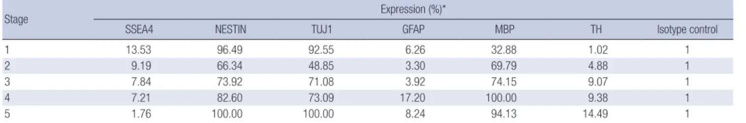
at stage 5. GFAP-positive cells made up 6.26% at stage 1 and the population did not show a significant change in number until stage 5. The initial MBP-positive cell population was 32.88% and reached 94.13% at the final stage. In the case of TH-positive cells, the initial expression level was very low (1.02%), but the popu- lation increased significantly during the experiment, giving a fi- nal value of 14.49% (Table 2). The relative expression percent- age of each value was compared with mouse IgG1 isotype as the control.
Genotypic analyses of hDPSCs by real-time PCR
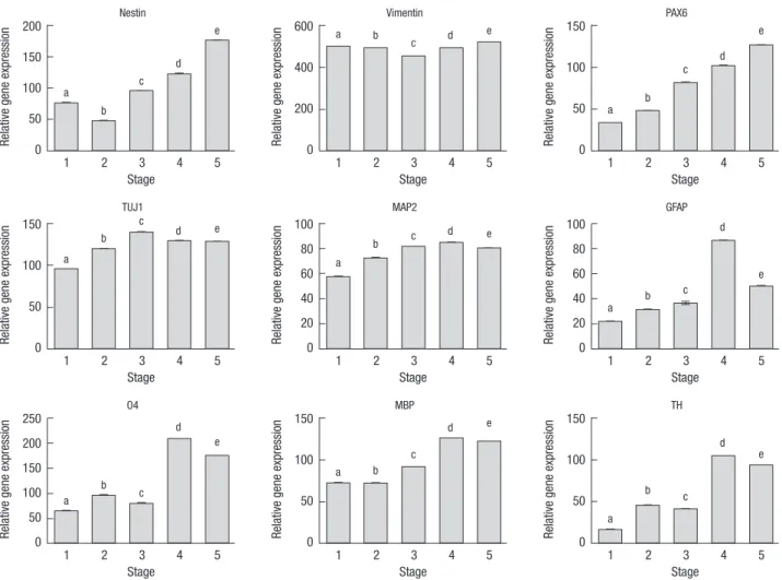
The expression of Vimentin (neural stem cell marker) was con- stant, whereas the expressions of NESTIN and PAX6 (neural lineage marker) increased gradually over the course of time.
TUJ1, MAP2, GFAP, MBP, O4 (oligodendrocyte marker), and TH showed maximized expressions at stage 4 (Fig. 2).
Identification of differentiated hDPSCs by TH ELISA The quantification of TH from cell lysates was evaluated with an ELISA kit (Fig. 3). As compared with stage 1 (0.110 ± 0.032 ng/mL), the TH value at stage 5 increased to 0.526 ± 0.033 ng/
mL. The P value for the two-tailed test was less than 0.0001. These results suggest that hDPSC-derived dopaminergic neurons pos- sess the ability to synthesize dopamine.
Table 2. Flow cytometric results of target genes through differentiational stages of hDPSCs
Stage Expression (%)*
SSEA4 NESTIN TUJ1 GFAP MBP TH Isotype control
1 13.53 96.49 92.55 6.26 32.88 1.02 1
2 9.19 66.34 48.85 3.30 69.79 4.88 1
3 7.84 73.92 71.08 3.92 74.15 9.07 1
4 7.21 82.60 73.09 17.20 100.00 9.38 1
5 1.76 100.00 100.00 8.24 94.13 14.49 1
*Each expression percentage is relative value, compared with mouse IgG1 isotype control.
DISCUSSION
Currently, hDPSCs have been shown to be able to differentiate into neural precursors or neurotrophic support for dopaminer- gic neurons, but not into mature dopaminergic neurons (13). In the present study, we have provided novel evidence that dopa- minergic neurons and several neuronal cells can be generated from hDPSCs by in vitro stimulation. To explore the differentia- tion of hDPSCs, specific markers of the various neuron devel- opmental stages were selected; namely, SSEA4 (a marker for mesenchymal stem cells), Nestin (a neural stem cell and pro- genitor cell marker), Pax6 (a neuronal lineage marker), TUJ1 (an early marker for neurons), MAP2 (a marker for mature neu- rons), GFAP (a mature neuron marker for astrocytes), MBP and O4 (mature neuron markers for oligodendrocytes), and TH (an enzyme required for the generation of dopamine and thus con- sidered as a marker for dopaminergic neurons) (14,15).
Protein level analysis of the differentiating hDPSCs was veri- fied by immunocytochemical analysis. The type and level of protein expression for neural-specific markers depend on the Fig. 2. Genotypic analyses of human dental pulp stem cells (hDPSCs) according to the various differentiation stages by real-time PCR. The different letters on top of the bars show significant differences at P < 0.05. P values are for the analysis of variance Tukey’s test, n = 4.
Relative gene expression
Stage
1 2 3 4 5 200
150 100 50 0
Nestin
a b
c d
e
Relative gene expression
Stage
1 2 3 4 5 600
400
200
0
Vimentin
a b
c d e
Relative gene expression
Stage
1 2 3 4 5 150
100
50
0
PAX6
a b
c d
e
Relative gene expression
Stage
1 2 3 4 5 150
100
50
0
TUJ1
a b
c d e
Relative gene expression
Stage
1 2 3 4 5 100
80 60 40 20 0
MAP2
a
b c d e
Relative gene expression
Stage
1 2 3 4 5 100
80 60 40 20 0
GFAP
a b c
d
e
Relative gene expression
Stage
1 2 3 4 5 250
200 150 100 50 0
O4
a
b c
d e
Relative gene expression
Stage
1 2 3 4 5 150
100
50
0
MBP
a b
c
d e
Relative gene expression
Stage
1 2 3 4 5 150
100
50
0
TH
a
b c
d e
Fig. 3. Tyrosine hydroxylase (TH) synthesis by human dental pulp stem cells (hDPSCs) at stage 1 and 5 through ELISA analysis. n = 4, t-test. *P < 0.001.
ng/mL
Stage
1 5
0.6
0.4
0.2
0
* TH ELISA
various differentiation stages. Expression of the stem cell maker was observed at the early stages, but the frequency decreased gradually. Expressions of the early neural stem cell marker and early neural marker remained steady throughout the various stages. The mature neural markers were highly expressed from stage 3 onward. Most importantly, expression of the dopami- nergic neuron marker was found at stage 5. To confirm the im- munocytochemistry results, we performed a flow cytometry analysis. Quantitative cell count analyses indicated that of the treated hDPSCs, 100% expressed Nestin and TUJ1, 8.24% ex- pressed GFAP, and 94.13% expressed MBP, and of this popula- tion, 14.49% expressed TH. This result was consistent with the immunocytochemistry experiments.
Real-time PCR analysis indicated that when hDPSCs were cultured in neuronal inductive medium, the Nestin transcript levels appeared to be upregulated from stage 3 onward, where- as there was only a minor alteration in the expression of VIMEN- TIN. The PAX6, TuJ1, MAP2, O4, MBP, and TH transcript levels were all increased in the differentiation medium, which indi- cated that hDPSC-derived spheres could generate mature neu- rons and a number of committed differentiated dopaminergic neurons. Interestingly, the induced hDPSCs expressed high lev- els of MBP and O4 transcripts, but low levels of GFAP, which is consistent with the ability of the induction cocktail to trans-dif- ferentiate the hDPSCs into an oligodendrocyte lineage. At stage 5, GFAP expression had decreased to about 8%, meaning that the astrocyte population was relatively low.
To determine whether the induced hDPSCs release TH, we measured the intracellular levels of TH at stages 1 and 5, using an ELISA kit. The TH level was significantly enhanced at stage 5, increasing to 0.526 ± 0.033 ng/mL from 0.110 ± 0.032 ng/mL at stage 1. This indicated that hDPSCs could potentially differenti- ate into dopaminergic neuronal-like cells in an appropriate en- vironment. This 5-step method produced about 1.02% TH-pos- itive neurons at an efficiency of about 92.55% of TUJ1-positive neurons in the presence of ITS in the medium. Moreover, when FGF basic, FGF-8b, Shh-N, and ascorbic acid were added to the medium, the production increased to about 14.5% TH-positive neurons at an efficiency of 100% of TUJ1-positive neurons. TH was expressed in hDPSCs even in the absence of the differenti- ation factors retinoic acid and brain-derived neurotrophic fac- tor (16).
Signaling with trophic factors plays an important role in the differentiation of stem cells (16). During the procedure, we sim- ulated the induction and differentiation of hDPSCs using sev- eral growth factors. For the preinduction stage 1 of hDPSCs, me- dium containing β-mercaptoethanol, serum, and LIF was used (17). β-Mercaptoethanol and serum are survival factors that support the viability and differentiation of neurons (18,19). LIF is required for appropriate changes in neuronal gene expres- sion to ensure the survival and maintenance of neurons (20), as
well as to promote neural stem cell self-renewal, preventing the emergence of more differentiated cell types (21). hDPSCs form- ed the neurosphere in the KO-ES medium without LIF/retinoic acid (RA) at stage 2. LIF impairs neurosphere formation in vitro, even at a very low dose (21). Moreover, the hDPSCs showed neurosphere formation without RA treatment. The avoidance of RA treatment is based on the fact that RA is a negative factor for dopaminergic neuron generation (22). The neurosphere is a nonadhesive aggregate that is composed of free-floating clus- ters of neural precursor cells of varying cell types (heterogene- ous), including neurons, astrocytes, and oligodendrocytes (23).
The neurosphere-like aggregates of stem cells are an important property of neural stem cells (24). This formation of floating spheres under the appropriate medium means that hDPSCs might possess the potential to differentiate into neural stem cells. In the neurosphere stage, the SSEA4, Nestin, TuJ1, and GFAP expression levels were decreased, while those of MBP and TH were increased, meaning that the neurosphere niche is different to that of the stage 1 environment.
After the neurosphere had attached to the plate, neural pro- genitor cells spread out into a monolayer. At stage 3, ITS and N-2 were used to select and enrich for neural stem cell popula- tions. The included fibronectin provides support for the cell at- tachment and spreading. The FGF and Shh-N contained in the medium induce effective neuronal differentiation because with- out these factors, neuronal morphology is not observed (25). In the late stages, hDPSC-derived spheres were induced into do- paminergic neurons with a cocktail of Shh-N, FGF-8b, FGF ba- sic, and ascorbic acid. These are thought to work in unison to mediate the induction of dopaminergic cells (26). Under this condition, the hDPSC-derived spheres generated a number of TuJ1- and MAP2-positive neurons, and at the final step, some of them were TH-positive cells, suggesting that the controlled dif- ferentiation of hDPSCs by the growth factor cocktail had led to the activation of active TH and the induction of functional neu- rons (24). Wang et al. (24) showed that TH was expressed only in neurospheres induced by a cocktail of these growth factors.
In this study, we have identified the potential dopaminergic differentiation of hDPSCs, which was confirmed by morpho- logical, immunocytochemical, flow cytometric, real-time PCR, and ELISA analyses. The induced TH-positive neurons were shown of TH. These results suggest that hDPSC-derived dopa- minergic neurons possess the ability to synthesize dopamine.
Most of the dopaminergic differentiation studies have used ES cells up to now. However, our study suggests that hDPSCs can be a promising autologous cell source for the treatment of PD.
For further study, it will be important to determine whether these cells possess the electrophysiological characteristics of neu- rons, as well as to investigate the therapeutic efficacy of hDPSCs in alleviating PD in an animal model through transplantation.
DISCLOSURE
The authors have no potential conflicts of interest to disclose.
AUTHOR CONTRIBUTION
Conception and design of study: Kwon TG, Chun SY. Perform- ing the experiments: Jang YJ. Data analysis: Choi SH. Drafting of the manuscript: Yoo ES, Chun SY. Critical revision of the manu- script for important intellectual content: Kwon TG, Soker S. Sta- tistical analysis: Jang YJ. Receiving grant: Yoo ES.
ORCID
So Young Chun http://orcid.org/0000-0003-4500-4956 Shay Soker http://orcid.org/0000-0002-8458-9232 Yu-Jin Jang http://orcid.org/0000-0001-5660-3001 Tae Gyun Kwon http://orcid.org/0000-0002-4390-0952 Eun Sang Yoo http://orcid.org/0000-0002-7442-6886 REFERENCES
1. Dauer W, Przedborski S. Parkinson’s disease: mechanisms and models.
Neuron 2003; 39: 889-909.
2. Mandel S, Grünblatt E, Riederer P, Gerlach M, Levites Y, Youdim MB.
Neuroprotective strategies in Parkinson’s disease: an update on progress.
CNS Drugs 2003; 17: 729-62.
3. Kriks S, Shim JW, Piao J, Ganat YM, Wakeman DR, Xie Z, Carrillo-Reid L, Auyeung G, Antonacci C, Buch A, et al. Dopamine neurons derived from human ES cells efficiently engraft in animal models of Parkinson’s disease. Nature 2011; 480: 547-51.
4. Hayashi T, Wakao S, Kitada M, Ose T, Watabe H, Kuroda Y, Mitsunaga K, Matsuse D, Shigemoto T, Ito A, et al. Autologous mesenchymal stem cell- derived dopaminergic neurons function in parkinsonian macaques. J Clin Invest 2013; 123: 272-84.
5. Swijnenburg RJ, Schrepfer S, Govaert JA, Cao F, Ransohoff K, Sheikh AY, Haddad M, Connolly AJ, Davis MM, Robbins RC, et al. Immunosuppres- sive therapy mitigates immunological rejection of human embryonic stem cell xenografts. Proc Natl Acad Sci U S A 2008; 105: 12991-6.
6. de Wert G, Mummery C. Human embryonic stem cells: research, ethics and policy. Hum Reprod 2003; 18: 672-82.
7. Wang Y, Chen S, Yang D, Le WD. Stem cell transplantation: a promising therapy for Parkinson’s disease. J Neuroimmune Pharmacol 2007; 2: 243- 50.
8. Lizier NF, Kerkis A, Gomes CM, Hebling J, Oliveira CF, Caplan AI, Ker- kis I. Scaling-up of dental pulp stem cells isolated from multiple niches.
PLoS One 2012; 7: e39885.
9. Gronthos S, Mankani M, Brahim J, Robey PG, Shi S. Postnatal human dental pulp stem cells (DPSCs) in vitro and in vivo. Proc Natl Acad Sci U S A 2000; 97: 13625-30.
10. Khanna-Jain R, Vanhatupa S, Vuorinen A, Sandor GK, Suuronen R, Man-
nerstrom B, Miettinen S. Growth and differentiation of human dental pulp stem cells maintained in fetal bovine serum, human serum and se- rum-free/xeno-free culture media. J Stem Cell Res Ther 2012; 2: 1-11.
11. Miura M, Gronthos S, Zhao M, Lu B, Fisher LW, Robey PG, Shi S. SHED:
stem cells from human exfoliated deciduous teeth. Proc Natl Acad Sci U S A 2003; 100: 5807-12.
12. Zhang W, Walboomers XF, Shi S, Fan M, Jansen JA. Multilineage differ- entiation potential of stem cells derived from human dental pulp after cryopreservation. Tissue Eng 2006; 12: 2813-23.
13. Nosrat IV, Smith CA, Mullally P, Olson L, Nosrat CA. Dental pulp cells provide neurotrophic support for dopaminergic neurons and differenti- ate into neurons in vitro; implications for tissue engineering and repair in the nervous system. Eur J Neurosci 2004; 19: 2388-98.
14. Klein C, Fishell G. Neural stem cells: progenitors or panacea? Dev Neu- rosci 2004; 26: 82-92.
15. Guo L, Yin F, Meng HQ, Ling L, Hu-He TN, Li P, Zhang CX, Yu S, Duan DS, Fan HX. Differentiation of mesenchymal stem cells into dopaminer- gic neuron-like cells in vitro. Biomed Environ Sci 2005; 18: 36-42.
16. Sanchez-Ramos J, Song S, Cardozo-Pelaez F, Hazzi C, Stedeford T, Will- ing A, Freeman TB, Saporta S, Janssen W, Patel N, et al. Adult bone mar- row stromal cells differentiate into neural cells in vitro. Exp Neurol 2000;
164: 247-56.
17. Woodbury D, Schwarz EJ, Prockop DJ, Black IB. Adult rat and human bone marrow stromal cells differentiate into neurons. J Neurosci Res 2000;
61: 364-70.
18. Katayama M, Ishii K. 2-Mercaptoethanol-independent survival of fetal mouse brain neurons cultured in a medium of human serum. Brain Res 1994; 656: 409-12.
19. Ishii K, Katayama M, Hori K, Yodoi J, Nakanishi T. Effects of 2-mercapto- ethanol on survival and differentiation of fetal mouse brain neurons cul- tured in vitro. Neurosci Lett 1993; 163: 159-62.
20. Sun Y, Zigmond RE. Involvement of leukemia inhibitory factor in the in- creases in galanin and vasoactive intestinal peptide mRNA and the de- creases in neuropeptide Y and tyrosine hydroxylase mRNA in sympathet- ic neurons after axotomy. J Neurochem 1996; 67: 1751-60.
21. Bauer S, Patterson PH. Leukemia inhibitory factor promotes neural stem cell self-renewal in the adult brain. J Neurosci 2006; 26: 12089-99.
22. Kawasaki H, Mizuseki K, Nishikawa S, Kaneko S, Kuwana Y, Nakanishi S, Nishikawa SI, Sasai Y. Induction of midbrain dopaminergic neurons from ES cells by stromal cell-derived inducing activity. Neuron 2000; 28:
31-40.
23. Bez A, Corsini E, Curti D, Biggiogera M, Colombo A, Nicosia RF, Pagano SF, Parati EA. Neurosphere and neurosphere-forming cells: morphologi- cal and ultrastructural characterization. Brain Res 2003; 993: 18-29.
24. Wang J, Wang X, Sun Z, Wang X, Yang H, Shi S, Wang S. Stem cells from human-exfoliated deciduous teeth can differentiate into dopaminergic neuron-like cells. Stem Cells Dev 2010; 19: 1375-83.
25. Trzaska KA, Kuzhikandathil EV, Rameshwar P. Specification of a dopa- minergic phenotype from adult human mesenchymal stem cells. Stem Cells 2007; 25: 2797-808.
26. Ye W, Shimamura K, Rubenstein JL, Hynes MA, Rosenthal A. FGF and Shh signals control dopaminergic and serotonergic cell fate in the anteri- or neural plate. Cell 1998; 93: 755-66.