38(8), 1003~1007(2009) DOI: 10.3746/jkfn.2009.38.8.1003
부추의 함황화합물이 인체 암세포 증식에 미치는 영향
박순영1․김재용2․박경욱1․강갑석3․박기훈4․서권일1†
1
순천대학교 식품영양학과,
2경북대학교 식품공학과
3
부산정보대학 호텔조리과,
4경상대학교 환경생명화학과
Effects of Thiosulfinates Isolated from Allium tuberosum L.
on the Growth of Human Cancer Cells
Sun-Young Park1, Jae-Yong Kim2, Kyung-Wuk Park1, Kap-Suk Kang3, Ki-Hun Park4, and Kwon-Il Seo1†
1Dept. of Food and Nutrition, Sunchon National University, Jeonnam 540-742, Korea
2Dept. of Food Science and Technology, Kyungpook National University, Daegu 702-701, Korea
3Dept. of Hotel Culinary Arts, Busan College of Information Technology, Busan 616-737, Korea
4Dept. of Environmental Biotechnology, Gyeongsang National University, Gyeongnam 660-701, Korea
Abstract
To develop Allium tuberosum L. as a cancer preventive food material, thiosulfinates and biological active components were isolated from Allium tuberosum L. and the apoptotic effects of thiosulfinates in human cancer cells were examined. Thiosulfinates decreased viable cell numbers in dose- and time-dependent manners.
Thiosulfinates at the 20 μg/mL concentration inhibited more than 60% cell proliferation in HepG2 and A549 human cancer cells, respectively. Also the morphology of cells treated with thiosulfinates of 30 μg/mL concentration was distorted with shrunken cell mass while the cell number was lower than that of control cells.
The IC
50values in the HepG2 cells were higher than those of the A549 cells. Thiosulfinates at the 30 μg/mL concentration showed the formation of apoptotic bodies and a nuclear condensation, and an increase in the cell populations of the sub-G1 phase in the HepG2 cells. These results indicate that thiosulfinates from Allium tuberosum L. inhibited cell proliferation in HepG2 via apoptosis.
Key words: Allium tuberosum L., thiosulfinates, cytotoxic activity, apoptosis
†
Corresponding author. E-mail: [email protected]
†
Phone: 82-61-750-3655, Fax: 82-61-752-3657
서 론
최근 우리나라는 서구화된 식생활, 환경오염, 운동부족 및 스트레스 등의 여러 가지 원인으로 암, 비만, 당뇨병 및 심혈 관질환 등과 같은 만성퇴행성질환이 증가하고 있다(1). 특히 이들 만성퇴행성질환 중에서 암은 2008년 통계자료에 의하 면 전체 인구 사망원인의 1위라고 보고하고 있다(2). 현재 암에 대한 치료는 대부분 항암제를 이용한 치료법이 이용되 고 있으나 면역기능 이상 및 암세포 이외의 독성을 나타내고 있어 선택적인 항암제 개발이 요구되고 있는 실정이다. 최근 에는 암세포에만 선택적으로 작용할 수 있는 항암제를 천연 물로부터 개발하려는 연구들이 진행되고 있다(3).
부추(
Allium tuberosum)는 백합과
Allium속에 속하는 다년생 초본으로 주로 우리나라 및 일본, 중국 등에 분포하 고 있다(4,5). 예로부터 부추는 특유의 맛과 향취에 의해 기 호도가 높은 향신채소로 재배되어져 왔으며, 한방에서는 맛
이 맵고 성질이 따뜻하여 소화관과 혈액순환에 좋으며, 민간
요법으로 소염, 해독작용 및 지혈작용이 있다고 알려져 약재
로도 많이 사용되어져 왔다(6). 부추의 주요 영양성분으로는
카로틴, 비타민 B
2, 비타민 C, 칼슘, 철 등을 함유하고 있고,
주요 성분으로는 allyl sulfide, pentose 및 allylthiamine, 여
러 가지 sulfide 유도체와 adenosine, alanine, glutamic acid,
aspartic acid, valine 등 아미노산, dimethyl disulfide와 di-
methyl trisulfide 같은 8가지 지방족 함황화합물 등이 함유
되어 있는 것으로 확인되었다(7,8). 최근 부추의 생리활성에
관한 연구로는 quinone reductase 유도활성을 이용한 항암
연구(9,10), 항균효과(11), 김해부추 및 포항 부추의 SOD유
사활성 측정(12) 및 항산화효과(13,14) 등에 대한 연구 결과
가 보고되고 있으나 부추의 유용성분을 암 예방 기능성식품
으로 이용하기 위해서는 더 많은 연구가 진행되어야 할 것이
며, 특히 부추의 생리활성물질인 thiosulfinate에 대한 연구
가 필수적이라 생각된다. 그러나 부추의 항암효과에 대한
O O
↑ ↑
H
3C-S-S-CH
3H
3C-S-S-CH
2-CH=CH
2(1) (2)
Fig. 1. Structures of S-methyl methanethiosulfinate (1) and S-methyl 2-propene-1-thiosulfinatepurified (2) isolated from Allium tuberosum L.
기존의 연구는 주로 추출물에 관한 연구가 진행되어 왔으며, 부추의 생리활성물질인 thiosulfinates에 대한 연구는 거의 없는 실정이다.
따라서 본 연구자는 이전의 연구에서 부추로부터 thio- sulfinates를 분리한 바 있으며(15,16)(Fig. 1), 본 연구에서는 부추의 항암효과를 규명하기 위하여 함황화합물을 이전의 연구 방법과 동일하게 분리하여 인체 암세포의 사멸이 apoptosis에 의해서 유도되는 지를 조사하였다.
재료 및 방법
실험재료 및 부추 함황화합물 분리
본 실험에 사용된 부추(
Allium tuberosum)는 전라남도 나주에서 수확한 것을 구입하여 4 kg 가식부만 잘 세정하여
± 1 cm 크기로 자른 후 7일 동안 CH
2Cl
2에 2회 침지 추출하 여 솜으로 여과하였으며, 엽록소 및 수분을 제거하기 위하여 lead(Ⅱ) acetate trihydrate 15%로 2~3회 분액한 후 CH
2Cl
2층만 취한 후 무수황산나트륨을 사용하여 잔여수분을 제거 하여 완전히 농축한 후 물을 첨가하여 용해하는 성분만을 취하여 시료로 사용하였다(17).
암세포 증식 억제효과
본 실험에 사용한 간암세포주인 HepG2(human liver cancer)와 폐암세포주인 A549(human lung cancer)를 한국 세포주은행(KCLB)에서 분양 받아 10% FBS(fetal bovine serum)를 첨가한 RPMI 1640(Gibco, California, USA)배지 를 첨가하여 37
oC, 5% CO
2incubator에서 계대배양하면서 실험에 사용하였다.
Monolayer로 자란 암세포주를 0.25% trypsin-EDTA용 액으로 처리하여 single cell로 만든 후 최종농도가 1×10
5cells/mL가 되도록 희석하여 24 well plate에 분주한 다음 37
oC, 5% CO
2incubator에서 24시간 동안 배양한 후, 부추 함황화합물을 1, 5, 10, 20 및 30 μg/mL 농도로 첨가하여 24, 48 및 72시간 반응시켜 암세포 증식 정도를 SRB(sulfo- rhodamine B)(Sigma, Missouri, USA)방법에 의하여 측정 하였다(18).
암세포 형태의 관찰
대조군과 실험군의 암세포 모양 변화를 관찰하기 위하여 암세포증식 억제능을 측정하기 전에 위상차 현미경으로 세
포의 형태학적 변화를 관찰하였다.
SubG1의 변화량 측정
Monolayer로 배양한 세포주를 0.25% trypsin-EDTA용 액으로 처리하여 single cell로 만든 후 최종농도가 1×10
6cells/mL가 되도록 희석하여 6 well plate에 분주한 후 37
oC, 5% CO
2incubator에서 24시간 배양하였다. 24시간 후 부추 함황화합물을 1, 5 및 10 μg/mL 농도로 처리하여 48시간 더 배양시켰다. 암세포를 PBS(phosphate buffered saline)로 2회 세척한 후 70% 에탄올을 첨가하고 4
oC에서 하루 동안 방치하였다. 고정된 암세포를 PBS로 세척하고 RNase(0.1 mg/mL)를 첨가하여 37
oC 30분 반응시킨 후 1 mg/mL의 PI(propidium iodide) 용액으로 30분간 염색한 후 Flow cy- tometer(EPICS XL, Backman Coulter, California, USA)를 이용하여 세포주기를 분석하였다(18).
부추 함황화합물 처리에 따른 암세포 핵의 형태변화 부추 함황화합물의 처리에 의한 간암세포의 apoptosis 유 발 여부를 확인하기 위한 핵의 형태변화 관찰을 위하여 부추 함황화합물을 30 μg/mL 농도를 처리하여 48시간 배양하였 다. 배양이 종료된 well에서 회수한 세포를 PBS로 3회 세척 하고 4% paraformaldehyde로 상온에서 10분간 고정하였다.
염색이 용이하도록 0.1 % Triton-X 100을 10분간 처리하여 세포막을 용해한 후 형광 염색물질인 DAPI(4,6-diamidino- 2-phenylindole, 2 μg/mL)용액을 이용하여 20분간 염색하였 다. 이들 세포를 다시 PBS로 2회 수세한 후 형광현미경을 이용하여 핵의 형태 변화를 관찰하였다(×400 배율)(18).
통계처리
실험결과는 평균(mean)±표준편차(SD)로 나타내었으며, Student's
t-test를 이용하여 통계처리한 후 p<0.05 수준에 서 유의성을 검증하였다.
결과 및 고찰
부추 함황화합물의 암세포 증식 억제효과
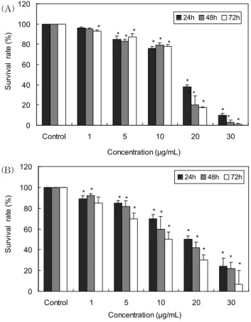
부추 함황화합물이 인체 간암세포(HepG) 및 폐암세포 (A549)의 증식을 억제하는지를 측정한 결과는 Fig. 2와 같 다. 즉 두 종류의 암세포들은 부추 함황화합물을 처리하지 않은 대조군에 비하여 부추 함황화합물을 20 μg/mL 농도로 처리하여 24시간 후 측정한 결과 암세포의 성장을 60% 이상 억제하였으며, 농도 및 시간 의존적으로 이들 암세포의 성장 을 억제하였다. 또한 이들 암세포에 대한 부추 함황화합물의 IC
50값을 측정한 결과는 Table 1과 같다. 즉 HepG2 세포의 경우 함황화합물의 IC
50값은 24, 48 및 72시간 처리 시 각각 16.98, 15.82 및 14.78 μg/mL로 나타났으며, A549세포의 경 우는 각각 40.15, 32.3 및 31.17 μg/mL로 나타났다.
Park 등(19)은 부추 메탄올 추출물 및 분획물이 HepG2
0 20 40 60 80 100 120
Control 1 5 10 20 30
Concentration (μg/mL)
S u rvi va l r a te ( % ) .
24h 48h 72h
*
* * *
* * *
*
*
*
*
* * (A)
*
*
0 20 40 60 80 100 120
Control 1 5 10 20 30
Concentration (μg/mL)
S u rvi va l r a te ( % ) .
24h 48h 72h
* * * *
* * *
* *
*
* * *
* (B)
Fig. 2. Effects of thiosulfinates isolated from Allium tuber- osum L. on the proliferation of human cancer cells for 24, 48, and 72 hr. (A) HepG2, (B) A549. Data values were expressed as mean±SD of triplicate determinations. Significant differences were compared with the control group at
*p<0.05 by Student's t-test.
Table 1. IC
50values of thiosulfinates isolated from Allium tuberosum L. on the HepG2 and A549 cells
Cells IC
50(μg/mL) of thiosulfinates
24 hr 48 hr 72 hr
HepG2
A549 16.98±0.38
40.15±6.28 15.82±1.08
32.3±2.02 14.78±0.27 31.17±5.77 Data values were expressed as mean±SD of triplicate de- termination.
및 4종의 암세포주 성장 억제 효과가 있다고 보고한 바 있다.
또한 본 연구자는 이전의 부추로부터 주요 생리활성 물질인 thiosulfinates를 분리하여 S-methylmethanthiosulfinates, S-methyl 2-propene-1-thiosulfinates 화합물들이 함유되 어 있음을 확인하였으며, 이들 thiosulfinates들은 각종 인체 암세포 성장을 억제하였을 뿐만 아니라 복수암을 유발시킨 마우스의 수명 연장효과가 있다고 보고하였다(15).
따라서 이전의 연구결과와 본 연구결과를 종합해 볼 때 부추 함황화합물에는 암을 억제할 수 있는 주요 생리활성 물질들이 함유되어 있으므로 추후 암 예방 식품으로 활용 가능성이 있을 것으로 사료되어진다.
암세포의 형태학적 변화
부추 함황화합물에 따른 인체 간암세포와 폐암세포의 형
Fig. 3. Inverted photomicrograph of human cancer cells treated with thiosulfinates isolated from Allium tuberosum L. for 48 hr (×200). (A) Not-treated (HepG2), (B) HepG2 cells treated with 30 μg/mL of thiosulfinates for 48 hr, (C) Not-treated (A549), (D) A549 cells treated with 30 μg/mL of thiosulfinates for 48 hr.
태학적 변화를 확인하기 위해 함황화합물을 30 μg/mL 농도 로 처리하여 48시간 후 위상차 현미경으로 관찰한 결과는 Fig. 3과 같다. 즉 무처리군 세포의 경우 culture plate에 안정 적으로 부착되어 정상적인 증식이 이루어진 모습인 반면, 부추 함황화합물 30 μg/mL을 처리 시 암세포의 증식이 감소 하였으며, culture plate로부터 분리되어 배양액에 부유한 세 포는 일정한 모양을 잃고 크기가 줄어 사멸되는 것으로 관찰 할 수 있었다.
부추 함황화합물 처리에 의한 암세포의 subG1 변화량 측정
암세포 중 부추 함황화합물에 의한 성장 억제효과가 높았 던 간암세포(HepG2)의 사멸이 apoptosis에 의해 유도되는 지를 확인하기 위하여 부추 함황화합물을 1, 5 및 10 μg/mL 농도로 처리하여 48시간 반응시킨 후 flow cytometer를 통 하여 subG1 변화량을 측정하였다(Fig. 4). 즉 대조군은 subG1의 분포가 0.87로 나타났으나, 부추 함황화합물을 처 리 시 subG1 변화량이 농도 의존적으로 증가하였으며, 간암 (HepG2)세포에 함황화합물을 10 μg/mL 농도로 첨가 시 34.03로 크게 증가되어 부추 함황화합물이 apoptosis를 유도 하여 간암세포의 증식을 억제하는 것을 추측할 수 있었다.
Park 등(15)은 부추 함황화합물을 5, 10, 20 및 30 μg/mL 농도로 유방암세포(MCF-7)처리하여 subG1 변화량을 측정 한 결과 농도 의존적으로 증가하였으며, 유방암세포의 사멸 이 apoptosis에 의해 유도되어진다고 보고하였다.
또한 Park 등(20)은 부추와 함께 allicin을 위암세포에 처
리하여 flow cytometry를 통해 세포주기를 분석한 결과 세
포주기 중 apoptosis와 관련한 subG1의 분포가 농도증가에
따라 증가한다고 보고하여 본 연구결과와 유사한 경향을 나
0.87 1.47
4.63
34.03
0 10 20 30 40 50
Control 1 5 10
Concentration (μg/mL)
su b G 1 P h a se ( % ) . *
Fig. 4. Sub-G1 population of HepG2 cells treated with thio- sulfinates isolated from Allium tuberosum L. for 48 hr. Data values were expressed as mean±SD of triplicate determinations.
Significant differences were compared with the control group at
*
p<0.05 by Student's t-test.
타내었다.
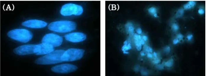
부추 함황화합물 처리에 따른 암세포 핵의 형태변화 부추 함황화합물에 의한 암세포의 사멸이 apoptosis에 의 하여 유도되는 지를 확인하기 위하여 간암암세포(HepG2)에 함황화합물을 30 μg/mL 농도로 처리하여 DAPI 염색을 이 용하여 핵의 형태변화를 확인하였다(Fig. 5). 즉 부추 함황화 합물을 처리하지 않은 대조군은 간암세포의 핵이 손상 없이 일정한 반면, 부추 함황화합물을 처리하였을 때 핵이 손상되 어 apoptosis의 특징 중 하나인 apoptotic bodies를 관찰할 수 있었다.
Park 등(20)은
Allium속인 마늘의 주요 성분인 allicin을 인체 위암세포(AGS)에 처리한 후 DAPI 염색을 통하여 핵 의 형태변화를 형광현미경으로 관찰한 결과 allicin의 농도 가 증가할수록 핵이 응축되고, DNA fragmentation 및 apoptotic body를 보여 allicin이 apoptosis를 유도한다고 보 고하였다. 또한 본 연구자의 이전 연구결과에 의하면 부추 함황화합물들(thiosulfinates)은 인체전립선 암세포(RC58T/
hSA/#4, LNcap, PC-3, DU145) 성장을 15 μg/mL 농도 이하 에서 강하게 억제하였으며, 이들 사멸이 caspase 의존 및 비의존 apoptosis 경로에 의해 유도된다고 보고한 바 있다 (16). 따라서
Allium속에 속하는 채소들의 주요성분인 함황
(B) (A)
Fig. 5. Nuclear fragmentation in the HepG2 cells treated with thiosulfinate isolated from Allium tuberosum L. for 24 hr.
(A) Not-treated, (B) HepG2 cells treated with 30 μg/mL of thio- sulfinate for 24 hr.
화합물들은 인체 암세포의 성장이 apoptosis에 의해 억제되 는 것을 확인할 수 있으며, 앞으로 본 연구 결과를 토대로 부추의 사멸기전에 대한 더 구체적인 연구들이 많이 진행되 어져 부추를 암 예방 식품으로 활용할 수 있는 연구 자료들 을 확립해야 할 것으로 사료된다.
요 약
부추를 암 예방 식품 소재로 활용하기 위하여 부추로부터 주요 생리활성 물질인 함황화합물을 분리하여 인체 암세포 성장 억제 및 간암세포의 사멸이 apoptosis에 의해 유도되는 지를 조사하였다. 부추 함황화합물을 1, 5, 10, 20 및 30 μg/
mL 농도로 24, 48 및 72시간별로 간암(HepG2) 및 폐암세포 (A549)에 처리하여 암세포 증식억제 효과를 측정한 결과 HepG2 및 A549 세포에서 농도 및 시간 의존적으로 그 성장 을 억제하였으며, 20 μg/mL 농도 이상에서 암세포 성장이 60% 이상 억제되었다. 또한 부추 함황화합물 30 μg/mL 농 도로 처리 시 대조군에 비하여 폐암 및 간암 세포수 감소 및 심한 형태학적 변화가 관찰되었다. 이들 암세포의 IC
50값을 측정한 결과 부추 함황화합물은 폐암세포(A549)보다 간암세포(HepG2)에 더 효과가 있었다. 한편 부추 함황화합 물은 30 μg/mL 농도에서 핵 응축 및 apoptotic body를 나타 내었으며, 농도 의존적으로 subG1 DNA 함량이 증가함으로 써 HepG2 암세포 사멸이 apoptosis에 의해 유도되는 것을 확인할 수 있었다.
감사의 글
본 연구는 2004년도 순천대학교 자체 연구과제 지원 사업 에 의하여 수행된 연구 결과의 일부이며, 그 지원에 감사드 립니다.
문 헌