• 검색 결과가 없습니다.

Cosmetic Potency of Puerariae Radix in Dermal Fibroblasts

N/A
N/A
Protected

Academic year: 2021

Share "Cosmetic Potency of Puerariae Radix in Dermal Fibroblasts"

Copied!
5
0
0

로드 중.... (전체 텍스트 보기)

전체 글

(1)

Introduction

Interaction between epidermis and dermis plays an important role in the functioning of the skin, including wound healing and hair follicle formation

1,2)

. Within skin dermis the most abundant cell type is fibroblasts, the primary role is to secrete components of the extracellular matrix

3)

. There are several types of skin fibroblast within the dermis, which can be defined by their spatial location, and exist as morphologically and functionally heterogenous subpopulations

3-5)

.

Dermal fibroblasts produce the protein molecules including collagen, elastin, laminin, and fibronectin which comprise extracellular matrix. Collagen is a main structural protein in the extracellular space of the body. Collagenases are zinc endopeptidases that digest nearly all collagen fibers in their insoluble triple helical form. Thus, skin wrinkle can be caused by an increase in collagen degradation by collagenase

6-8)

.

Hair loss is a distressing disorder and androgenetic alopecia is the common form of hair loss in both men and women. Circulating testosterone is converted into the more potent dehydrotestosterone (DHT) by 5α-reductase. Thus, inhibiting of 5α-reductase activity is one of the therapeutic

targets for androgenic alopecia

9-11)

.

Radix Puerariae Lobatae is the dried root of Pueraria lobata (Wild.) Ohwi belonging to the family of Fabaceae or Leguminosae, which is a twining perennial herb with woody base native to South East Asia regions, such as Korea, China and Japan

12)

. Puerarin, the first isoflavone isolated from the root of Pueraria lotaba , has been shown to play a pharmacological actions in improving cardiovascular system

12,13)

. Puerariae Radix was first described in the Sinnongbonchogyeong. It is known to have strong antipyretic and slightly sweating efficacy

14,15)

. This herbal medicine has traditionally been used for improving the body function, such as promoting circulation and increasing the blood flow

12,13)

.

In the previous study, ethanol extract of Puerariae Radix (EPR) increased the proliferation of human hair dermal papilla cell (HHDPCs) through phosphorylation of ERK and Akt

16)

. Although mRNA expressions of signaling molecules, such as FGF7, BMP7, and CTNNB1, were induced by water extract of Puerariae Radix (EPR) in HHDPCs

17)

, other pharmacological effects are still unclear. Here, we evaluated whether EPR is able to modulate the activity of collagenase and 5α-reductase. Additionally, it has been shown that EPR promotes cell proliferation through

Cosmetic Potency of Puerariae Radix in Dermal Fibroblasts

Jae Yun Lee

1

, Seo A Park

2

, Won Hong Woo

3,4

, Yeun Ja Mun

1,5

*

1 : Department of Herbal Resources, Graduate School of Oriental Medicine, Wonkwang University, 2 : Department of Beauty Design Graduate School, Wonkwang University,

3 : Department of Anatomy, School of Oriental Medicine, Wonkwang University,

4 : Research Center of Traditional Korean Medicine, Wonkwang University, 5 : Institute of Environmental Science, Wonkwang University Interaction between epidermis and dermis plays an important role in wound healing and hair follicle formation.

This study focused on investigating the potency of ethanol extract of Puerariae Radix (EPR) as cosmetic ingredient using human dermal fibroblasts (hDFn). Our results revealed that EPR suppressed collagenase activity dose-dependently. EPR inhibited activity of 5α-reductase I and II at the final concentration of 25 μg/ml in hDFn cells.

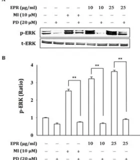
Also, EPR promoted the proliferation and the ERK activation of cells. ERK phosphorylation by EPR was blocked by specific inhibitor of ERK, PD98059. EPR-induced cell proliferation was blocked by PD98059. This means that EPR could promote the proliferation of hDFn cells via the activation ERK. Collectively, these results suggest that EPR may be used as a new cosmetic ingredient.

keywords : Pureraria Lobata , Collagenase, 5α-reductase, ERK, Dermal fibroblast

* Corresponding author

Yeun Ja Mun, Department of Herbal Resources, Graduate School of Oriental Medicine, Wonkwang University, 344-2, Sinyong-dong, Iksan-si, Jeollabuk-do, Korea

·E-mail : [email protected] ·Tel : +82-63-850-6942

·Received : 2019/01/07 ·Revised : 2019/02/15 ·Accepted : 2019/02/20

ⓒ The Society of Pathology in Korean Medicine, The Physiological Society of Korean Medicine

pISSN 1738-7698 eISSN 2288-2529 http://dx.doi.org/10.15188/kjopp.2019.02.33.1.63

Available online at https://kmpath.jams.or.kr

(2)

phosphorylation of ERK in hDFn cells.

Materials and Methods

1. Preparation of herb extract

Puerariae Radix were purchased from Human herbs Inc. (Korea). The Pureraria Lobata (200 g) was soaked in 2 L of 100% ethanol for 3 days at room temperature with sonication for 30 min once a day. The solution was filtrated using a standard sieve (No. 270, 53 μm; Korea), evaporated to dryness at 40℃ under vacuum (Eyela N-11, Japan), and freeze-dried (PVTFD10RS, IlShin BioBase, Korea). The amount of ethanol extract was 8.23 g (yield: 1.65%) and stored in a freezer of Professional Graduate School of Oriental Medicine, Wonkwang University. The extract was dissolved in DMSO, and DMSO was used in the concentration range of less than 0.01% final.

2. Cell culture and Drug treatment

Human dermal fibroblast neonatal (hDFn) cells were obtained from Cascade Biologics (MA, USA). hDFn cells were cultured in DMEM supplemented with 10% FBS, 100 μg/mL streptomycin and 100 units/mL penicillin. The cells were cultured at 37 ℃ in a humidified chamber with 95% air and 5% CO

2

.

3. Cell viability assay using MTT

Cytotoxity was determined by 3-(4,5-dimethylthiazol-2yl) -2,3-diphenyl-tetrazolium bromide (MTT) assay. hDFn cells were seeded onto 24-well plates at a density of 4×10

3

of and 2×10

3

cells/well, respectively. After incubation with various concentrations of EPR for 24 h, 20 μl of MTT (0.1 mg/mL) was added to each culture well and further incubated at 37 ℃ for 3 h. After incubation for 3 h at 37

℃, the MTT solution was removed and 200 μl of DMSO was added and mixtures were shaken, the formazan was dissolved for 10 min. The absorbance of the solubilized formazan was measured at 570 nm using a spectrofluorometer(F-2500, Hitachi, Tokyo, Japan).

4. Western blot analysis

After the indicated EPR, the medium was removed, and the cells were rinsed with PBS twice. After the addition of 0.6 ml of cold RIPA buffer (10 mM Tris pH 7.5, 100 mM NaCl, 1 mM EDTA, 0.5% Na-deoxycholate, 0.1% SDS, 1%

Triton X 100) and protease inhibitors, cells were scraped followed at 4℃. Cell lysate was then subjected to a centrifugation of 14,000 × g for 15 min at 4℃. Resultant

protein samples were separated by a SDS-PAGE gel and transferred onto a polyvinylidine difloride membrane (Millipore, USA). Membrane was stained by ponceu to confirm uniform transfer of all samples and then incubated in blocking solution (TBS with 0.05% tween 20) containing 5% non-fat dry-milk or 3% bovine serum albumin for 1 h at room temperature. The membrane was incubated with 1:1000 diluted primary antibodies and 1:2000 diluted secondary antibodies coupled to horseradish peroxidase (HRP).

Immuno- reactive proteins were detected with the ECL Kit.

The bands were analyzed densitometrically by using a Chemi-doc image analyzer (Bio-Rad, Hercules, CA, USA).

5. Statistical analysis

Values were shown as a mean ± S.D., and the data was performed using analysis of variance followed by the Student's t-test and one-way ANOVA. Values of *p < 0.05, and **p < 0.01 were considered statistically significant.

Results

1. EPR inhibits collagenase activity

Collagen is a key component of extracellular matrix.

Collagenases are enzymes that break the peptide bonds in collagen. Therefore, the effect of EPR on this enzyme activity was evaluated. Collagenase activity was inhibited by EPR treatment in dose-dependently(Fig. 1).

Fig. 1. EPR inhibits collagenase activity. Collagenase activity was measured as described in Materials and Methods. The values shown represent the mean ± SD of triplicate. *p < 0.05, **p < 0.01.

2. EPR decreases 5α-reductase activity in hDFn cells

5α-reductase is responsible for the conversion of testosterone into namely 5α-DHT causing androgen alopecia. The 5α-reductase analysis revealed that 5α -reductase I and II were expressed in hDFn cells (Fig. 2).

Amount of 5α-reductase I was significantly decreased in

EPR treated cells, while amount of 5α-reductase II was

slightly decreased by EPR. The level of 5α-reductase

(3)

expression was reduced by minoxidil (MI) and finasteride (FI) used as a positive control. These results suggest that EPR acts as a 5α-reductase I inhibitor in hDFn cells.

Fig. 2. EPR inhibits 5α-reductases expression in hDFn cells. Cells were treated with EPR (10, 25 μg/ml), MI (10 μM), or FI (10 μM). After 48 h, the cytosol was collected, and the level of 5α-reductase I was analyzed by Western blot.

3. EPR facilitates the proliferation of hDFn cells

To examine the effect of EPR on proliferation, hDFn cells were treated with different doses of EPR (5-25 μg/ml).

After 48 h and 72 h, EPR facilitated the proliferation of cultured hDFn cells in a dose-dependent manner (Fig. 3).

On 72 h, EPR (5, 10 and 25 μg/ml) had increased the cell proliferation by 105.2%, 109.8% and 115.6%, respectively, compared to untreated control. MI (10 μM), used as a positive control, increased the cell proliferation by 110.2%.

In particular, cell proliferation by EPR treatment at 25 μ g/ml concentration was more effective than by MI.

Fig. 3. EPR facilitates the proliferation of hDFn cells. Cells were treated with EPR (5–25 μg/ml) or MI (10 μM) for 48 h or 72 h. The proportion of survival cells were measured by MTT assay. Experiments were repeated in triplicate and the results were expressed as the mean ± SD. *p < 0.05, **p

< 0.01.

4. EPR promotes the proliferation via activation of ERK The ERK pathway is also important in cell proliferation;

the inhibition of ERK has been shown to result in a decrease in cell proliferation

18,19)

. In order to elucidate pathways involved in the cell proliferation of EPR on hDFn cells, ERK response to EPR treatment was investigated. ERK phosphorylation was induced by the treatment with EPR

(Fig. 4A). EPR (10 and 25 μg/ml) facilitated the expression of phosphor-ERK by 2.7 fold and 3.2 fold, respectively. MI (10 μM) also significantly increased phosphor-ERK by 2.5 fold (Fig. 4B).

Fig. 4. EPR promotes phosphorylation of ERK in hDFn cells. (A) Cells were treated with EPR (10, 25 μg/ml) or MI (10 μM) for 30 min. The level of phosphorylation of ERK was analyzed by Western blot. (B) Quantification of active ERK expression in hDFn cells. Experiments were repeated in triplicate and the results were expressed as the mean ± SD. *p

< 0.05, **p < 0.01.

Fig. 5. Activation of ERK by EPR blocks by ERK inhibitor. (A) Immunoblotting analysis of active ERK expression in hDFn cells. Cells were pre-treated with MEK/ERK inhibitor PD98059 (20 μM) for 1 h and incubated in EPR (10, 25 μg/ml) or MI (10 μM) for 30 min. (B) Quantification of active ERK expression in hDFn cells. Experiments were repeated in triplicate and the results were expressed as the mean ± SD. *p

< 0.05, **p < 0.01.

(4)

To confirm the effect of ERK activation by EPR, the inhibitor of ERK, PD98059 (20 μM), was pre-treated in EPR treated hDFn cells. As shown in Fig. 5, ERK phosphorylation by EPR was blocked specific ERK inhibitor. These results suggest that EPR promoted transient activation of ERK in cell.

Next, we investigated whether ERK activation is involved in the cell proliferation effect by EPR. The EPR-induced cell proliferation was reduced by treatment with PD98059(Fig. 6).

These results indicate that EPR facilitated the proliferation via activation of ERK in hDFn cells.

Fig. 6. EPR facilitates the proliferation via activation of ERK in hDFn cells. Cells were pre-treated with ERK inhibitor (PD98059, 20 μM) for 1 h and incubated with EPR (10, 25 μg/ml) or MI (10 μM) for 72 h. The proportion of survival cells were measured by MTT assay. Experiments were repeated in triplicate and the results were expressed as the mean ± SD. *p < 0.05, **p < 0.01.

Discussion

Dermal fibroblasts are cells within the dermis layer in the skin which are responsible for generating connective tissue and allowing the skin to recover from injury. Skin aging is clinically characterized by wrinkles, rough skin texture, and blotchy dyspigmentation

6-8)

. In this study, we demonstrated that EPR can inhibit collagenase activity in hDFn cells.

Although numerous products claim to be useful for treating hair loss, they have sexual-related side-effects and unpredictable efficacy

20,21)

. It has been reported that there are three subtypes of 5α-reductase in prostate: namely 5α -reductase I, 5α-reductase II, and 5α-reductase III. At least two subtypes of 5α-reductase, 5α-reductase I and 5α -reductase II, have been identified in the skin

22,23)

. Various therapeutic options for androgenic alopecia have been introduced, including oral or topical 5α-reductase inhibitors. FI and dutasteride are well known as 5α -reductase II inhibitors, which have been approved for the treatment and prevention of androgenic alopecia. Despite their promising efficacy, FI has been reported to alter

systemic steroid metabolism, leading to conditions such as metabolic syndrome, insulin resistance, and diabetes

24)

. In this study, we examined the expression pattern of 5 α-reductase isoform in hDFn cells and investigated whether EPR regulates the 5α-reductase activity in cells. We have confirmed that 5α-reductase I is a major isoform in hDFn cells. In addition, EPR has an inhibitory activity of 5α -reductase in the cells.

The ERK pathway is important in cell proliferation; the inhibition of ERK has been shown to result in a decrease in cell proliferation

25,26)

. Our study revealed that EPR increased ERK phosphorylation in the cells. ERK phosphorylation by EPR was blocked by specific inhibitor of ERK, PD98059.

Furthermore, EPR promoted cell proliferation was blocked by PD98059. These results suggest that EPR has the potential to increase the proliferation of hDFn cells through the activation of ERK.

Our results revealed that EPR suppressed collagenase activity dose-dependently. EPR inhibited activity of 5α -reductase I and II in cells. Also, EPR promoted the proliferation and the ERK activation of cells. This means that EPR promoted the proliferation of hDFn cells via the activation ERK. Collectively, these results suggest that EPR may be used as a new cosmetic ingredient.

Acknowledgements

This paper was supported by Wonkwang university in 2017.

References

1. Hogan BL. Morphogenesis. Cell. 1999;96:225-33.

2. Hogan BL, Kolodziej PA. Organogenesis: Molecular Mechanisms of Tubulogenesis. Nat Rev Genet.

2002;3:513-23.

3. Tracy LE, Minasian RA, Caterson EJ. Extracellular matrix and dermal fibroblast function in the healing wound.

Adv Wound Care (New Rochelle). 2016;5:119-36.

4. Sorrell JM, Caplan AI. Fibroblast heterogeneity: more than skin deep. J Cell Sci. 2004;117:667-75.

5. Driskell RR, Lichtenberger BM, Hoste E et al. Distinct fi broblast lineages determine dermal architecture in skin development and repair. Nature. 2013;504:277-81.

6. Imokawa, G. Mechanism of UVB-induced wrinkling of the

skin: Paracrine cytokine linkage between keratinocytes

and fibroblasts leading to the stimulation of elastase. J

Investig Dermatol Symp Proc. 2009;14:36-43.

(5)

7. Imokawa, G. Ishida, K. Biological mechanisms underlying the ultraviolet radiation-induced formation of skin wrinkling and sagging I: Reduced skin elasticity, highly associated with enhanced dermal elastase activity, triggers wrinkling and sagging. Int J Mol Sci.

2015;16:7753-75.

8. Bossi O. Gartsbein M. Leitges M. Kuroki T. Grossman S.

Tennenbaum T. UV irradiation increases ROS production via PKCdelta signaling in primary murine fibroblasts. J Cell Biochem. 2008:105:194-207.

9. Uemura M, Tamura K, Chung S, Honma S, Okuyama A, Nakamura Y, Nakagawa H. Novel 5alpha-steroid reductase (SRD5A3, type-3) is overexpressed in hormone-refractory prostate cancer. Cancer Sci. 2008;99:81-6.

10. Chen W, Orfanos C. The 5α-reductase system and its inhibitors. Dermatology. 1996;193:177-84.

11. Bayne E, Flanagan J, Einstein M, Ayala J, Chang B, Azzolina B, Whiting D, Mumford R, Thiboutot D, Singer I. Immunohistochemical localization of types 1 and 2 5-reductase in human scalp. Br J Dermatol.

1999.141:481-91.

12. The Pharmacopoeia Commission of PRC. Pharmacopoeia of the People’s Republic of China Version 2000;1 Beijing:

Chemical Industry Press; 2000; b The Pharmacopoeia Commission of PRC. Pharmacopoeia of the People’s Republic of China Version 2005;1. Beijing: Chemical Industry Press; 2005;c The Pharmacopoeia Commission of PRC. Pharmacopoeia of the People’s Republic of China Version 2010;1 Beijing: Chemical Industry Press;

2010.

13. Luo Q, Hao J, Yang Y, Yi H. Herbalogical textual research on “Gegen.” Zhongguo Zhongyao Zazhi.

2007;32:1141-4.

14. Kim IR, Kim HC, Kuk YB, Park SJ, Park YK, Park JH, Seo BI, Seo YB, Shin MK, Lee YJ, Lee YC, Lee JH, Leem KH, Cho SI, Chung JK, Joo YS, Choi HY. Boncho-Hak.

Young-Lim Press, Seoul. 2007;184-5.

15. Yun YG. Oriental prescription & Explanation.

Euiseongdang Publishing, Seoul. 2004. pp 241-2.

16. Park SA, Ko KS, In MH, Mun YJ, Woo WH. Effect of Puerariae Radix Ethanol Extract on the Proliferation of Human Dermal Papilla Cells. Journal of Physiology &

Pathology in Korean Medicine. 2017;31(3):167-72.

17. Adlercreutz H. Phyto-oestrogens and cancer. The Lancet Oncology. 2002;3:364-73.

18. Molavi O, Narimani F, Asiaee F, Sharifi S, Tarhriz V, Shayanfar A, Hejazi M, Lai R. Silibinin sensitizes chemo-resistant breast cancer cells to chemotherapy.

Pharm Biol. 2017;55:729-39.

19. Whelchel A, Evans J, Posada J. Inhibition of ERK activation attenuates endothelin-stimulated airway smooth muscle cell proliferation. Am J Respir Cell Mol Biol. 1997;16:589-96.

20. Mella JM, Perret MC, Manzotti M, Catalano HN, Guyatt G.

Efficacy and safety of finasteride therapy for androgenetic alopecia: a systematic review. Arch Dermatol. 2010;146:1141-50.

21. Lee S, Lee YB, Choe SJ, Lee WS. Adverse Sexual Effects of Treatment with Finasteride or Dutasteride for Male Androgenetic Alopecia: A Systematic Review and

Meta-analysis. Acta Derm Venereol

doi:10.2340/00015555-3035. Epub ahead of print. 2019.

22. Azzouni F, Godoy A, Li Y, Mohler J. The 5 alpha-reductase I isozyme family: A review of basic biology and their role in human diseases. Adv Urol doi:10.1155/2012/530121. 2011.

23. Jain R, Monthakantirat O, Tengamnuay P, De-Eknamkul W. Avicequinone C isolated from Avicennia marina exhibits 5α-reductase-Type I inhibitory activity using an androgenic alopecia relevant cell-based assay system.

Molecules. 2014;19:6809-21.

24. Gupta AK, Charrette A. The efficacy and safety of 5α -reductase inhibitors in androgenetic alopecia: a network meta-analysis and benefit–risk assessment of finasteride and dutasteride. J Dermatolog Treat.

2014;25:156-61.

25. Whelchel A, Evans J, Posada J. Inhibition of ERK activation attenuates endothelin-stimulated airway smooth muscle cell proliferation. Am J Respir Cell Mol Biol. 1997;16:589-96.

26. Molavi O, Narimani F, Asiaee F, Sharifi S, Tarhriz V, Shayanfar A, Hejazi M, Lai R. Silibinin sensitizes chemo-resistant breast cancer cells to chemotherapy.

Pharm Biol. 2017;55:729-39.

수치

Fig.  1.  EPR  inhibits  collagenase  activity.  Collagenase  activity  was  measured  as  described  in  Materials  and  Methods
Fig.  4.  EPR  promotes  phosphorylation  of  ERK  in  hDFn  cells.  (A)  Cells  were treated with EPR (10, 25 μg/ml) or MI (10 μM) for 30 min
Fig.  6.  EPR  facilitates  the  proliferation  via  activation  of  ERK  in  hDFn  cells

참조

관련 문서

In addition, the expression of REDD1 was increased when Huh7 cells were treated with PA, and overexpression of REDD1 affected cell survival and lipid

In this study, 3D PCL scaffolds were fabricated by using notch type nozzle via fused deposition modeling (FDM) to improve the cell adhesion and

The untreated surfaces and AA plasma treated with 3D-PCL surfaces were also examined for their in vitro pre-osteoblast (MC3T3-E1) cells proliferation and

Periodontal ligament cells were embedded in acellular dermal matrix(Alloderm Ⓡ ). Roots of unextracted teeth were classified into the positive control group, in

protein levels in control or PIG3 depleted HCT116 and HeLa cells were tested. by

Glutamate excitotoxicity induced by excessive activation of NMDA receptor causes various damage to cells, which leads to cell death.. In previous studies, increased ROS

4 &gt; Effects of Taro on COX-2 expression and iNOS expression(hot water) in human thyroid cancer cells. The cells were pretreated for 48hours with either

Activated Foxo3a can inhibit the activation of p53 by DNA damage [46] and Resulted in a significant down-regulation of both p21 and p53 expression in Foxo3a- siRNA U2OS cells