DOI: https://doi.org/10.3339/jkspn.2020.24.1.42
ISSN 2384-0250 (online)Coexistence of Central Diabetes Insipidus and
Prolonged Cerebral Salt Wasting Syndrome after Brain Tumor Surgery: A Case Report
Disturbances in water and salt balances are relatively common in children after brain tumor surgery. However, the coexistence of different diseases of water and sodium homeostasis is challenging to diagnose and treat. The coexistence of combined central diabetes insipidus (CDI) and cerebral salt wasting syndrome (CSWS) is rare and may impede accurate diagnosis. Herein, we report the case of an 18-year-old girl who underwent surgery for a germinoma and who presented prolonged coexistence of CDI and CSWS. The patient was diagnosed with panhy- popituitarism with CDI at presentation and was treated with hydrocortisone, levo- thyroxine, and desmopressin. Postoperatively, she developed polyuria of more than 3L/day, with a maximum daily urine output of 7.2 L/day. Her serum sodium level decreased from 148 to 131 mEq/L. Polyuria was treated with desmopressin at incremental doses, and hyponatremia was managed with fluid replacement. At 2 months after surgery, she presented with hyponatremia-induced seizure. Polyuria and hyponatremia combined with natriuresis indicated CSWS. Treatment with flud- rocortisone were initiated; then, her electrolyte level gradually normalized. CSWS is self-limiting and generally resolves within 2 weeks. However, the patient in this study still required treatment with vasopressin and fludrocortisone at 16-months after sur gery. Hyponatremia in a patient with CDI may be erroneously interpreted as ina de quate CDI control or syndrome of inappropriate antidiuretic hormone secretion, leading to inappropriate treatment. The identification of the potential combination of CDI and CSWS is important for early diagnosis and treatment.
Key words: Diabetes insipidus, Cerebral salt wasting, Hyponatremia, Polyuria
Ji Sun Lee, M.D.
1Hee Jo Baek, M.D., Ph.D.
2Chan Jong Kim, M.D., Ph.D.
1Eun Mi Yang, M.D.
1Department of Pediatrics
1, Chonnam National University Hospital, Chonnam National University Medical School, Gwangju, Korea, Department of Pediatrics
2, Hwasoon Chonnam National University Hospital,
Chonnam National University Medical School, Hwasoon, Korea
Corresponding author:
Eun Mi Yang, M.D.
Department of Pediatrics, Chonnam National University Hospital, 42 Jebong- ro, Dong-gu, Gwangju 61469, Republic of Korea
Tel: +82- 62-220-6647 Fax: +82- 62-222-6103
E-mail: [email protected] Received: 29 January 2020 Revised: 27 February 2020 Accepted: 25 March 2020
This is an open-access article distributed under the terms of the Creative Commons Attribu tion Non-Commercial License (http://
crea tivecom mons.org/licenses/by-nc/4.0/) which permits unrestricted non-commercial use, distribution, and reproduction in any medium, provided the original work is properly cited.
Copyright © 2020 The Korean Society of Pediatric Nephrology
Introduction
Imbalance in fluid and electrolytes homeostasis is not rare in children after acute central nervous system (CNS) injury that particularly involves the pi
tuitary gland
1). Conditions, such as central diabetes insipidus (CDI), which can be transient or permanent and partial or complete, syndrome of inappro
priate antidiuretic hormone secretion (SIADH), and cerebral salt wasting syndrome (CSWS) can occur in children with CNS injury
2,3). CDI is associ
ated with hypernatremia, whereas SIADH and CSWS are correlated to hypo
natremia
4). Patients with CDI and CSWS can lead to polyuria; CDI causes free
water loss, CSWS causes both water and salt loss. In contrast, patients with
SIADH experience free water retention. Thus, a differential diagnosis is important due to varying treatments and prog
noses for different conditions. Delayed and incorrect diag
nosis and management of these patients are likely to result in severe mortality and morbidity.
As two different water homeostasis abnormalities can coexist, diagnosis is sometimes extremely challenging.
Concomitant CDI and CSWS is a rare clinical finding
58). Polyuria caused by CDI can obscure polyuria that results from CSWS, and numerous clinicians still consider SIADH as the primary differential diagnosis of hyponatremia in patients with acute illness
5,9). When this condition occurs, the prognosis is unfavorable due to frequent misdiagnosis and/or incorrect sodium correction, thereby posing a diag
nostic and therapeutic challenge
8,10). Herein, we report the case of a patient with CDI and prolonged CSWS who was successfully treated.
Case report
A 18yearold girl with amenorrhea for 2 years, galac
torrhea, and progressive weight loss (from 63 to 45 kg) visited the hospital. Brain magnetic resonance imaging (MRI) was conducted for the evaluation of amenorrhea, and results revealed a 6.3 cm multiloculated cystic and solid enhancing mass involving the right basal ganglia, right hy
pothalamus, pituitary stalk, pituitary gland, 3
rdventricle, right anterior thalamus, and right frontal lobe and a 1.1 cm additional enhancing mass in the pineal gland, indicating offmidline and pineal germinoma (Fig. 1). She presented
with polyuria, and her laboratory examination results were as follows: sodium (Na) level, 159 mEq/L; plasma osmol
ality, 323 mOsm/kg; urine specific gravity, ≤1.005; urine osmolality, 216 mOsm/kg. Moreover, the patient complained of thirst. The anterior pituitary hormonal profile showed low free T4 (0.546 [range 0.7–1.8] ng/dL), normal serum TSH (2.12 [range 0.2–4.2] ng/dL), low cortisol (3.6 [range 8–19] µg/dL), normal serum adrenocorticotropic hormone (34 [range 10–60] pg/mL), prepubertal gonadotropin (LH level: <0.01 mIU/mL, FSH level: 0.61 mIU/mL) and elevated serum prolactin levels (41.3 [range 4.8–23.3] ng/mL). The patient was diagnosed with CDI and panhypopituitarism and was treated with lthyroxine, hydrocortisone, and desmopressin, and replacement therapy was begun 5 days before surgery. Fifteen hours after surgery, the patient pre
sented with increased urine output, from a diuresis of 700 cc over 6 h to 1,900 cc over 4 h (9 cc/kg/h). Thus the patient was treated with 200 mg of hydrocortisone for 24 h, and a total of 3,300 cc of intravenous plasma solution was admi
nistered within 24 h. During this time, the patient’s Na level increased from 148 to 153 mEq/L. The patient was considered as inadequately controlled CDI; thus, a part from oral desmopressin, 15 U of vasopressin was admini
stered subcutaneously. On the second postoperative day, the patient presented with diuresis of 5,000 cc over 24 h (4.7 cc/kg/h). Thus, she received treatment with desmo
pressin, 200mg of hydrocortisone for 24 h, as well as 1,200 cc of intravenous fluids and 2,500 cc of oral fluids. The pa
tient presented with hypernatremia, with a Na level at 147 mEq/L. After several days, the Na levels normalized, and intravenous isotonic saline infusion was continually admi
Fig. 1. Brain MRI in diagnosis. A 6.3 cm, multiloculated cystic and solid enhancing mass involving right
basal ganglia, right hypothalamus, pituitary stalk, pituitary gland, 3rd ventricle, right anterior thalamus,
and right frontal lobe and a 1.1 cm additional enhancing mass in pineal gland, sugge sting off-midline
germinoma (white arrow) and pineal germinoma (white arrow).
nistered. On day 8, the patient underwent 1
stchemotherapy for 3 days. Massive vomiting occurred, and the clinical symptoms of dehydration persisted. Although the dose of desmopressin was increased, she still presented with poly
uria (7,210 cc, 6.0 cc/kg/h) with a decrease in Na level from 148 to 125 mEq/L on day 13. Hypernatremia improved after increasing the intravenous hypertonic fluid rate and hydrocortisone dose. Furthermore, the dose of oral desmo
pressin was increased up to 0.8 mg per day with nasal des
mopressin as needed. Two months after surgery, the patient received routine prechemotherapy hydration that is twice her maintenance rate and started 3
rdchemotherapy (intra
venous carboplatin, 450 mg/m
2for 1 day, etoposide 150 mg/m
2for 3 days). The patient presented with nausea and polyuria with slight dehydration. Ongoing treatment was required with high amounts of intravenous sodium supple
mentation. However, her serum Na level continually de
creased below 135 mEq/L with high urinary losses (urine Na level: 34–201 mEq/L). With symptoms, such as polyuria and hyponatremia, the diagnosis of CDI combined with CSWS was considered. A plasma aldosterone level <14.8 (range: 30–160) pg/mL and renin activity at 0.06 [range:
0.32–1.84 (supine)] ng/mL per hour were suppressed. At
tempts at weaning the concentration and rate of hypertonic saline replacement were thwarted by hyponatremia and polyuria of up to 7 L per day (6.9 mL/kg/h). Fludrocorti
sone (0.05 mg daily) treatment was initiated because it is effective as an adjunct treatment for CSWS. However, the patient still presented with hyponatremia, polyuria and natriuresis after the 4
thchemotherapy period. Despite the initiation of fludrocortisone, the patients’ serum sodium level was 128 mmol/L; urine output, was 5.5 mL/kg per h;
and urine sodium level, 38 mEql/L. She presented a hypo
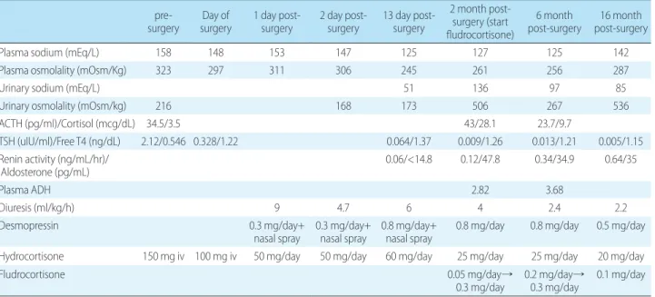
natremic seizure which was treated with hypertonic saline (characterized by generalized tonic movement, 3 times a day, lasting about 2 to 3 minutes). In addition, the dose of fludrocortisone was gradually increased to a maximum of 0.3 mg daily. Shortly thereafter, the serum Na level improved (145 mEq/L), and the urinary Na level decreased (Table 1).
Her urinary volume subsequently normalized (4 mL/kg/h).
The hypertonic fluid dose was gradually decreased. She was discharged from the hospital 19 days after the initiation of fludrocortisone and was treated with fludrocortisone 0.3 mg a day, orally desmopressin 0.8 mg/day, hydrocortisone 25 mg/day, and levothyroxine 50 mcg/day. Treatment with fludrocortisone was continued at a dose of 0.3 mg daily for another 5 months and was then decreased. Sixteen months after surgery, the patient still required oral desmopressin (0.5 mg/day) and fludrocortisone 0.1 mg per day along, with corticosteroids and, levothyroxine (50 mcg/day). Thus, the electrolyte level of the patient was maintained at normal.
Followup brain MRI revealed that the germinoma had a
Table 1. The Related Data of the Patient before and after Surgery surgery pre- Day of
surgery 1 day post-
surgery 2 day post-
surgery 13 day post- surgery
2 month post- surgery (start fludrocortisone)
6 month
post-surgery 16 month post-surgery
Plasma sodium (mEq/L) 158 148 153 147 125 127 125 142
Plasma osmolality (mOsm/Kg) 323 297 311 306 245 261 256 287
Urinary sodium (mEq/L) 51 136 97 85
Urinary osmolality (mOsm/kg) 216 168 173 506 267 536
ACTH (pg/ml)/Cortisol (mcg/dL) 34.5/3.5 43/28.1 23.7/9.7
TSH (uIU/ml)/Free T4 (ng/dL) 2.12/0.546 0.328/1.22 0.064/1.37 0.009/1.26 0.013/1.21 0.005/1.15 Renin activity (ng/mL/hr)/
Aldosterone (pg/mL) 0.06/<14.8 0.12/47.8 0.34/34.9 0.64/35
Plasma ADH 2.82 3.68
Diuresis (ml/kg/h) 9 4.7 6 4 2.4 2.2
Desmopressin 0.3 mg/day+
nasal spray 0.3 mg/day+
nasal spray 0.8 mg/day+
nasal spray 0.8 mg/day 0.8 mg/day 0.5 mg/day Hydrocortisone 150 mg iv 100 mg iv 50 mg/day 50 mg/day 60 mg/day 25 mg/day 25 mg/day 20 mg/day
Fludrocortisone 0.05 mg/day→
0.3 mg/day 0.2 mg/day→
0.3 mg/day 0.1 mg/day
Abbreviations: ACTH, Adrenocorticotropic hormone; TSH, Thyroid stimulating hormone; Free T4, Free thyroxine; ADH, Antidiuretic hormone; iv, intravenous
stable status for 16 months.
Discussion
Water and electrolyte imbalance are not rare in children with intracranial pathologies, such as tumors and head in
jury, that particularly involve the pituitary gland. Hypona
tremia is common electrolyte disorder and is found in 8 to 35% of patients following pituitary surgery
4,7). It can be as
sociated with the SIADH, CSWS, treatment of CDI, and excessive fluid administration in patients with adipsia
11). Identical acute brain damage may cause either SIADH or CSWS. The clinical and biochemical features of both dis
eases are identical and the only discriminative feature is the volume status: it tends to be expanded in SIADH and low in CSWS
12). CSWS is true hypernatremia and extracellular water loss due to inappropriate Na wasting in the urine, whereas SIADH is a dilutional hyponatremia caused by increased antidiuretic hormone secretion
13). Symptoms of volume depletion (hypotension, increased heart rate, and weight loss) occur with CSWS, whereas symptoms of volume expansion occur with SIADH (decreased urine output and weight gain). Although determination of volume state is challenging to identify, CSWS can be differentiated from SIADH based on urine production
12). However, SIADH can coexist with CDI and in this case the excessive urine production secondary to natriuresis can be misinterpreted as poor control of CDI.
The different water homeostasis abnormalities may occur in isolation or may coexist with other conditions. Conco
mitant CDI and CSWS is rare and has been associated with a poor prognosis and increased mortality
7,11). This unfavor
able prognosis is mainly attributed to the frequent misdiag
nosis or incorrect correction of Na level. In our case, po
lyuria secondary to natriuresis is thought to be as inade quate CDI control and dose of desmopressin was increased which cause further deterioration in the degree of hyponatremia.
Hyponatraemia in a patient with DI can be caused by water intoxication secondary to excessive desmopressin replace
ment, to coexistent CSW, to concurrent untreated or under
treated cortisol deficiency
4), anticonvulsant treatment, or to a different source of sodium loss
11). Excessive exogenous antidiuretic hormone causes free water retention and
results in dilutional hyponatremia due to oliguria
12). The patient developed extensive polyuria, indicating that the excessive dose of desmopressin was not the cause. And, panhypopituitarism was controlled with appropriate hor
mone replacement therapy, indicating that the cortisol de
ficiency was not the cause. Our patient receiving nephro
toxic chemotherapeutics in the treatment of brain tumor, it can be very difficult to distinguish CSWS from renal tu
bulopathy. The excessive degree of natriuresis compared to the degree of altered renal tubular threshold for phosphate and potassium reabsorption, and tubular protein leak should suggest that tubulopathy is not major cause
11). In addition, aldosterone levels are typically suppressed in CSWS and normal or high in SIADH with plasma renin suppressed in both
1). Decreased plasma renin and aldoste
rone secretion in our patient could be suspected of CSWS.
The presence of hyponatraemia and natriuresis in urinalysis collected while the patient develops polyuria (prior to des
mopressin dose) with dehydration feature and recovery of hyponatremia after administration of water and sodium will also suggest CSWS. Finally, we diagnosed the patient with coexistence of CSWS and CDI.
The treatment of CSWS is to restore intravascular vo
lume and serum sodium and normal saline is frequently used as an initial fluid
1,12). Once euvolemia is achieved, the degree of hyponatremia should be reevaluated. If serum sodium levels are severely decreased (<125 mEq/L) or a large volume of intravenous fluid is required to maintain euvolemia, intravenous hypertonic saline can also be used
13)