Sanguinarine Induces Apoptosis in Human Hepatocellular Carcinoma HepG2 Cells through the Generation of ROS and Modulation of Akt/ERK Signaling Pathways
Ju Yeong Hwang
1and Yung Hyun Choi
1,2*
1Anti-Aging Research Center, Dongeui University, Busan 614-714, Korea
2Department of Biochemistry, Dongeui University College of Korean Medicine, Busan 614-052, Korea
Received July 13, 2015 /Revised September 3, 2015 /Accepted September 7, 2015
Sanguinarine is a benzophenanthridine alkaloid originally isolated from the roots of Sanguinaria canadensis.
It has multiple biological activities (e.g., antioxidant and antiproliferative) and immune-enhancing potential. In this study, we explored the proapoptotic properties and modes of action of sanguinarine in human hepatocellular carcinoma HepG2 cells. Our results revealed that sanguinarine inhibited HepG2 cell growth and induced apoptosis in a dose-dependent manner. The induction of apoptosis by sanguinarine was associated with the up-regulation of Fas and Bax, the release of cytochrome c from the mitochondria to the cytosol, and the loss of the mitochondrial membrane potential. In addi- tion, sanguinarine activated caspase-9 and -8, initiator caspases of the intrinsic and death extrinsic pathways, respectively, and caspase-3, accompanied by proteolytic degradation of poly (ADP-ribose) polymerase. Sanguinarine also triggered the generation of reactive oxygen species (ROS). The elimi- nation of ROS by N-acetylcysteine reversed sanguinarine-induced apoptosis. Furthermore, sanguinar- ine induced the dephosphorylation of Akt and the phosphorylation of mitogen-activated protein kin- ases, including extracellular signal-regulated kinase (ERK), c-jun N-terminal kinase (JNK), and p38.
The growth inhibition was enhanced by the combined treatment of sanguinarine with a phosphatidyli- nositol 3'-kinase (PI3K) inhibitor and an ERK inhibitor but not JNK and p38 inhibitors. Overall, our data indicate that the proapoptotic effects of sanguinarine in HepG2 cells depend on ROS production and the activation of both intrinsic and extrinsic signaling pathways, which is mediated by blocking PI3K/Akt and activating the ERK pathway. Thus, our data suggest that sanguinarine may be a natural compound with potential for use as an antitumor agent in liver cancer.
Key words : Apoptosis, extracellular signal-regulated kinase (ERK), PI3K/Akt, reactive oxygen species
(ROS), sanguinarine
*Corresponding author
*Tel : +82-51-850-7413, Fax : +82-51-853-4036
*E-mail : [email protected]
This is an Open-Access article distributed under the terms of the Creative Commons Attribution Non-Commercial License (http://creativecommons.org/licenses/by-nc/3.0) which permits unrestricted non-commercial use, distribution, and reproduction in any medium, provided the original work is properly cited.
Journal of Life Science 2015 Vol. 25. No. 9. 984~992 DOI : http://dx.doi.org/10.5352/JLS.2015.25.9.984
서 론
항암치료의 가장 중요한 전략 중 하나가 apoptosis 경로를 통해 암세포만을 특이적으로 사멸시키는 방법이다. 현재까지 알려진 apoptosis는 크게 extrinsic (death receptor-mediated) 및 intrinsic (mitochondrial-mediated) pathway로 구분된다 [16, 27]. Extrinsic pathway는 세포막에 존재하는 death re- ceptor (DR)에 death ligand가 결합하여 caspase-8과 같은 ini- tiator caspase의 활성을 유도하여 이루어진다[16, 17]. 반면, intrinsic pathway는 미토콘드리아의 기능 조절과 연관된 ini- tiator caspase인 caspase-9의 활성 증가를 동반한다[12, 28].
비록 initiator caspase가 각 경로에서 다르지만, 이들은 최종적
으로 effector caspase (caspase-3/-7)의 활성을 통하여 다양한 기질 단백질의 분해와 DNA 단편화를 유도하여 apoptosis를 유발한다[14, 16, 27].
항균, 항염증, 항산화, 항암활성, 면역 증강을 포함한 다양한 약리적 효능을 지니고 있는 것으로 알려져 있는 sanguinarine 은 전통 한의학에서 오랜 동안 사용되어 온 혈근초(Sanguina-
ria canadensis) 및 현호색과(Family Fumaria) 식물의 뿌리에 존재하는 benzophenanthridine alkaloid계열 물질의 일종이다 [18, 31, 33, 40]. 최근까지의 연구에 의하면 sanguinarine은 다 양한 암세포에서 apoptosis를 유발하는 것으로 보고되어 있으 며, 간암, 유방암, 방광암, 대장암 및 구강상피암세포 등을 포 함한 많은 암세포에서 reactive oxygen species (ROS) 생성 의 존적 미토콘드리아 기능 손상과 연계되어 있었다[7, 13, 19, 23, 24]. 또한 sanguinarine은 약재 내성 극복에 탁월한 효능이 있을 것으로 기대되며[17, 30, 42], endoplasmic reticulum 경 로가 동반될 가능성도 제시되었다[19].
아울러, sanguinarine에 의한 암세포의 apoptosis 유도 과정
에는 세포 내 신호전달계 중, nuclear factor kappaB [15, 43],
phosphatidylinositol 3'-kinase (PI3K)/Akt [8, 21, 30] 및 mi-
togen activated protein kinases (MAPKs) [21, 24, 41] 경로 등이 관여하는 것으로 보고되고 있다. 그리고 extrinsic path- way의 주요 개시 인자에 해당되는 caspase-8의 활성 증가뿐 만 아니라 intrinsic pathway에 관여하는 caspase-9의 활성화 [1, 8, 22, 24, 32, 34]가 sanguinarine에 의한 암세포의 apopto- sis 유도에 관여하며, 특히 Bid의 truncation이 관찰되는 것[1, 2, 7, 8, 23, 24]으로 보아, sanguinarine에 의한 apoptosis 유발 에는 두 가지 경로의 동시 활성화가 관여할 가능성이 매우 높음을 알 수 있다. 이들의 결과는 모두 Bcl-2 family 단백질의 발현 변화 및 caspase 의존적 apoptosis 유발이 동반되는 현상 들이며, 이 과정에서 mitochondrial membrane potential (MMP, Δψm)의 소실이 관찰되었다.
또한 sanguinarine은 암세포의 전이(metastasis)를 억제하 며[9, 35, 38], 혈관 신생과정(angiogenesis)을 차단[3, 11, 20, 36, 44]하는 등의 효능도 가지는 것으로 보고되고 있다. 그러나 이러한 sanguinarine의 다양한 항암작용 효능이 보고되었음 에도 불구하고 간암세포를 대상으로 이루어진 연구는 매우 미비한 실정이다. 따라서 본 연구에서는 sanguinarine의 항암 활성에 대한 추가적인 자료를 제시하기 위하여 인체 간암세포 의 증식억제와 연관된 apoptosis 유도 기전을 HepG2 세포를 대상으로 조사하였으며, 유의적인 결과를 얻었기에 이를 보고 하고자 한다.
재료 및 방법
실험재료 및 항체
본 실험에 사용된 sanguinarine (13-methylbenzodi-ox- olo[5,6-]-1,3-dioxolo[4,5-I] phenanthridinium)은 Sigma-Aldrich Co. (St. Louis, MO, USA)에서 구입하였으며, 10 mM의 농도 로 methanol에 희석한 후, 적정 농도로 배지에 희석하여 처리 하였다. 세포의 배양을 위한 RPMI-1640 배지, 우태아혈청(fe- tal bovine serum, FBS) 및 1%의 penicillin/streptomycin은 Gibco BRL (Gaithersburg, NY, USA)에서 구입하였다. 세포증 식 측정을 위해 사용된 3-(4,5-dimethyl-2 thiazolyl)-2,5- diphnyl-2H-tetrazolium bromide (MTT)와 MMP (Δψm) 값의 변화 측정을 위한 5,5′,6,6′-tetrachloro-1,1′,3,3′-te- traethyl-imidacarbocyanine iodide (JC-1) 및 ROS의 생성을 억제하기 위하여 사용된 N-ace-tylcysteine (NAC)은 Sigma- Aldrich Co.에서 구입하였다. 단백질 분석을 위하여 사용된 Fas, Fas ligand (FasL), Bcl-2, Bcl-xL, Bax, poly(ADP-ribose) polymerase (PARP), cytochrome c 및 actin 항체는 Santa Cruz Biotechnology Inc. (Santa Cruz, CA, USA)에서 구입하 였으며, cytochrome c oxidase IV (COX IV), Akt, p-Akt, ex- tracellular signal-regulated kinase (ERK), p-ERK, c-jun N-ter- minal kinase (JNK), p-JNK, p38, p-p38 항체는 Cell Signaling Technology (Danvers, MA, USA)에서 구입하였다. Immuno-
blotting을 위해 2차 항체로 사용된 horseradish peroxidase (HRP)-conjugated anti-mouse 및 anti-rabbit 항체와 en- hanced chemiluminoesence (ECL) 용액은 Amersham Life Science Corp. (Arlington Heights, IL, USA)에서 구입하였다.
Caspases 활성 측정을 위한 colorimetric assay kit는 R&D Systems (Minneapolis, MN, USA)에서 구입하였으며, ROS 생 성 변화를 확인하기 위한 fluorescent probe 인 2`,7`-di-chlor- odihydrofluorescein diacetate (H
2DCFDA)는 Molecular Probes (Leiden, Netherlands)에서 구입하였다. PI3K/Akt 및 MAPKs 신호전달계 각각에 해당되는 억제제들은 CalBiochem (San Diego, CA, USA)에서 구입하였다.
세포배양 및 MTT assay
본 실험에 사용한 HepG2 인체 간암세포는 American Type Culture Collection (Rockville, MD, USA)에서 분양 받았으며, 세포의 배양을 위해 10% FBS 및 1%의 penicillin/streptomy- cin이 포함된 RPMI-1640 배지를 사용하였고, 37℃, 5% CO
2조건 하에서 배양하였다. MTT assay를 이용한 세포 생존율의 측정을 위하여 세포 배양용 6 well plate에 1×10
5cells/ml로 HepG2 세포를 분주하고 적정 농도의 sanguinarine 단독 및 다양한 신호전달 조절제를 각 well에 처리하여 배양하였다.
24시간 후 배지를 제거하고 MTT 용액(0.5 mg/ml)을 200 μl씩 분주하고 37℃에서 반응시켰다. 2시간 후 MTT 시약을 제거하 고 dimethyl sulfoxide (DMSO, Sigma-Aldrich Co.)를 1 ml 씩 각 well에 분주하여 생성된 formazan을 녹인 후 96 well plate에 200 μl씩 옮겨서 enzyme-linked immunosorbent as- say (ELISA) reader (Molecular Devices, Sunnyvale, CA, USA)로 540 nm에서 흡광도를 측정하였다.
Annexin V-Fluorescein Isothiocyanate (FITC) 염색 에 의한 apoptosis의 정량적 측정
Apoptosis 유발의 정량적 분석을 위해 준비된 세포들을 PBS를 이용하여 2~3회 정도 세척하고 10 mM HEPES/NaOH, pH7.4, 140 mM NaCl 및 2.5 mM CaCl
2가 포함된 annexin V binding buffer (Becton Dickinson, San Jose, CA, USA)에 부유시킨 다음 annexin V-FITC 및 propidium iodide (PI)를 처리하여 암실에서 15분 동안 반응을 시켰다. 반응이 끝난 후 35-mm mesh를 이용하여 단일세포로 분리하고 flow cy- tometer (FACS Calibur, Becton Dickinson, San Jose, CA, USA)를 적용시켜 apoptosis가 유발된 세포(V
+/PI
–)를 형광반 응에 따라 분석하였다.
Western blot analysis에 의한 단백질 발현의 분석
준비된 세포들에 적당량의 lysis buffer [25 mM Tris-Cl (pH
7.5), 250 mM NaCl, 5 mM ethylenediaminetetraacetic acid
(EDTA), 1% Nonidet-P40 (NP-40), 1 mM pheny-methyl sul-
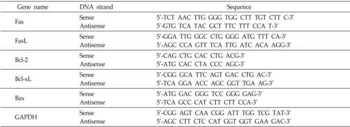
Table 1. Sequences of the primer pairs employed in the RT-PCR reactions
Gene name DNA strand Sequence
Fas Sense
Antisense
5'-TCT AAC TTG GGG TGG CTT TGT CTT C-3' 5'-GTG TCA TAC GCT TTC TTT CCA T-3'
FasL Sense
Antisense
5'-GGA TTG GGC CTG GGG ATG TTT CA-3' 5'-AGC CCA GTT TCA TTG ATC ACA AGG-3'
Bcl-2 Sense
Antisense
5'-CAG CTG CAC CTG ACG-3' 5'-ATG CAC CTA CCC AGC-3'
Bcl-xL Sense
Antisense
5'-CGG GCA TTC AGT GAC CTG AC-3' 5'-TCA GGA ACC AGC GGT TGA AG-3'
Bax Sense
Antisense
5'-ATG GAC GGG TCC GGG GAG-3' 5'-TCA GCC CAT CTT CTT CCA-3'
GAPDH Sense
Antisense
5'-CGG AGT CAA CGG ATT TGG TCG TAT-3' 5'-AGC CTT CTC CAT GGT GGT GAA GAC-3'
fonyl fluoride (PMSF) 및 5 mM dithiothreitol (DTT)]를 첨가
하여 4℃에서 1시간 동안 반응시킨 후, 14,000 rpm으로 30분간 원심분리하여 상층액에 있는 단백질을 분리하였다. 세포질과 미토콘드리아 단백질의 분리를 위해서는 mitochondria/cy- tosol fractionation kit (Biovision, Milpitas, CA, USA)를 사용 하였다. 적정 양의 단백질을 동량의 Laemilni sample buffer (Bio-Rad, Herculs, CA, USA)와 혼합하여 sodium dodecyl sulphate (SDS)-polyacrylamide gel을 이용하여 전기영동을 실시하였다. 이를 다시 nitrocellulose membrane (Schleicher and Schuell, Keene, NH, USA)으로 전이시키고 5% skim milk 를 1시간 처리하여 비특이적인 단백질들에 대한 blocking을 실시하였다. 그리고 적정 1차 항체를 처리하여 상온에서 2시 간 이상 반응시킨 다음 PBS-T (PBS with Tween 20)로 세척하 고 해당 2차 항체를 사용하여 상온에서 1시간 정도 반응시켰 다. 반응이 끝난 후 암실에서 ECL 용액에 적용시킨 후 X-ray film에 감광시켜 특정 단백질의 발현 양을 분석하였다.
RNA의 분리 및 reverse transcription-polymerase chain reaction (PCR)
전사 수준에서 특정 유전자의 발현 변화를 조사하기 위하여 적정 조건에서 배양된 세포들의 RNA를 RNeasy kit (Qiagen, La Jolla, CA, USA)를 이용하여 분리한 후, AMV reverse tran- scriptase (Amersham Life Science Corp.)를 이용하여 cDNA 를 합성하였다. 분석 대상 유전자들에 적절한 primer (Table 1)를 이용한 PCR은 Mastercycler (Eppendorf, Hamburg, Germany)를 사용하여 수행하였다. PCR 반응 산물을 1%
agarose을 이용하여 분리한 후, ethidium bromide (EtBr, Sigma-Aldrich Co.) 염색을 실시한 후 ultra violet (UV)하에서 해당 유전자의 발현 차이를 확인하였으며, 동일 조건에서 glyceraldehyde-3-phosphate dehydrogenase (GAPDH) 유전 자를 internal control로 사용하였다.
ROS 생성 변화 및 MMP 값의 측정
세포 내 ROS 생성 변화를 확인하기 위하여 준비된 세포들 을 10 μM의 H
2DCFDA로 20분간 염색하였으며, ROS 생성 억 제를 위한 ROS scavenger인 NAC 처리는 sanguinarine을 처 리하기 1시간 전에 처리하였다. 세포의 MMP 변화 정도를 측 정하기 위해서는 10 μM의 JC-1 용액을 처리하여 20분 동안 상온에서 반응시켰다. 반응이 끝난 후 원심분리하여 상층액을 제거하고 PBS를 첨가하여 세포를 부유시킨 다음 단일세포로 분리하였다. 이렇게 준비된 세포를 flow cytometer에 적용시 켜 ROS 및 MMP 값의 변화를 분석하였다.
Caspases 활성 변화의 측정
Caspase-3, -8 및 -9의 활성 정도는 colorimetric assay kit에 제시된 방법에 준하여 조사하였다. 이를 위하여 준비된 세포 를 모은 뒤 단백질을 추출하고 정량하여 각각 20 μg의 단백질 을 fluorogenic peptide 기질[caspase-3, Asp-Clu-Val-Asp (DEVD)-p-nitroaniline (pNA); caspase-8, Ile-Glu-Thr-Asp (IETD)-pNA; caspase-9, Leu-Glu-His-Asp (LEHD)-pNA] 100 μM이 함유된 extraction buffer [40 mM HEPES (pH 7.4), 20%
glycerol (v/v), 1 mM EDTA, 0.2% NP-40 및 10 mM DTT]
50 μl에 혼합하였으며, micro plate에 다시 extraction buffer에 희석하여 각 시료당 총 부피가 100 μl가 되게 하였다. 준비된 plate를 37℃에서 2시간 동안 반응시킨 후 ELISA reader를 이 용하여 405 nm의 흡광도를 이용하여 반응의 정도를 측정하였 다.
통계처리
실험 결과들의 유의성을 검정하기 위하여 분산분석을 실
시한 후 p<0.05 수준에서 Duncan's multiple range tests를
실시하였다. 이때 사용한 모든 통계분석은 SPSS 17.0 (IBM
SPSS Inc, New York, USA) 통계 프로그램을 이용하여 처리
하였다.
A
B
C
Fig. 1. Inhibition of cell viability and induction of apoptosis by sanguinarine treatment in human hepatocellular carcino- ma HepG2 cells. HepG2 cells were seeded in 6 well plate at 1×10⁵ cells/ml and treated with the indicated concen- trations of sanguinarine for 24 hr. (A) Cell viability was estimated by the metabolic-dye-based MTT assay. (B) To quantify the degree of apoptosis induced by sanguinar- ine, the cells were stained with FITC-conjugated Annexin V and PI for DNA flow cytometry analysis. Apoptotic cells are determined by counting the % of annexin V+/PI
– cells. Each point represents the mean ± SD of three independent experiments (*p<0.05 vs. untreated control).
(C) The cells were lysed and then equal amounts of cell lysates were separated on SDS-polyacrylamide gels and transferred to nitrocellulose membranes. The membranes were probed with anti-PARP and actin antibodies, and the proteins were visualized using an ECL detection system. Actin was used as an internal control.
A
B
Fig. 2. Effects of sanguinarine treatment on the expression of FasL and Fas in HepG2 cells. (A) Cells were treated with various concentrations of sanguinarine for 24 hr, and to- tal RNAs were isolated and reverse-transcribed. The re- sulting cDNAs were then subjected to PCR with FasL and Fas primers. The reaction products were separated in 1.0% agarose gel and visualized by EtBr staining.
GAPDH was used as an internal control for the RT-PCR.
(B) Aliquots containing total proteins isolated from cells grown under the same conditions as (A) were subjected to SDS-polyacrylamide gels followed by immunoblot analysis with specific antibodies against FasL and Fas proteins, and an ECL detection system. Actin was used as an internal control.
결과 및 고찰
Sanguinarine 처리에 의한 HepG2 간암세포의 증식 억 제 및 apoptosis 유발
HepG2 간암세포의 증식에 미치는 sanguinarine의 영향을 조사하기 위하여 정상세포에서는 세포독성이 없는 범위의 농 도[37]로 희석된 배지에서 24시간 동안 배양된 세포들을 대상 으로 MTT assay를 실시하였다. Fig. 1A에 나타낸 결과에서 알 수 있듯이, sanguinarine의 처리 농도가 증가할수록 HepG2 세포의 증식이 유의적으로 감소되었다. 이러한 증식 억제가
apoptosis 유발과 관련성이 있는지의 여부를 조사하기 위하여 annexin V-FITC/PI 염색을 이용한 flow cytometry 분석을 실 시한 결과, sanguinarine의 농도가 증가할수록 apoptosis가 유 발된 세포(V
+/PI
–)의 빈도가 유의적으로 증가되어 sanguinar- ine 처리에 의한 HepG2 간암세포의 증식억제가 apoptosis 유 도와 연관성이 있음을 알 수 있었다(Fig. 1B). Sanguinarine에 의한 apoptosis가 caspase 의존적으로 일어나는지를 조사하기 위하여 effector caspase (caspase-3/7)의 대표적인 기질 단백 질[14]에 해당되는 PARP의 단편화 여부를 조사한 결과, san- guinarine의 처리 농도가 증가할수록 단편화된 85 kD PARP 의 발현이 점차 증가되었다(Fig. 1C). 이는 sanguinarine 처리 에 의한 HepG2 세포의 증식 억제가 apoptosis 유발과 연관되 어 있음을 보여주는 결과이다.
HepG2 간암세포에서 DR 및 Bcl-2 family 유전자들의 발현에 미치는 sanguinarine의 영향
이상에서 관찰된 sanguinarine 처리에 의한 apoptosis 유발
현상과 연관된 몇 가지 기본적인 기전 해석의 일환으로 DR
및 Bcl-2 family에 속하는 주요 인자들의 발현 변화를 조사하
였다. 대표적인 apoptosis 유발경로 중, extrinsic pathway의
A
B
Fig. 3. Effects of sanguinarine treatment on the expression of Bcl-2 family members in HepG2 cells. (A) Cells were treated with various concentrations of sanguinarine for 24 hr, and total RNAs were isolated and re- verse-transcribed. The resulting cDNAs were then sub- jected to PCR with the indicated primers. The reaction products were separated in 1.0% agarose gel and vi- sualized by EtBr staining. (B) Aliquots containing total proteins isolated from cells grown under the same con- ditions as (A) were subjected to SDS-polyacrylamide gels followed by immunoblot analysis with the in- dicated antibodies, and an ECL detection system.
GAPDH and actin were used as internal controls for the RT-PCR and Western blot analysis, respectively.
개시에 관여하는 Fas/FasL 경로[16, 27]에 미치는 sanguinar- ine의 영향을 조사한 결과, sanguinarine 처리 농도의 증가에 따라 Fas의 발현이 전사 및 번역 수분에서 모두 증가하였다 (Fig. 2). Fas의 발현 증가는 최소한 FasL와의 결합 촉진 가능 성을 높여주는 결과로 해석이 가능하기 때문에 extrinsic path- way의 활성 가능성을 간접적으로 보여주지 것이라 생각된다.
또한 Fig. 3의 결과에서 알 수 있듯이, sanguinarine의 처리에 따라, anti-apoptotic Bcl-2 및 Bcl-xL의 발현은 큰 변화가 없었 으나, pro-apoptotic Bax의 발현이 전사 및 번역 수준에서 모두 증가되었다. 특히 intrinsic pathway에서 미토콘드리아 막 투 과성을 조절하는 단백질로 알려진 Bcl-2 family에 속하는 단백 질 중, Bcl-2 및 Bcl-xL과 같은 anti-apoptotic Bcl-2 family 단백 질은 미토콘드리아 막의 탈분극을 억제하여 막 투과성을 감소 시킨다[6, 12]. 이는 결과적으로 내막과 외막 사이에 존재하는 cytochrome c의 방출을 억제하여 apoptosis를 억제하지만, pro-apoptotic Bax는 미토콘드리아 막으로 이동하여 막 투과 성을 증가시킴으로써 cytochrome c의 방출을 촉진하여 apop- tosis를 유도하기 때문에 sanguinarine 처리에 의한 apoptosis 유발에 extrinsic 및 intrinsic pathway 모두가 관여할 가능성 을 보여주는 결과이다.
HepG2 간암세포에서 sanguinarine 처리에 의한 cytochrome c의 유리 및 ROS 생성의 증가
Fig. 3의 결과에서 관찰된 sanguinarine 처리에 따른 Bax의 상대적 발현 증가는 미토콘드리아 막 투과성 증가를 일으켜 세포질로의 cytochrome c의 방출을 촉진할 가능성이 높기 때 문에[6, 12, 26], 미토콘드리아와 세포질 단백질을 분리하여 cy- tochrome c 발현의 차이를 비교하였다. 그 결과, sanguinarine 처리 농도 증가에 따라 미토콘드리아 내 cytochrome c의 발현 은 감소된 반면, 세포질에서의 발현 증가가 관찰되었다(Fig.
4A). 한편 다양한 세포 내, 외 신호에 의하여 발생하는 ROS는 DNA 및 미토콘드리아의 손상을 야기하여 apoptosis 유발 촉 진 신호로 작용한다[10, 29]. 따라서 sanguinarine 처리에 의한 HepG2 간암세포의 apoptosis 유발이 ROS 생성과 연관성이 있는지의 여부를 조사한 결과, sanguinarine의 처리 15 분부터 ROS의 생성이 증가되어 30 분 처리군에서 ROS의 생성이 가장 높게 나왔으며, 그 이후부터는 점차 감소되는 경향성을 보였 다(Fig. 4B). 아울러, sanguinarine 처리에 의한 ROS의 생성은 대표적인 ROS scavenger인 NAC의 전처리에 의하여 현저하 게 감소되었다. 이상의 결과는 sanguinarine 처리에 의한 HepG2 간암세포의 apoptosis 유발에는 ROS의 생성과 미토콘 드리아에서 cytochrome c의 유리가 동반되어 있으며, 이는 미 토콘드리아의 기능 이상과 연관이 있음을 보여주는 것이다.
HepG2 간암세포에서 sanguinarine 처리에 의한 MMP 의 소실과 caspase 활성의 증가
이상의 결과에서 sanguinarine 처리에 의한 HepG2 간암세
포의 미토콘드리아 기능 상실 가능성을 보여주었기에, MMP
값에 미치는 sanguinarine의 영향을 JC-1 염색에 의한 flow
cytometry 분석[5, 28]으로 조사한 결과, sanguinarine이 함유
된 배지에서 배양된 HepG2 세포에서 MMP의 소실이 매우
증가되었다(Fig. 5A). 이러한 MMP의 소실이 ROS 생성의 증
가에 의한 것인지를 조사하기 위하여 NAC를 선처리 한 후
sanguinarine이 처리한 배지에서 배양된 세포의 MMP 값을
조사한 결과 정상 배지에서 배양된 세포의 값에 매우 근접하
게 나타나(Fig. 5A), sanguinarine 처리에 의한 MMP 값의 소
실, 즉 미토콘드리아의 기능 소실은 ROS 생성 의존적인 현상
임을 알 수 있었다. 아울러 sanguinarine 처리에 의한 apopto-
sis 유발에 caspase의 활성이 관여하는지의 여부를 조사한 결
과, sanguinarine이 처리된 배지에서 배양된 HepG2 세포에서
extrinsic 및 intrinsic pathway의 각 initiator caspase에 해당되
는 caspase-8과 -9뿐 만 아니라, 두 pathway의 effector caspase
인 caspase-3의 활성 또한 유의적으로 증가되었다(Fig. 5B). 그
러나 NAC의 전처리에 의한 ROS의 생성을 차단한 경우 cas-
pase의 활성은 거의 완벽하게 차단되었으며, PARP의 단편화
A
B
C
Fig. 5. Loss of MMP and activation of caspases by sanguinarine treatment in HepG2 cells. (A) Cells were treated with sanguinarine (1.5 μM) for 24 hr after 1 hr pre-treatment with or without NAC (10 mM). Then the cells were in- cubated with JC-1 dye (10 μM) for 30 min in the dark and the loss of MMP was measured by flow cytometry.
Each point represents the mean of two independent experiments. (B) The cells grown under the same con- ditions as (A) were lysed and aliquots containing total proteins (150 μg) were incubated with substrates, DEVD-pNA, IETD-pNA and LEHD-pNA, for in vitro cas- pase-3, -8 and- 9 activity, respectively, at 37℃ for 2 hr.
The released fluorescent products were measured.
Results shown are expressed as the mean of three in- dependent experiments (*, p<0.05 vs. untreated control;
#, p<0.05 vs. sanguinarine-treated cells). (C) Aliquots con- taining total proteins (50 μg) were subjected to SDS-poly- acrylamide gels followed by immunoblot analysis with anti-PARP antibody, and an ECL detection system. Actin was used as an internal control.
A
B
C
Fig. 4. Release of cytochrome c and generation of ROS by san- guinarine treatment in HepG2 cells. (A) Cells were treat- ed with various concentrations of sanguinarine for 24 hr, and cytosolic and mitochondrial proteins were sepa- rated on SDS-polyacrylamide gels and transferred to ni- trocellulose membranes. The membranes were probed with cytochrome c antibody and the proteins were vi- sualized using an ECL detection system. COX IV and actin were used as internal controls of mitochondrial (M.F.) and cytosolic fractions (C.F.), respectively. (B and C) HepG2 cells were treated with sanguinarine (1.5 μM) for the indicated times (B) or 30 min (C) after 1 hr pre-treatment with or without NAC (10 mM), a ROS scavenger. The medium was discarded and cells were incubated at 37°C in the dark for 20 min with new cul- ture medium containing 10 μM H2DCFDA and then used to evaluate ROS generation by flow cytometry.
Each point represents the mean of two independent experiments.
도 일어나지 않았다(Fig. 5C). 이들 결과에서 sanguinarine에 의한 HepG2 간암세포의 apoptosis 유발은 ROS 생성에 의한 미토콘드리아 기능 손상과 이에 따른 extrinsic 및 intrinsic pathway가 동시에 활성화되어 일어나는 현상임을 알 수 있었 다.
Sanguinarine 처리에 의한 apopotosis 유발에서 PI3K/Akt 및 MAPKs 신호 전달계의 역할
세포 내 특정 조절 단백질들의 인산화 상태는 apoptosis를 포함한 세포의 운명 결정에 중요한 현상으로 인식되고 있다.
다양한 세포 내 신호 전달계 중, PI3K/Akt 경로는 세포의 증식
및 성장에 핵심적인 역할을 하며, 대부분의 종양조직에서 활
성이 증대되어 있다[25, 39]. 그리고 MAPKs 경로에 포함되는
다양한 kinase 들은 세포의 내, 외 환경 변화에 따라 다양한
반응에 관여하며, 일반적으로 ERK가 세포의 증식 촉진에 관
여하며 JNK나 p38은 반대적인 역할을 한다고 알려져 있으나,
세포 활성물질의 생성 및 다양한 생물학적 기능은 세포가 처
한 환경이나 동일 조건에서도 세포의 종류에 따라 현저한 차
이를 보인다[4, 45]. 따라서 이들 신호계의 제어 현상에 대한
이해는 항암제 발굴 과정에서 해당 후보물질의 세포 내 역할
A
B
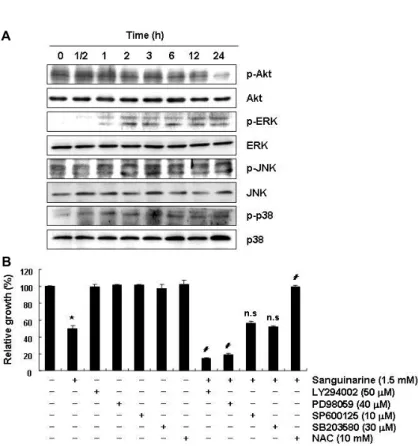
Fig. 6. Effects of sanguinarine treatment on the PI3K/
Akt and MAPKs signalling pathways in HepG2 cells. (A) Cells were treated with sanguinarine (1.5 μM) for the indicated times, and aliquots containing total proteins (30~50 μg) were sub- jected to SDS-polyacrylamide gels followed by immunoblot analysis with the indicated anti- bodies and an ECL detection system. (B) Cells were treated with sanguinarine (1.5 μM) for 24 hr after 1 hr pre-treatment with or without the indicated inhibitors (LY294002, PI3K inhibitor;
PD98059, ERK inhibitor; SP600125; JNK in- hibitor; SB203580; p38 inhibitor) or NAC (10 mM). Then the growth inhibition was meas- ured by an MTT assay. The data shown are means ± SD of three independent experiments (*, p<0.05 vs. untreated control; #, p<0.05 vs. san- guinarine-treated cells; n.s., non significant).
에 대한 많은 정보를 제공하여 준다. 따라서 sanguinarine 처 리에 따른 HepG2 세포의 apoptosis 유도 과정에서 이들 신호 전달계의 관여 여부를 부가적으로 조사한 결과, sanguinarine 의 처리 시간의 증가에 따라 인산화된 Akt 단백질의 발현은 점차 감소되었으며, MAPKs에 속한 3가지 인자들(ERK, JNK 및 p38)은 다소 시간 차이는 있었으나, sanguinarine 처리에 따라 활성이 증가되었다(Fig. 6A). 이러한 sanguinarine 처리 에 따른 각 kinase의 활성 변화가 apoptosis 유도와 어떤 연관 성이 있는지를 조사한 결과, PI3K 및 ERK 저해제(LY294002 및 PD98059)를 처리하여 PI3K/Akt 및 ERK 경로를 인위적으 로 차단한 경우 sanguinarine에 의한 증식 억제는 더욱 증가되 었지만, JNK 및 p38 저해제(SP600125 및 SB203580)는 sangui- narine에 의한 증식 억제에는 유의적인 영향을 미치지 못하였 다. 따라서 대표적인 세포 내 증식억제 신호계인 PI3K/Akt 및 ERK의 활성 저하가 sanguinarine 처리에 의한 HepG2 세포 의 증식억제 및 apoptosis 유도에 관련이 있음을 알 수 있었다.
결론적으로 sanguinarine에 의한 HepG2 간암 세포의 증식 억제는 apoptosis 유발과 밀접한 연관이 있었으며, 여기에는 extrinsic 및 intrinsic pathway 동시에 관여할 것으로 추정된 다. 아울러 이 과정에는 ROS 생성에 따른 미토콘드리아의 기 능 손상이 동반되었으며, 세포 내 PI3K/Akt 및 ERK 신호 전달 경로 변화가 동반되었음을 확인할 수 있었다.
감사의 글
이 논문은 2015학년도 동의대학교 교내연구비에 의해 연구 되었음(과제번호: 2015AA006).
References
1. Adhami, V. M., Aziz, M. H., Mukhtar, H. and Ahmad, N.
2003. Activation of prodeath Bcl-2 family proteins and mi- tochondrial apoptosis pathway by sanguinarine in im- mortalized human HaCaT keratinocytes. Clin. Cancer Res.
9, 3176-3182.
2. Ahsan, H., Reagan-Shaw, S., Breur, J. and Ahmad, N. 2007.
Sanguinarine induces apoptosis of human pancreatic carci- noma AsPC-1 and BxPC-3 cells via modulations in Bcl-2 family proteins. Cancer Lett. 249, 198-208.
3. Basini, G., Santini, S. E., Bussolati, S. and Grasselli, F. 2007.
Sanguinarine inhibits VEGF-induced Akt phosphorylation.
Ann. N. Y. Acad. Sci. 1095, 371-376.
4. Bassi, R., Heads, R., Marber, M. S. and Clark, J. E. 2008.
Targeting p38-MAPK in the ischaemic heart: kill or cure?
Curr. Opin. Pharmacol. 8, 141-146.
5. Bedner, E., Li, X., Gorczyca, W., Melamed, M. R. and Darzynkiewicz, Z. 1999. Analysis of apoptosis by laser scan- ning cytometry. Cytometry 35, 181-195.
6. Brenner, D. and Mak, T. W. 2009. Mitochondrial cell death effectors. Curr. Opin. Cell Biol. 21, 871-877.
7. Choi, W. Y., Kim, G. Y., Lee, W. H. and Choi, Y. H. 2008.
Sanguinarine, a benzophenanthridine alkaloid, induces
apoptosis in MDA-MB-231 human breast carcinoma cells through a reactive oxygen species-mediated mitochondrial pathway. Chemotherapy 54, 279-287.
8. Choi, W. Y., Jin, C. Y., Han, M. H., Kim, G. Y., Kim, N.
D., Lee, W. H., Kim, S. K. and Choi, Y. H. 2009. Sanguinarine sensitizes human gastric adenocarcinoma AGS cells to TRAIL-mediated apoptosis via down-regulation of AKT and activation of caspase-3. Anticancer Res. 29, 4457-4465.
9. Choi, Y. H., Choi, W. Y., Hong, S. H., Kim, S. O., Kim, G.
Y., Lee, W. H. and Yoo, Y. H. 2009. Anti-invasive activity of sanguinarine through modulation of tight junctions and matrix metalloproteinase activities in MDA-MB-231 human breast carcinoma cells. Chem. Biol. Interact. 179, 185-191.
10. Circu, M. L. and Aw, T. Y. 2001. Reactive oxygen species, cellular redox systems, and apoptosis. Free. Radic. Biol. Med.
48, 749-762.
11. De Stefano, I., Raspaglio, G., Zannoni, G. F., Travaglia, D., Prisco, M. G., Mosca, M., Ferlini, C., Scambia, G. and Gallo, D. 2009. Antiproliferative and antiangiogenic effects of the benzophenanthridine alkaloid sanguinarine in melanoma.
Biochem. Pharmacol. 78, 1374-1381.
12. Degli Esposti, M. and Dive, C. 2003. Mitochondrial mem- brane permeabilisation by Bax/Bak. Biochem. Biophys. Res.
Commun. 304, 455-461.
13. Dong, X. Z., Zhang, M., Wang, K., Liu, P., Guo, D. H., Zheng, X. L. and Ge, X. Y. 2013. Sanguinarine inhibits vas- cular endothelial growth factor release by generation of re- active oxygen species in MCF-7 human mammary ad- enocarcinoma cells. Biomed. Res. Int. 2013, 517698.
14. Duriez, P. J. and Shah, G. M. 1997. Cleavage of poly(ADP-ri- bose) polymerase: a sensitive parameter to study cell death.
Biochem. Cell. Biol. 75, 337-349.
15. Duvoix, A., Delhalle, S., Blasius, R., Schnekenburger, M., Morceau, F., Fougère, M., Henry, E., Galteau, M. M., Dicato, M. and Diederich, M. 2004. Effect of chemopreventive agents on glutathione S-transferase P1-1 gene expression mechanisms via activating protein 1 and nuclear factor kappaB inhibition. Biochem. Pharmacol. 68, 1101-1111.
16. Fulda, S. and Debatin, K. M. 2006. Extrinsic versus intrinsic apoptosis pathways in anticancer chemotherapy. Oncogene 25, 4798-4811.
17. Gatti, L., Cossa, G., Tinelli, S., Carenini, N., Arrighetti, N., Pennati, M., Cominetti, D., De Cesare, M., Zunino, F., Zaffaroni, N. and Perego, P. 2014. Improved apoptotic cell death in drug-resistant non-small-cell lung cancer cells by tumor necrosis factor-related apoptosis-inducing ligand- based treatment. J. Pharmacol. Exp. Ther. 348, 360-371.
18. Godowski, K. C. 1989. Antimicrobial action of sanguinarine.
J. Clin. Dent. 1, 96-101.
19. Gu, S., Yang, X. C., Xiang, X. Y., Wu, Y., Zhang, Y., Yan, X. Y., Xue, Y. N., Sun, L. K. and Shao, G. G. 2015.
Sanguinarine-induced apoptosis in lung adenocarcinoma cells is dependent on reactive oxygen species production and endoplasmic reticulum stress. Oncol. Rep. 34, 913-919.
20. Gupta, S. C., Kim, J. H., Prasad, S. and Aggarwal, B. B. 2010.
Regulation of survival, proliferation, invasion, angiogenesis,
and metastasis of tumor cells through modulation of in- flammatory pathways by nutraceuticals. Cancer Metastasis Rev. 29, 405-434.
21. Han, M. H., Kim, S. O., Kim, G. Y., Kwon, T. K., Choi, B.
T., Lee, W. H. and Choi, Y. H. 2007. Induction of apoptosis by sanguinarine in C6 rat glioblastoma cells is associated with the modulation of the Bcl-2 family and activation of caspases through downregulation of extracellular sig- nal-regulated kinase and Akt. Anticancer Drugs 18, 913-921.
22. Han, M. H., Yoo, Y. H. and Choi, Y. H. 2008. Sanguinarine-in- duced apoptosis in human leukemia U937 cells via Bcl-2 downregulation and caspase-3 activation. Chemotherapy 54, 157-165.
23. Han, M. H., Park, C., Jin, C. Y., Kim, G. Y., Chang, Y. C., Moon, S. K., Kim, W. J. and Choi, Y. H. 2013. Apoptosis induction of human bladder cancer cells by sanguinarine through reactive oxygen species-mediated up-regulation of early growth response gene-1. PLoS One 8, e63425.
24. Han, M. H., Kim, G. Y., Yoo, Y. H. and Choi, Y. H. 2013.
Sanguinarine induces apoptosis in human colorectal cancer HCT-116 cells through ROS-mediated Egr-1 activation and mitochondrial dysfunction. Toxicol. Lett. 220, 157-166.
25. Hu, L., Hofmann, J., Lu, Y., Mills, G. B. and Jaffe, R. B.
2002. Inhibition of phosphatidylinositol 3'-kinase increases efficacy of paclitaxel in in vitro and in vivo ovarian cancer models. Cancer Res. 62, 1087-1092.
26. Jeong, S. Y. and Seol, D. W. 2008. The role of mitochondria in apoptosis. BMB Rep. 41, 1122.
27. Jin, Z. and El-Deiry, W. S. 2005. Overview of cell death sig- naling pathways. Cancer Biol. Ther. 4, 139-163.
28. Jourdain, A. and Martinou, J. C. 2009. Mitochondrial out- er-membrane permeabilization and remodelling in apopto- sis. Int. J. Biochem. Cell Biol. 41,1884-1889.
29. Kadenbach, B., Arnold, S., Lee, I. and Hüttemann, M. 2004.
The possible role of cytochrome c oxidase in stress-induced apoptosis and degenerative diseases. Biochim. Biophys. Acta.
1655, 400-408.
30. Kim, S., Lee, T. J., Leem, J., Choi, K. S., Park, J. W. and Kwon, T. K. 2008. Sanguinarine-induced apoptosis: gen- eration of ROS, down-regulation of Bcl-2, c-FLIP, and syn- ergy with TRAIL. J. Cell. Biochem. 104, 895-907.
31. Laster, L. L. and Lobene, R. R. 1990. New perspectives on Sanguinaria clinicals: individual toothpaste and oral rinse testing. J. Can. Dent. Assoc. 56, 19-30.
32. Lee, J. S., Jung, W. K., Jeong, M. H., Yoon, T. R. and Kim, H. K. 2012. Sanguinarine induces apoptosis of HT-29 human colon cancer cells via the regulation of Bax/Bcl-2 ratio and caspase-9-dependent pathway. Int. J. Toxicol. 31, 70-77.
33. Mackraj, I., Govender, T. and Gathiram, P. 2008. Sanguinar- ine. Cardiovasc. Ther. 26, 75-83.
34. Park, H., Bergeron, E., Senta, H., Guillemette, K., Beauvais, S., Blouin, R., Sirois, J. and Faucheux, N. 2010. Sanguinarine induces apoptosis of human osteosarcoma cells through the extrinsic and intrinsic pathways. Biochem. Biophys. Res.
Commun. 399, 446-451.
35. Park, S. Y., Jin, M. L., Kim, Y. H., Lee, S. J. and Park, G.
초록:HepG2 인체 간암세포의 ROS 생성 및 ERK/Akt 신호전달 경로 조절을 통한 sanguinarine의 apoptosis 유도
황주영
1․최영현
1,2*
(1동의대학교 항노화연구소, 2동의대학교 한의과대학 생화학교실)
혈근초(Sanguinaria canadensis)에서 처음 분리된 sanguinarine은 항산화, 항암 및 면역 증강 등의 효능이 있는 것으로 알려진 alkaloid 계열 물질 중의 하나이다. 본 연구에서는 인체간암 HepG2 세포를 대상으로 sanguinarine 의 apoptosis 유도 효능 및 관련 기전 해석을 시도하였다. 본 연구의 결과에 의하면 sanguinarine은 HepG2 간암세 포의 증식을 처리 농도 의존적으로 억제하였으며, 이는 apoptosis 유도와 연관성이 있었다. Sanguinarine에 의한 apoptosis 유도에는 Fas 및 Bax의 발현 증가, 미토콘드리아에서 세포질로의 cytochrome c 유리 및 MMPl (Δψm) 의 소실을 동반하였다. Sanguinarine은 intrinsic 및 extrinsic apoptosis pathway의 활성에 관여하는 initiator cas- pase인 caspase-9와 -8의 활성과 effector caspase인 caspase-3의 활성 및 PARP 단백질의 단편화를 유발하였다.
Sanguinarine은 또한 ROS의 생성을 촉진시켰으며, N-acetylcysteine 처리에 의한 ROS의 생성을 차단하였을 경우, sanguinarine에 의한 apoptosis 효능이 완벽하게 차단되었다. 아울러 sanguinarine은 Akt의 인산화를 억제한 반 면, MAPKs의 인산화를 촉진시켰으며, 특히 PI3K와 ERK의 선택적 억제제는 sanguinarine에 의한 HepG2 간암세 포의 증식을 더욱 억제시켰다. 따라서 sanguinarine에 의한 HepG2 간암세포의 apoptosis 유발에는 ROS 생성 의 존적인 intrinsic 및 extrinsic signaling pathway가 동시에 활성화되며, PI3K/Akt 및 ERK 신호계가 관여함을 알 수 있었다.
2014. Sanguinarine inhibits invasiveness and the MMP-9 and COX-2 expression in TPA-induced breast cancer cells by inducing HO-1 expression. Oncol. Rep. 31, 497-504.
36. Pica, F., Balestrieri, E., Serafino, A., Sorrentino, R., Gaziano, R., Moroni, G., Moroni, N., Palmieri, G., Mattei, M., Garaci, E. and Sinibaldi-Vallebona, P. 2012. Antitumor effects of the benzophenanthridine alkaloid sanguinarine in a rat synge- neic model of colorectal cancer. Anticancer Drugs 23, 32-42.
37. Slunská, Z., Gelnarová, E., Hammerová, J., Táborská, E. and Slaninová, I. 2010. Effect of quaternary benzo[c]phenan- thridine alkaloids sanguilutine and chelilutine on normal and cancer cells. Toxicol. In Vitro 24, 697-706.
38. Sun, M., Liu, C., Nadiminty, N., Lou, W., Zhu, Y., Yang, J., Evans, C. P., Zhou, Q. and Gao, A. C. 2012. Inhibition of Stat3 activation by sanguinarine suppresses prostate can- cer cell growth and invasion. Prostate 72, 82-89.
39. Toker, A. and Yoeli-Lerner, M. 2006. Akt signaling and can- cer: surviving but not moving on. Cancer Res. 66, 3963-3966.
40. Vlachojannis, C., Magora, F. and Chrubasik, S. 2012. Rise and fall of oral health products with Canadian bloodroot extract. Phytother. Res. 26, 1423-1426.
41. Vogt, A., Tamewitz, A., Skoko, J., Sikorski, R. P., Giuliano,
K. A. and Lazo, J. S. 2005. The benzo[c]phenanthridine alka- loid, sanguinarine, is a selective, cell-active inhibitor of mi- togen-activated protein kinase phosphatase-1. J. Biol. Chem.
280, 19078-19086.
42. Weerasinghe, P., Hallock, S., Tang, S. C., Trump, B. and Liepins, A. 2006. Sanguinarine overcomes P-glycoprotein- mediated multidrug-resistance via induction of apoptosis and oncosis in CEM-VLB 1000 cells. Exp. Toxicol. Pathol. 58, 21-30.
43. Xu, J. Y., Meng, Q. H., Chong, Y., Jiao, Y., Zhao, L., Rosen, E. M. and Fan, S. 2012. Sanguinarine inhibits growth of hu- man cervical cancer cells through the induction of apoptosis.
Oncol. Rep. 28, 2264-2270.
44. Xu, J. Y., Meng, Q. H., Chong, Y., Jiao, Y., Zhao, L., Rosen, E. M. and Fan, S. 2013. Sanguinarine is a novel VEGF in- hibitor involved in the suppression of angiogenesis and cell migration. Mol. Clin. Oncol. 1, 331-336.
45. Yang, Y., Zhu, X., Chen, Y., Wang, X. and Chen, R. 2007.
p38 and JNK MAPK, but not ERK1/2 MAPK, play im- portant role in colchicine-induced cortical neurons apoptosis.
Eur. J. Pharmacol. 576, 26-33.