769
모야모야병을 가진 파열된 지속성 삼차신경동맥 동맥류 *
- 증 례 보 고 -
울산대학교 의과대학 서울중앙병원 신경외과학교실
윤병민·안재성·김준수·권 양·권병덕
= Abstract =
Ruptured Persistent Trigeminal Artery Aneurysm Associated with Moyamoya Disease
- -
- - Case Report - - - -
Byung Min Yun, M.D., Jae Sung Ahn, M.D., Joon Soo Kim, M.D., Yang Kwon, M.D., Byung Duk Kwun, M.D.
Department of Neurological Surgery, Asan Medical Center, College of Medicine, University of Ulsan, Seoul, Korea
oyamoya disease is defined as the development of collateral pathways, associated with bilateral chronic progre- ssive stenosis of the carotid fork. Persistent trigeminal artery is the vessel most frequently observed to persist into adult life among persistent carotid-basilar and carotid-vertebral anastomotic vessels. The authors present a man who had a sudden, severe headache and brain CT showed subarachnoid hemorrhage in left interpeduncular and prepontine cistern. Four-vessel angiogram revealed moyamoya disease associated with aneurysm arising from the junction of persistent trigeminal artery aneurysm and basilar artery. As a treatment, coil embolization was tried but it was failed because of anatomical difficulty of aneurysm. The aneurysm was successfully treated with clipping surgery 10 days later. To our knowledge, this is the first case being reported.
KEY WORDS:SAH・Moyamoya disease・Persistent Trigeminal artery・Aneurysm.
서 론
모야모야병과 지속성 삼차신경동맥(Persistent Trigemi- nal Artery:이하 PTA)이 공존하는 경우는 드물며, 이 경 우 대부분은 모야모야병에 의해 뇌출혈이나 뇌허혈 등의 증 상으로 발현되었고, 동맥류가 있는 경우라도 파열이 안된 경 우였다
17). 문헌상 처음으로 동맥류의 파열로 인해 증상이 발현된 경우를 치험하였기에 문헌 고찰과 함께 보고하고자 한다.
증 례
37세 남자환자로 내원 하루전 발생한 갑작스런 심한 두
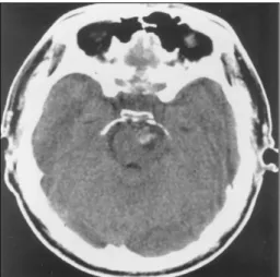
통을 주소로 내원하였다. 환자는 과거력상 특이 병력이 없 었으며, 입원 당시 시행한 신경학적 검사 소견에서 의식은 가면상태(drowsy)였으며, 경부 강직이 있었고, 운동, 감각 및 심부건 반사는 정상이었고, 병적 반사는 없었다. 입원 당 시의 뇌단층촬영(CT)에서 각간조(interpeduncular cistern) 및 전교조(prepontine cistern)에 출혈을 보였다(Fig. 1).
혈관 조영에서 전방순환(anterior circulation)의 경우 양 측의 내경동맥이 안구동맥 기시부의 바로 위 원위부에서 시 작하여 후방교통동맥이 분지하기 전부분까지 협착이 있었고, 우측은 Fisher의 경동맥 분류에 따르면, C5 부분(segment) 에서 PTA를 통해 후방순환계와 통하고 있었으며, 뇌저동맥 (basilar artery)과 만나는 부위에 내・후방으로 향한 6×
12mm 크기의 동맥류가 있었고, 후방혈관계에서 후방 교통 동맥을 통해 중뇌혈관으로 순환이 이루어지고 있었고, 전뇌 혈관의 분포영역에는 외경동맥을 통해 이루어지고 있었다.
M M M M
*본 논문은 1999년 대한신경외과학회 추계학술대회 포스터로 발표되었음.
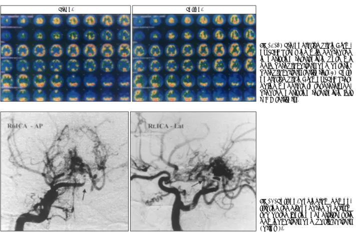
양측에서 렌즈핵선조체성 모야모야혈관(lenticulostriate moyamoya collateral)이 관찰되었고, 좌측은 PTA가 없었 으며, 나머지는 우측과 거의 동일 하였다. 후방순환(poste- rior circulation)의 경우, 좌측 추골기저동맥혈관조영에서 는 후방교통동맥을 통하여 양쪽 중뇌혈관이 조영되고 있었 고, 양측 후뇌혈관은 주로 우경동맥의 PTA를 통해 조영되 있었다(Fig. 2A, B). 모야모야병이 동반되어 뇌혈관 예비 능(perfusion reservoir)을 보기 위해 시행한 뇌핵의학적 단층 검사(Brain Diamox SPECT)에서 양측 전대뇌・중 대뇌동맥혈관 분포영역에 약간의 혈관예비능이 감소되어 있었다(Fig. 3). 치료로는 먼저 혈관내 시술을 시도하였다.
우측 내경동맥과 좌측 추골동맥을 통해 접근을 하였으나 동 맥류의 위치가 PTA와 뇌기저동맥이 만나는 곳에 있고, 동
Fig. 1. Pre-operative brain CT showing hemorrhage in the inter- peduncular cistern.
Fig. 2. A, B:Pre-operative angiogram showing a saccular aneurysm from the junction of right per- sistent trigeminal artery and basilar artery(arrow), bilateral distal ICA occulusion, and transdural blood supply through the middle meningeal artery and
A A A A
B
B B
B
맥류의 목이 넓어서, 시술후 뇌저동맥에 혈류가 가지 않을 가능성이 있다고 판단되어 코일을 이용한 혈관내 시술을 중 지하고 10일후 수술적 치료로 동맥류 결찰술을 시행하였 다. 수술은 좌측 Orbitofrontal craniotomy와 Zygomatic osteotomy를 시행하였다. Fundus는 매끄럽지 않았으며, 동맥류 주변에 유착이 있었고 출혈의 소견이 남아 있는 것 으로 보아 파열한 것으로 보이며 이를 결찰하였다. 수술후 시행한 뇌혈관 조영술상 남아 있는 동맥류는 없었고(Fig. 5), 뇌핵의학적 단층 검사상 좌측 중대뇌동맥혈관분포영역에 혈 류량(perfusion)과 혈관 예비능이 수술전에 비해 감소되었 다(Fig. 3). 수술 후에 환자는 우측반신 부전 마비가 왔으 며, 이는 개두술시 기존에 유지되었던 경막측부순환(trans- dural circulation)이 손상을 입었기 때문으로 생각되며, 환 자는 재활 치료를 받아서 퇴원 시에는 부축 없이도 3층 높 이의 건물을 혼자서 오르내릴 수 있었다.
고 찰
모야모야병은 뇌기저의 혈관들이 점차로 협착되면서, 부행 혈관(collateral circulation)이 발달되는 병으로 현재까지도 원인이 정확히 밝혀지지 않았다.혈관기형이 일반인에서 보
다 더 많이 동반된다고 보고되며, Yabumoto 등
18)은 뇌동 맥류가 약 5.6%에서 동반된다고 하였으며, Mawad 등
13)은 뇌동정맥 기형은 3%에서 동반된다고 하였다. 정상적으로 원 시적 삼차신경동맥은 태아기 4mm에 발생하여 14mm 태 아기에 퇴화하여야 하는데, 퇴화하지 않고 남아있는 경우에 PTA라 하며, 1844년 Quain의 부검 예에서 처음 기술되었 고, 1922년 Oertel에 의해 명명되었으며, 성인의 경우 혈 관 조형상 약 0.1~0.6%로 발견된다고 한다
4). PTA도 모 야모야병과 마찬가지로 약 25%의 환자에서 혈관 기형이 동 반된다고 보고되는데
2), 이 중 PTA의 동맥류는 소수에서 보고되고 있다
3). 한편 모야모야병과 PTA가 함께 있는 경 우는 더욱 극소수의 경우에서 보고되었다
7)8)17). Kwak과 Kadoya
11)는 모야모야병과 PTA가 동반된 때의 원인을 선 천적으로 보았으며, 태생학적으로 5~14mm 크기의 태아기 에 PTA는 정상적으로 사라지는 시기이고, 이 시기에 모야 모야병과 비슷한 혈관이 형성되는 시기이므로 이들은 연관 이 있다고 보고하였다. Suzuki 등
17)은 모야모야병과 PTA 를 동시에 갖고있는 일란성 쌍생아의 예를 들면서, 선천적 인 요인이 있지만 쌍생아에서 병의 발현시기가 서로 틀림을 보아 후천적 요인이 있다고 주장하였다. 한편 이들 경우에 도 모야모야병으로 인해 허혈이나 뇌출혈이 생긴 경우이거
Preop. Postop.
Fig. 3. 1) Pre-operative brain Diam- ox SPECT showing mild decreased in perfusion reservoir in both mi- ddle cerebral artery and anterior cerebral artery territories. 2) Post- operative brain Diamox SPECT rev- ealing moderate to severerely de- creased perfusion reservoir in left MCA territory.
Fig. 4. Post-op. angiogram demon-
strating the disappeared aneurysm
sac at the juntion of persistent tri-
geminal artery and basilar artery
(arrow).
나, 뇌혈관 촬영상 파열되지 않은 동맥류가 우연히 동반된 경우이며, 본 예처럼 PTA의 동맥류가 파열되서 증상을 나 타난 경우는 문헌상 처음이다.
모야모야병의 경우 동맥류가 발생하는 위치에 따라 크게 말초동맥 동맥류와 주동맥 동맥류로 나뉜다
6). 말초동맥 동 맥류는 주로 기저핵에 위치하며 모야모야 혈관 근처의 비 정상적인 미세한 망상혈관 근처에 호발하며, 조직학적으로 는 가성 동맥류로 판명되었으며
16), 일시적으로 나타났다가 사라질 수도 있다는 보고가 있다
12). Willis환 주위에서 발생 하는 주동맥 동맥류는 진성동맥류로 기형적이거나 혈역학적 원인으로 발생한다고 주장되고 있다
16). 일반적인 동맥류의 호발부위는 전방순환이 많으나(86.5%)
5)모야모야병의 경 우는 43%에서 후방순환계에서 발생한다고 Moriyama 등
14)은 보고하였다. 이의 원인으로 여러저자들은 지속적이고 진 행성인 경동맥의 폐쇄로 인해 후방순환계에 혈류가 증가하 여 뇌동맥류가 발생한다고 보고하였다
10)16).
수술적 치료에 있어서 모야모야병과 동맥류가 함께 있는 경우는 주의해야 할 점이 있다. 개두술시에 측부순환(tra- nsdural collateral vessel)이 가능한 한 손상이 안되어야 하며, 동맥류 주위에 모야모야 혈관이 있을 경우 이들은 손 상이 안되어야 하며, 수술시 저혈압이나 저산소혈증을 예방 하여 뇌허혈을 방지해야 한다
14). 이에 Adams 등
1)은 측부 순환문제에 있어서 표재성 측두엽동맥과 중대뇌동맥간의 직 접문합술(external-internal arterial bypass)을 먼저 시행 한 후에 동맥류 치료를 해야 한다고 주장하였다. 또한 동맥 류 주위에 모야모야 혈관이 있는 경우는 가급적 손상을 주 지않도록 수술하여야 하며, 저혈압이나 저산소혈증을 피하 기 위하여 마취과와 긴밀한 협조관계가 필요하다. 이런 수 술적인 장애요인을 극복하기 위해 Kwon 등
8)은 혈관내 시 술(coil embolization)도 한 방법이라 보고하였다. 수술시 기의 결정에 있어서 Nagai 등
15)은 급성기의 조기 수술은 뇌부종, 출혈로 인해 수술시야가 좋지 않으므로 지연 수술 이 좋다고 주장하였으나, Kodama 등
9)은 모야모야병과 파 열된 동맥류가 함께 있는 경우는 수술 후 혈관 연축이 발 생하는 경우가 일반적인 동맥류 파열 후에 오는 경우와 같 이 높으므로 재출혈과 혈관 연축의 치료를 고려하여 급성기 에 조기 수술을 한 후 혈관 연축에 대한 치료를 하는 것이 필요하다고 하였다. 본 환자의 경우 수술에 따른 모야모야 병의 합병증을 피하고자 동맥류에 대한 혈관내시술을 시도 하였으나, 이도 합병증이 따를 가능성이 높아서 개두술로 방법을 수정하였다. 먼저 직접문합술 시행후 결찰술을 시행 하는 것이 좋겠으나 재출혈과 혈관연축을 고려하여, 문합술 시행없이 개두술을 시행후 동맥류 결찰술을 시행하였다.
결 론
본 교실에서는 뇌지주막하 출혈로 발현된 지속성 삼차신 경동맥과 뇌저동맥 사이에 발생한 동맥류를 가진 모야모야 병의 환자를 성공적으로 치료할 수 있어서 문헌 고찰과 더 불어 보고하는 바이다.
•
논문접수일:1999년 7월 22일•
심사완료일:2001년 5월 8일•
책임저자:권 병 덕138-736 서울 송파구 풍납동 388-1
울산대학교 의과대학 서울중앙병원 신경외과학교실 전화:02) 3010-3550, 전송:02) 476-6738 E-mail:[email protected]
References
1) Adams HP, Kassell NF, Wisoff HS:Intracranial saccular
aneurysm and moyamoya disease. Stroke 10
:174-179, 1979
2) Agnoli AL:Vascular anomalies and subarachnoid hemorrh-age associated with persisting embryonic vessels. Acta Neuro- chir 60
:183-199, 1982
3) Ahmad I, Tominaga T, Suzuki M:Primitive trigeminal artery
associated with cavernous aneurysm -Case report- . Surg Neurol 41
:75-79, 1994
4) Cheng WC, Wang AD:Carotid-cavernous sinus fistula asso-
ciated with a primitive trigeminal artery. Neurosurgery 27
:802-805, 1990
5) Ferguson GG:Natural history of intracranial aneurysm. N
Engl J Med 305
:99, 1981
6) Grabel JC, Levine M, Ragland R:Moyamoya-like disease
associated with a lenticulostriate region aneurysms. J Neuro- surgery 70
:802-803, 1989
7) Kinjo T, Mukawa J, Takara E:Moyamoya disease associated
with primitive trigeminal artery. No Shinkei Geka 16
:1107- 12, 1988
8) Kwon TH, Moon SH, Lee HK:Intracranial aneurysm associ-
ated with moyamoya disease. J Korean Neurosurgical Society 28
(4
)(Suppl
):111, 1999
9) Kodama N, Sato M, Sasaki T:Treatment of ruputed cerebral
aneurysm assocaited with moyamoya disease. Surg Neurol 46
:62-66, 1996
10) Kodama N. Suzuki J:Moyamoya disease associated with
aneurysm. J Neurosurg 48
:565-569, 1978
11) Kwak R, Kadoya S:Moyamoya disease associated with per-
sitetnt trigeminal artery. J Neurosurgery 59
:166-171, 1983
12) Locksley HB:Report on the cooperative study of intracranialaneurysms and subarachnoid hemorrhage. J Neurosurg 25
:219-259, 1965
13) Mawad ME:Occulusive Vascular disease with cerebral Ar-
teriovenous malformation. Radiology 153
:401-408, 1984
14) Moriyama T, Teramoto N, Kitajima H:Moyamoya vesselsassociated with multiple cerebral aneurysm. Neurol Med Chir 26
:160-166, 1986
15) Nagai A, Miyazaki S, Tamagawa T:Two cases of ruptured
cerebral aneurysm associated with moyamoya phenomenon.
Neurol Med Chir 28
:94, 1985
16) Nagamine Y, Takahashi S, Sonobe M:Multiple intracranial
aneurym associated with moyamoya disease. J Neurosurg 54
:673-676, 1981
17) Suzuki S, Morioka T, Matsushima T:Moyamoya disease as-
sociated with persitetnt trigeminal artery variant in identical twins. Surg Neurology 45
:236-249, 1996
18) Yabumoto M:Moyamoya disease associated with intracranial