Copyright © 2018 Korean Stroke Society
This is an Open Access article distributed under the terms of the Creative Commons Attribution Non-Commercial License (http://creativecommons.org/licenses/by-nc/4.0/) which permits unrestricted non-commercial use, distribution, and reproduction in any medium, provided the original work is properly cited.
Original Article
Background and Purpose Little is known about prognosis after endovascular therapy (EVT) for acute large artery occlusion (LAO) caused by underlying intracranial atherosclerotic stenosis (ICAS).
Therefore, we investigated the prognosis following EVT according to the underlying etiology of LAO.
Methods Patients from the Acute Stroke due to Intracranial Atherosclerotic occlusion and Neurointervention-Korean Retrospective (ASIAN KR) registry (n=720) were included if their occlusion was in the intracranial anterior circulation and their onset-to-puncture time was <24 hours. Occlusion was classified according to etiology as follows: no significant stenosis after recanalization (Embolic group), and fixed significant focal stenosis in the occlusion site with flow impairment or re-occlusion observed during EVT (ICAS group). Patients were excluded when significant extracranial carotid lesions existed, and when the intracranial occlusion was intractable to EVT so that the etiology was undetermined. The effect of angiographic etiologic classification on outcomes was evaluated using multivariable analysis that was adjusted for potential confounders.
Results Among eligible patients (n=520), 421 and 99 were classified in the Embolic and ICAS groups, respectively. Patients in the Embolic and ICAS groups had similar successful reperfusion rates with EVT (79.6% vs. 76.8%, P=0.537) and 3-month functional independence (54.5% vs.
45.5%, P=0.104). In multivariable analysis, ICAS-related occlusion (odds ratio, 0.495; 95%
confidence interval, 0.269 to 0.913; P=0.024) showed poorer 3-month functional independence compared to embolic occlusion.
Conclusions After EVT, patients with acute ICAS-related occlusion have relatively poor functional outcomes compared to those with embolic occlusion. Novel strategies need to be developed to improve EVT outcomes for ICAS occlusion.
Keywords Cerebral infarction; Atherosclerosis; Embolism; Thrombectomy; Reperfusion; Treatment outcome
Prognosis of Acute Intracranial Atherosclerosis- Related Occlusion after Endovascular Treatment
Jin Soo Lee,a Seong-Joon Lee,a Joon Sang Yoo,b Jeong-Ho Hong,b Chang-Hyun Kim,c Yong-Won Kim,d,e Dong-Hun Kang,e,f Yong-Sun Kim,e Ji Man Hong,a Jin Wook Choi,g Bruce Ovbiagele,h
Andrew M. Demchuk,i Sung-Il Sohn,b Yang-Ha Hwangd
aDepartment of Neurology, Ajou University Medical Center, Ajou University School of Medicine, Suwon, Korea
bDepartment of Neurology, Keimyung University Dongsan Medical Center, Keimyung University School of Medicine, Daegu, Korea
cDepartment of Neurosurgery, Keimyung University Dongsan Medical Center, Keimyung University School of Medicine, Daegu, Korea
dDepartment of Neurology, Kyungpook National University Hospital, School of Medicine, Kyungpook National University, Daegu, Korea
eDepartment of Radiology, Kyungpook National University Hospital, School of Medicine, Kyungpook National University, Daegu, Korea
fDepartment of Neurosurgery, Kyungpook National University Hospital, School of Medicine, Kyungpook National University, Daegu, Korea
gDepartment of Radiology, Ajou University Medical Center, Ajou University School of Medicine, Suwon, Korea
hDepartment of Neurology, Medical University of South Carolina, Charleston, SC, USA
iDepartment of Clinical Neurosciences and Radiology, Hotchkiss Brain Institute, University of Calgary, Calgary, AB, Canada
Correspondence: Yang-Ha Hwang Department of Neurology, Kyungpook National University Hospital, School of Medicine, Kyungpook National University, 130 Dongdeok-ro, Jung-gu, Daegu 41944, Korea
Tel: +82-53-420-5758 Fax: +82-53-422-4265 E-mail: [email protected] Co-correspondence: Sung-Il Sohn Department of Neurology, Keimyung University Dongsan Medical Center, Brain Research Institute, Keimyung University School of Medicine, 56 Dalseong-ro, Jung-gu, Daegu 41931, Korea
Tel: +82-53-250-7075 Fax: +82-53-250-7830 E-mail: [email protected] Received: June 2, 2018 Revised: September 3, 2018 Accepted: September 4, 2018
Introduction
Several clinical trials have shown that endovascular treatment (EVT) using newer thrombectomy devices within an onset-to- puncture time of 6 to 12 hours has efficacy and safety superior to those of intravenous thrombolysis.1-5 Trials conducted more recently have revealed that EVT has substantial efficacy even within an onset-to-randomization time of 16 to 24 hours in selected patients.6,7 Although clinical practice guidelines have now been updated to reflect these advancements,8-11 the out- comes of EVT for acute large artery occlusion (LAO) presumably due to intracranial atherosclerotic stenosis (ICAS), which is en- countered more frequently in Asian patients, have not yet been adequately addressed.
In acute occlusion of the posterior circulation, the prognosis of ICAS-related occlusion is reportedly worse than that of em- bolic occlusion.12 In the anterior circulation, results of single- center anecdotal studies have shown comparable prognosis in ICAS-related occlusion.13,14 However, the ICAS population in these studies was younger with lower initial severity; therefore, large multicenter studies that include patients with ICAS-re- lated occlusion from endemic regions and those that control for confounding factors are warranted.
Therefore, in the present study, we retrospectively analyzed the prognosis of EVT for ICAS-related versus embolic occlusion of the anterior circulation by using prospectively collected data from a registry of information on patients at three comprehen- sive stroke centers in Korea.
Methods
Patient enrollment
The Acute Stroke due to Intracranial Atherosclerotic occlusion and Neurointervention-Korean Retrospective (ASIAN KR) regis- try was assembled for an observational study of consecutive patients aged ≥18 years who received EVT for the treatment of acute ischemic stroke due to intracranial and/or extracranial large vessel occlusion. Patient data were collected from three comprehensive stroke centers in Korea between January 2011 and February 2016.15 Some patients in the registry might have been included in previous pilot studies performed by each hos- pital.13,14 All clinical data were de-identified and allocated study identification numbers. The data collection protocol was approved by the Institutional Review Board of each respective hospital and was implemented in accordance with the ethical standards of the 1964 Declaration of Helsinki and its later amendments.
Etiologic classification of target occlusive lesions
The etiology of target large vessel occlusion (i.e., embolic or ICAS-related occlusion) was determined by core laboratory im- aging analyses based on angiographic diagnosis according to previous reports (Y.H.H. & J.S.L.).12,14,16 In brief, after confirma- tion of arterial occlusion, uncommon cerebral arterial diseases such as dissection, moyamoya disease, and vasculitis were evaluated. If the occluded vessel was completely recanalized after primary thrombectomy, the etiology was classified as em- bolic occlusion (Embolic group). A remnant stenosis of >70%, or a lesser degree of stenosis with a tendency toward reocclu- sion and/or flow impairment during the procedure, was classi- fied as ICAS-related occlusion (ICAS group). If grading was dif- ficult to determine or discordant, consensus was reached by the two graders (Y.H.H and J.S.L). In addition, this mechanism was further evaluated and could be amended by repeat angi- ography following EVT during admission (J.S.Y.). Consequently, Embolic and ICAS groups were included in the analyses.
Inclusion and exclusion criteria
For the current study, the following inclusion criteria were ap- plied: (1) acute intracranial LAO of the anterior circulation and (2) onset-to-puncture time of <1,440 minutes. The onset time was defined as last normal seen. Internal carotid artery (ICA) T, middle cerebral artery (MCA) M1, and MCA M2 occlusions were included in this study. Patients were excluded if (1) extra- cranial balloon angioplasty or stenting was performed; (2) an- giographic etiology was classified as dissection, extracranial ICA stenosis-related, or undetermined; or (3) the occlusion was intractable to mechanical thrombectomy so that the etiology could not be differentiated.
Evaluations
Premorbid and 3-month modified Rankin Scale (mRS) scores and National Institutes of Health Stroke Scale (NIHSS) scores at admission were analyzed. A good outcome was defined as a 3-month mRS score of 0 to 2 or no change compared to the premorbid mRS. Results of routine laboratory tests were also collected. After de-identification and blinding of clinical data, stroke neurologists, neuroradiologists, and neurointervention- ists with expertise in acute stroke management performed core laboratory imaging analyses to ensure consistent grading and to eliminate possible bias. The locations of initial large vessel occlusions were identified on baseline computed tomography (CT) or magnetic resonance angiography (S.J.L.). Alberta Stroke Program Early CT Scores (ASPECTS) were determined using noncontrast CT (S.I.S.). Final reperfusion was evaluated accord- ing to the modified treatment in cerebral ischemia (mTICI)
grade.17 Successful reperfusion was defined as mTICI grades 2b–3 (J.S.L.). Final recanalization was evaluated with arterial occlusive lesion grade.17 Post-procedural intracerebral hemor- rhages were classified in accordance with the criteria defined by the European Cooperative Acute Stroke Study (S.I.S.).18 Sub- arachnoid hemorrhage (SAH) was classified using the modified Fisher scale (S.I.S.).19 Serious hemorrhagic complications were defined as parenchymal hematoma type 2 and/or SAH grades 3 to 4 (thick SAH). Reocclusion was evaluated using repeat angi- ography performed during admission by comparing it with the last angiography performed during EVT (J.S.Y.).
Endovascular procedures
The type of EVT procedure was selected at the discretion of the treating physician. The direct aspiration method and stent re- trieval were routinely used. The direct aspiration method refers to a forced arterial suction thrombectomy that uses the Penum- bra system (Penumbra Inc., Alameda, CA, USA).20,21 Stent retrieval refers to clot removal by capturing and removing the thrombus with a stent retriever such as the Solitaire AB/FR (Medtronic, Ir- vine, CA, USA) or Trevo (Stryker, Kalamazoo, MI, USA).22-24 Balloon guide catheters, adjuvant lytic infusion, intracranial or extracra- nial angioplasty, and/or stenting were implemented as needed.
Statistical analysis
Comparative analyses between the Embolic and ICAS groups were performed for clinical characteristics, imaging findings, and treatment outcomes. Differences among the groups were
analyzed using chi-square tests for categorical variables, Mann-Whitney tests for ordinal variables, or t-tests for contin- uous variables. To evaluate the effect of underlying etiology on good outcomes, we performed a multivariable analysis that was adjusted for the following potential confounders: age, sex, premorbid mRS, initial NIHSS score, pretreatment ASPECTS, in- travenous tissue plasminogen activator use, onset-to-puncture time (minute), procedure time (minute), final successful reper- fusion, and serious hemorrhagic complications. An interaction between underlying etiology and some influential variables on patient outcomes was also evaluated with adjustment of the same confounding variables except procedure time. Regression lines of embolic occlusion and ICAS-related occlusion were drawn for the probability of good outcomes versus onset-to- puncture time, age, NIHSS score, and ASPECTS (unadjusted). A P<0.05 was considered significant. Statistical analysis was per- formed using the SPSS statistical package version 22.0 (IBM Co., Armonk, NY, USA).
Results
Classification process and final grouping
Among the 720 patients enrolled in the registry, 520 were in- cluded in the current study (Figure 1). We performed a blinded evaluation of the underlying etiology, using EVT angiographies combined with the evaluation of repeat angiographies during admission following EVT (608 of 720 patients). After exclusion criteria were applied regarding onset-to-puncture time, occlu-
Figure 1. Study flow chart. ASIAN KR, Acute Stroke due to Intracranial Atherosclerotic occlusion and Neurointervention-Korean Retrospective; ICA, internal carotid artery; ICAS, intracranial atherosclerotic stenosis; MCA, middle cerebral artery; ACA, anterior cerebral artery.
720 ASIAN KR registry
Inclusion
711 Onset-to-puncture time ≤1,440 minutes 9 Onset-to-puncture time >1,440 minutes
83 Other occlusion location (posterior circulation, MCA M3, ACA, etc.) 628 Anterior large artery occlusion
(ICA T, MCA M1, M2)
61 Extracranial ICA balloon/stenting 567 Absence of extracranial balloon/stenting
47 Other underlying mechanism (intractable, extracranial disease, dissection) 520 Angiographical mechanism differentiation
(embolic & ICAS-related occlusion)
Exclusion
7 Extracranial disease 2 Dissection 38 Intractable
421 Embolic group 99 ICAS group
sion location, and extracranial ICA treatment, 567 patients were included. Among these, the classification was changed into other category from etiology based on EVT angiography in 19 patients after evaluation of repeat angiography. Among the 19 patients, two were changed from ICAS etiology to embolism while 11 were changed from embolism to ICAS. Among intrac- table cases, one patient had proven ICAS while five had embo- lism. Finally, the Embolic and ICAS-related groups comprised 421 and 99 patients, respectively.
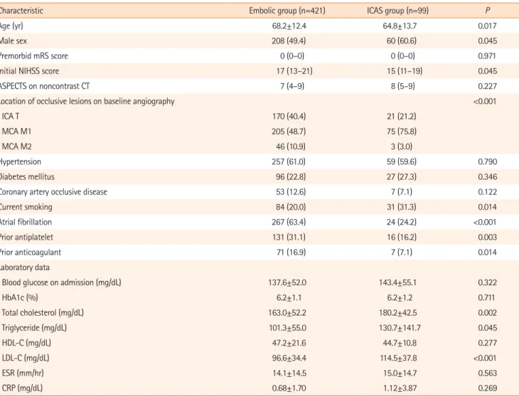
Differences in baseline characteristics
Table 1 describes the baseline clinical and laboratory data of the groups. Compared with the Embolic group patients, the ICAS group patients were significantly younger and mostly men, with a higher prevalence of a history of smoking. In the ICAS group, occlusive lesions were relatively more frequent in the MCA M1
portion, and these patients presented with significantly lower initial NIHSS scores than those in the Embolic group. In terms of laboratory data, the patients with ICAS presented with signifi- cantly higher levels of total cholesterol, triglycerides, and low density lipoprotein cholesterol than those in the Embolic group, while levels of glucose, glycosylated hemoglobin, and inflamma- tion markers did not differ between groups.
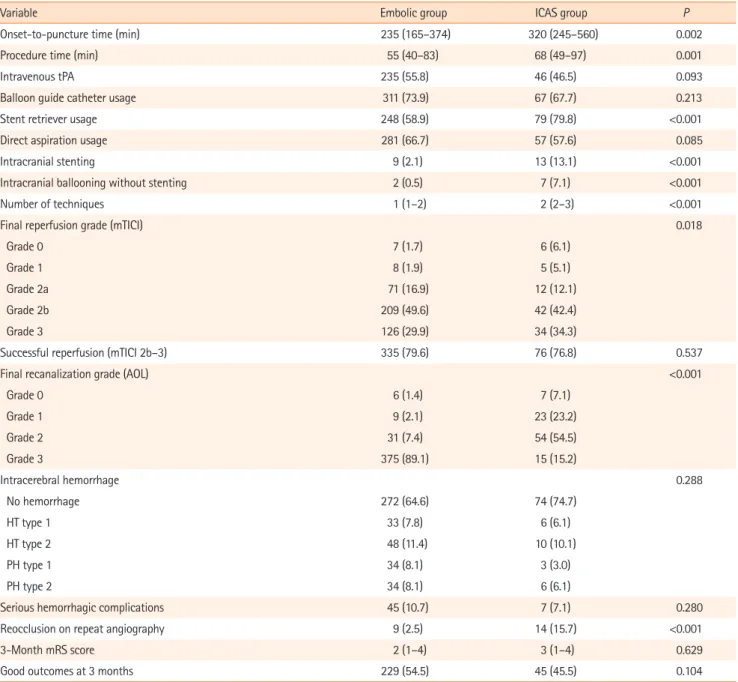
Differences in treatment and outcomes
Table 2 shows the comparison of treatments and outcomes ac- cording to etiological classification. Compared to the Embolic group, the ICAS group had a significantly longer median onset- to-puncture time (median, 235 and 320 minutes, P=0.002), and median total procedure time (median, 55 and 68 minutes, P=0.001), reflecting the complexity of the procedure. Concern- ing the final endovascular outcomes, the rates of successful
Table 1. Comparison of baseline characteristics and risk factors among groups
Characteristic Embolic group (n=421) ICAS group (n=99) P
Age (yr) 68.2±12.4 64.8±13.7 0.017
Male sex 208 (49.4) 60 (60.6) 0.045
Premorbid mRS score 0 (0–0) 0 (0–0) 0.971
Initial NIHSS score 17 (13–21) 15 (11–19) 0.045
ASPECTS on noncontrast CT 7 (4–9) 8 (5–9) 0.227
Location of occlusive lesions on baseline angiography <0.001
ICA T 170 (40.4) 21 (21.2)
MCA M1 205 (48.7) 75 (75.8)
MCA M2 46 (10.9) 3 (3.0)
Hypertension 257 (61.0) 59 (59.6) 0.790
Diabetes mellitus 96 (22.8) 27 (27.3) 0.346
Coronary artery occlusive disease 53 (12.6) 7 (7.1) 0.122
Current smoking 84 (20.0) 31 (31.3) 0.014
Atrial fibrillation 267 (63.4) 24 (24.2) <0.001
Prior antiplatelet 131 (31.1) 16 (16.2) 0.003
Prior anticoagulant 71 (16.9) 7 (7.1) 0.014
Laboratory data
Blood glucose on admission (mg/dL) 137.6±52.0 143.4±55.1 0.322
HbA1c (%) 6.2±1.1 6.2±1.2 0.711
Total cholesterol (mg/dL) 163.0±52.2 180.2±42.5 0.002
Triglyceride (mg/dL) 101.3±55.0 130.7±141.7 0.045
HDL-C (mg/dL) 47.2±21.6 44.7±10.8 0.277
LDL-C (mg/dL) 96.6±34.4 114.5±37.8 <0.001
ESR (mm/hr) 14.1±14.5 15.0±14.7 0.563
CRP (mg/dL) 0.68±1.70 1.12±3.87 0.269
Values are presented as mean±standard deviation, number (%), or median (interquartile range).
ICAS, intracranial atherosclerotic stenosis; mRS, modified Rankin Scale; NIHSS, National Institutes of Health Stroke Scale; ASPECTS, Alberta Stroke Program Early CT Scores; CT, computed tomography; ICA, internal carotid artery; MCA, middle cerebral artery; HbA1c, glycosylated hemoglobin; HDL-C, high density li- poprotein cholesterol; LDL-C, low density lipoprotein cholesterol; ESR, erythrocyte sedimentation rate; CRP, C-reactive protein.
reperfusion were similar in the Embolic and ICAS groups (79.6% vs. 76.0%, P=0.484).
No significant difference was observed between the groups in the grades and frequency of post-procedural intracerebral hemorrhage and the rate of serious hemorrhagic complica- tions. On repeat angiography, reocclusion occurred in nine of 362 patients (2.5%) in the Embolic group and 14 of 89 patients (15.7%) in the ICAS group, based on the last angiography per- formed during EVT (P<0.001).
Good outcomes at 3 months were achieved in 54.5% and 45.5% of patients in the Embolic and ICAS groups, respectively
(P=0.104), and the ordinal distribution of mRS scores was not significantly different between these groups (P=0.629) (Figure 2).
However, when angiographic etiology was included along with potential confounders in a logistic regression model to predict good outcomes at 3 months, the prognosis of ICAS-related oc- clusion was worse than that of embolic occlusion (odds ratio, 0.495; 95% confidence interval, 0.269 to 0.913; P=0.024) (Table 3). An interaction of underlying etiology on patient outcomes was observed with procedure time per 30 minutes (P=0.046), but not with the number of EVT techniques (P=0.214).
Graphs of onset-to-puncture time versus the probability of
Table 2. Comparison of treatments and outcomes among groups
Variable Embolic group ICAS group P
Onset-to-puncture time (min) 235 (165–374) 320 (245–560) 0.002
Procedure time (min) 55 (40–83) 68 (49–97) 0.001
Intravenous tPA 235 (55.8) 46 (46.5) 0.093
Balloon guide catheter usage 311 (73.9) 67 (67.7) 0.213
Stent retriever usage 248 (58.9) 79 (79.8) <0.001
Direct aspiration usage 281 (66.7) 57 (57.6) 0.085
Intracranial stenting 9 (2.1) 13 (13.1) <0.001
Intracranial ballooning without stenting 2 (0.5) 7 (7.1) <0.001
Number of techniques 1 (1–2) 2 (2–3) <0.001
Final reperfusion grade (mTICI) 0.018
Grade 0 7 (1.7) 6 (6.1)
Grade 1 8 (1.9) 5 (5.1)
Grade 2a 71 (16.9) 12 (12.1)
Grade 2b 209 (49.6) 42 (42.4)
Grade 3 126 (29.9) 34 (34.3)
Successful reperfusion (mTICI 2b–3) 335 (79.6) 76 (76.8) 0.537
Final recanalization grade (AOL) <0.001
Grade 0 6 (1.4) 7 (7.1)
Grade 1 9 (2.1) 23 (23.2)
Grade 2 31 (7.4) 54 (54.5)
Grade 3 375 (89.1) 15 (15.2)
Intracerebral hemorrhage 0.288
No hemorrhage 272 (64.6) 74 (74.7)
HT type 1 33 (7.8) 6 (6.1)
HT type 2 48 (11.4) 10 (10.1)
PH type 1 34 (8.1) 3 (3.0)
PH type 2 34 (8.1) 6 (6.1)
Serious hemorrhagic complications 45 (10.7) 7 (7.1) 0.280
Reocclusion on repeat angiography 9 (2.5) 14 (15.7) <0.001
3-Month mRS score 2 (1–4) 3 (1–4) 0.629
Good outcomes at 3 months 229 (54.5) 45 (45.5) 0.104
Values are presented as median (interquartile range) or number (%).
ICAS, intracranial atherosclerotic stenosis; tPA, tissue plasminogen activator; mTICI, modified treatment in cerebral ischemia; AOL, arterial occlusive lesion; HT, hemorrhagic transformation; PH, parenchymal hematoma; mRS, modified Rankin Scale.
good outcomes at 3 months revealed that the pattern of re- gression lines varied between embolic and ICAS-related occlu- sion (Figure 3A). While the probability of good outcomes in pa- tients with embolic occlusion declined as onset-to-puncture time increased, the probability of good outcomes in patients with ICAS-related occlusion did not decline but tended to in- crease with an increase in onset-to-puncture time. As for age, NIHSS score, and ASPECTS, the probability tended to be lower in the ICAS group than in the Embolic group, although the dif- ferences were not statistically significant (Figure 3B-D).
Supplementary Document, in which patients with atrial fi- brillation were excluded from the ICAS group, are shown in the
online site. The results from patients with ICAS-related occlu- sion but without atrial fibrillation are similar to the above re- sults obtained from patients with ICAS occlusion with or with- out atrial fibrillation.
Discussion
The major finding of the current study was that the prognosis of EVT for ICAS-related occlusion in the anterior circulation was relatively worse than that of EVT for embolic occlusion, in- dicating that better therapeutic approaches are required for patients with ICAS-related occlusion. Compared to patients Table 3. Results of logistic regression model for evaluating the association between occlusion etiology and good outcome
Variable Odds ratio (95% CI) P
Age (yr) 0.945 (0.925–0.965) <0.001
Male sex 0.989 (0.613–1.596) 0.964
Premorbid mRS score 0.578 (0.412–0.810) 0.001
Initial NIHSS score 0.897 (0.853–0.942) <0.001
Baseline intracranial occlusion 0.398
ICA T Reference
MCA M1 1.264 (0.757–2.111) 0.371
MCA M2 1.732 (0.747–4.018) 0.201
ASPECTS 1.313 (1.200–1.436) <0.001
Intravenous tPA use 0.963 (0.566–1.636) 0.889
Onset to puncture (min) 0.999 (0.997–1.000) 0.021
Procedure time (min) 0.981 (0.974–0.988) <0.001
Successful reperfusion 2.451 (1.359–4.418) 0.003
PH2 or SAH 3–4 0.242 (0.094–0.619) 0.003
Underlying etiology (ICAS vs. embolic occlusion) 0.495 (0.269–0.913) 0.024
CI, confidence interval; mRS, modified Rankin Scale; NIHSS, National Institutes of Health Stroke Scale; ICA, internal carotid artery; MCA, middle cerebral ar- tery; ASPECTS, Alberta Stroke Program Early CT Scores; tPA, tissue plasminogen activator; PH, parenchymal hematoma; SAH, subarachnoid hemorrhage; ICAS, intracranial atherosclerotic stenosis.
Figure 2. Modified Rankin Scale (mRS) scores at 3 months. Without adjustments, the distribution of mRS scores and the frequency of good outcomes did not differ between the Embolic and intracranial atherosclerotic stenosis (ICAS) groups.
3-Month modified Rankin Scale
0% 20% 40% 60% 80% 100%
Embolic group
ICAS group
0 1 2 3 4 5 6
with embolic occlusion, those with ICAS-related occlusion had lower initial NIHSS score and milder baseline ASPECTS, and were younger; these factors are known to be associated with good outcomes after EVT. Although the absolute rates of suc- cessful reperfusion were similar between the ICAS and Embolic groups, the outcomes of ICAS-related occlusion were worse than expected after adjustment for major confounders.
The relatively poor outcome in the ICAS group is mainly at- tributable to longer procedure time, reflecting the procedure complexity, and the higher rate of reocclusion. From our analy- sis, an interaction of underlying etiology and procedure time (per 30 minutes) was observed with patient outcomes. A meta- analysis from recent endovascular trials showed that time in- terval is more important in the hospital (door to reperfusion or image to reperfusion) than before hospital arrival (symptom onset to hospital arrival).25 Procedure time is a major portion of the in-hospital time; thus, the longer procedure time in the ICAS group could have contributed to the worse outcomes.
Another factor is reocclusion tendency in ICAS-related occlu- sion. Reocclusion after EVT can lead to early neurological dete- rioration.14,26 Reocclusion was more frequently seen in the ICAS group than in the Embolic group. Moreover, during EVT, ICAS-
related occlusion tends to have a high rate of refractoriness to mechanical thrombectomy due to repeat reocclusion, which necessitates additional rescue treatments such as intracranial stenting or lytic treatments, and eventually delays final reper- fusion time.27-31
Nevertheless, considering the relatively high rate of good outcomes (45.3%) in the ICAS group, which is similar to the 46% (33% to 71%) in pooled data from five early-window tri- als (HERMES) and 47% (45% to 49%) in two late-window tri- als,32,33 EVT using contemporary devices and methods can achieve substantial performance and satisfactory outcomes even for ICAS-related occlusions. This is particularly relevant to clinical practice in Asian countries, because Asian patients, who have a higher incidence of ICAS-related occlusions,16 are significantly underrepresented in successful EVT trials.1-7
The probability of good outcomes did not decline in these patients with an onset-to-puncture time of up to 24 hours, whereas it declined in patients with embolic occlusion and an increase in onset-to-puncture time. ICAS-related occlusion may have more opportunities for EVT because the ICAS prog- nosis patterns are similar to those associated with the “late window paradox” observed in recent trials conducted to dem- Figure 3. Relationship between good outcomes and well-known predictors in endovascular treatment for acute ischemic stroke and comparison between embolic and intracranial atherosclerotic stenosis (ICAS)-related occlusion. (A) The probability of good 3-month outcomes tended to decline in the Embolic group but stayed stable in the ICAS group as onset-to-puncture time increased up to 24 hours. (B-D) The probability of good outcomes appeared to be some- what less in ICAS-related occlusion than in embolic occlusion; however, this was not statistically significant. NIHSS, National Institutes of Health Stroke Scale;
ASPECTS, Alberta Stroke Program Early CT Scores.
1.00 0.75
0.50 0.25
500 1,000 1,500
Onset-to-puncture time (min)
Probability of good outcomes
Embolic occlusion ICAS-related occlusion
0
1.00
0.75 0.50
0.25
10 20 30 40
NIHSS score
Probability of good outcomes
Embolic occlusion ICAS-related occlusion
0
0 1.00 0.75
0.50 0.25
25 50 75 100
Age (yr)
Probability of good outcomes
Embolic occlusion ICAS-related occlusion
1.00
0.75 0.50
0.25
5 10
ASPECTS
Probability of good outcomes
Embolic occlusion ICAS-related occlusion
0
B A
C D
onstrate the efficacy of EVT in the late window (up to 24 hours onset-to-randomization time).6,7,33 Previous studies have indi- cated that good outcomes were unlikely in patients with evi- dently poor collaterals;34,35 patients with both rapidly and slowly failing collaterals can be ideal candidates for EVT within the usual early window (less than 60 to 300 minutes of onset- to-puncture time).35,36 In patients with universally good collat- erals, tiny-to-small infarcts are expected; therefore, these pa- tients were not considered good candidates for EVT.35 However, recent late-window trials have shown an absolute increase of 32% in good outcome rates compared to 19% in early window trials.32,33 Patients with preformed ICAS can have universally good collaterals, which are developed by hypoperfusion with stenosis.37 In an anecdotal study, the baseline infarct core vol- ume was significantly lower in patients with ICAS-related oc- clusion compared to those with embolic occlusion.38 Compared to early window trials using target artery occlusion and small core volume mostly based on noncontrast CT such as ASPECTS, these late window trials are based on infarct core volume with clinical or perfusion mismatch. Further imaging studies are warranted to incorporate this concept of “late window para- dox” in the treatment of ICAS-related occlusion.
Although imaging predictors should be studied further, dif- ferences in baseline characteristics including demographics and risk factors may differentiate ICAS-related occlusion from embolic occlusion. Besides younger age and male predomi- nance, MCA M1 occlusion was more frequent among locations of occlusive lesions in patients with ICAS. Well-known risk fac- tors for atherosclerosis were also documented in our patients with ICAS, as current smokers were more frequent, and levels of total and low-density cholesterol and triglycerides were higher. Our study members plan to further investigate differen- tial factors of various imaging tools. In addition, better treat- ment methods for ICAS-related occlusion will be further evalu- ated using the ASIAN KR registry.
The study has several limitations. First, this was a retrospec- tive study with data from three Korean centers and included patients treated with somewhat outdated devices such as the first-generation Penumbra system. Thus, the results of this study are not generalizable to the entire Korean or Asian popu- lation or to contemporary treatments. Second, based on the analyses of a current real-world study, patients whose occlu- sion failed to open were excluded. If the intractable cases ac- counted for more patients with embolic occlusion, i.e., those with greater age and higher initial NIHSS scores, the poor out- comes in the Embolic group might be underestimated. Howev- er, those with ICAS-related occlusion had longer onset-to- puncture and procedure times and these important confound-
ing factors had been adjusted by logistic regression analysis, so it is believed that the current results are not significantly devi- ated. Lastly, although etiological classification was based on an evaluation of angiography during EVT and was confirmed by further analysis with repeat angiography during admission, this assessment might still be incomplete. In patients with ICAS-re- lated occlusion, the frequency of atrial fibrillation was 24%.
However, the baseline characteristics and risk factors in the ICAS group were significantly different from those in the Em- bolic group, and the analytic results were not significantly changed when atrial fibrillation was excluded from the ICAS group (shown in the Supplementary Tables 1-3); thus, our main outcome, i.e., prognosis, is believed to be accurately reflected in our results.
In conclusion, our study showed that following EVT, patients with acute ischemic stroke due to ICAS-related occlusion had a relatively poorer prognosis than patients with acute stroke due to embolic occlusion.
Supplementary materials
Supplementary materials related to this article can be found online at https://doi.org/10.5853/jos.2018.01627.
Disclosure
The authors have no financial conflicts of interest.
Acknowledgments
This work was partly supported by the National Research Foundation of Korea (NRF) Grant funded by the Korea Government (MSIP) (No.
2014R1A5A2010008: Sung-Il Sohn; NRF-2018R1A2B6007094: Jin Soo Lee).
References
1. Berkhemer OA, Fransen PS, Beumer D, van den Berg LA, Lings- ma HF, Yoo AJ, et al. A randomized trial of intraarterial treat- ment for acute ischemic stroke. N Engl J Med 2015;372:11-20.
2. Goyal M, Demchuk AM, Menon BK, Eesa M, Rempel JL, Thornton J, et al. Randomized assessment of rapid endovas- cular treatment of ischemic stroke. N Engl J Med 2015;372:
1019-1030.
3. Saver JL, Goyal M, Bonafe A, Diener HC, Levy EI, Pereira VM, et al. Stent-retriever thrombectomy after intravenous t-PA vs. t-PA alone in stroke. N Engl J Med 2015;372:2285-2295.
4. Jovin TG, Chamorro A, Cobo E, de Miquel MA, Molina CA,
Rovira A, et al. Thrombectomy within 8 hours after symptom onset in ischemic stroke. N Engl J Med 2015;372:2296-2306.
5. Campbell BC, Mitchell PJ, Kleinig TJ, Dewey HM, Churilov L, Yassi N, et al. Endovascular therapy for ischemic stroke with perfu- sion-imaging selection. N Engl J Med 2015;372:1009-1018.
6. Nogueira RG, Jadhav AP, Haussen DC, Bonafe A, Budzik RF, Bhuva P, et al. Thrombectomy 6 to 24 hours after stroke with a mismatch between deficit and infarct. N Engl J Med 2018;
378:11-21.
7. Albers GW, Marks MP, Kemp S, Christensen S, Tsai JP, Ortega- Gutierrez S, et al. Thrombectomy for stroke at 6 to 16 hours with selection by perfusion imaging. N Engl J Med 2018;
378:708-718.
8. Powers WJ, Derdeyn CP, Biller J, Coffey CS, Hoh BL, Jauch EC, et al. 2015 American Heart Association/American Stroke As- sociation focused update of the 2013 guidelines for the early management of patients with acute ischemic stroke regard- ing endovascular treatment: a guideline for healthcare pro- fessionals from the American Heart Association/American Stroke Association. Stroke 2015;46:3020-3035.
9. Casaubon LK, Boulanger JM, Blacquiere D, Boucher S, Brown K, Goddard T, et al. Canadian stroke best practice recommen- dations: hyperacute stroke care guidelines, update 2015. Int J Stroke 2015;10:924-940.
10. Hong KS, Ko SB, Yu KH, Jung C, Park SQ, Kim BM, et al. Up- date of the Korean clinical practice guidelines for endovas- cular recanalization therapy in patients with acute ischemic stroke. J Stroke 2016;18:102-113.
11. Powers WJ, Rabinstein AA, Ackerson T, Adeoye OM, Bambaki- dis NC, Becker K, et al. 2018 Guidelines for the early manage- ment of patients with acute ischemic stroke: a guideline for healthcare professionals from the American Heart Association/
American Stroke Association. Stroke 2018;49:e46-e110.
12. Kim YW, Hong JM, Park DG, Choi JW, Kang DH, Kim YS, et al.
Effect of intracranial atherosclerotic disease on endovascular treatment for patients with acute vertebrobasilar occlusion.
AJNR Am J Neuroradiol 2016;37:2072-2078.
13. Lee JS, Hong JM, Lee KS, Suh HI, Demchuk AM, Hwang YH, et al. Endovascular therapy of cerebral arterial occlusions:
intracranial atherosclerosis versus embolism. J Stroke Cere- brovasc Dis 2015;24:2074-2080.
14. Hwang YH, Kim YW, Kang DH, Kim YS, Liebeskind DS. Impact of target arterial residual stenosis on outcome after endo- vascular revascularization. Stroke 2016;47:1850-1857.
15. Lee JS, Lee SJ, Hong JM, Choi JW, Hong JH, Chang HW, et al.
Temporal changes in care processes and outcomes for endo- vascular treatment of acute ischemic stroke: retrospective registry data from three Korean centers. Neurointervention
2018;13:2-12.
16. Lee JS, Hong JM, Kim JS. Diagnostic and therapeutic strate- gies for acute intracranial atherosclerosis-related occlusions.
J Stroke 2017;19:143-151.
17. Tomsick T, Broderick J, Carrozella J, Khatri P, Hill M, Palesch Y, et al. Revascularization results in the Interventional Manage- ment of Stroke II trial. AJNR Am J Neuroradiol 2008;29:582- 587.
18. Fiorelli M, Bastianello S, von Kummer R, del Zoppo GJ, Larrue V, Lesaffre E, et al. Hemorrhagic transformation within 36 hours of a cerebral infarct: relationships with early clinical de- terioration and 3-month outcome in the European Coopera- tive Acute Stroke Study I (ECASS I) cohort. Stroke 1999;30:
2280-2284.
19. Frontera JA, Claassen J, Schmidt JM, Wartenberg KE, Temes R, Connolly ES Jr, et al. Prediction of symptomatic vasospasm after subarachnoid hemorrhage: the modified fisher scale.
Neurosurgery 2006;59:21-27.
20. Kang DH, Hwang YH, Kim YS, Park J, Kwon O, Jung C. Direct thrombus retrieval using the reperfusion catheter of the penumbra system: forced-suction thrombectomy in acute ischemic stroke. AJNR Am J Neuroradiol 2011;32:283-287.
21. Turk AS, Frei D, Fiorella D, Mocco J, Baxter B, Siddiqui A, et al. ADAPT FAST study: a direct aspiration first pass technique for acute stroke thrombectomy. J Neurointerv Surg 2014;6:
260-264.
22. Roth C, Papanagiotou P, Behnke S, Walter S, Haass A, Becker C, et al. Stent-assisted mechanical recanalization for treat- ment of acute intracerebral artery occlusions. Stroke 2010;
41:2559-2567.
23. Saver JL, Jahan R, Levy EI, Jovin TG, Baxter B, Nogueira RG, et al. Solitaire flow restoration device versus the Merci Retriever in patients with acute ischaemic stroke (SWIFT): a ran- domised, parallel-group, non-inferiority trial. Lancet 2012;
380:1241-1249.
24. Nogueira RG, Lutsep HL, Gupta R, Jovin TG, Albers GW, Walker GA, et al. Trevo versus Merci retrievers for thrombectomy re- vascularisation of large vessel occlusions in acute ischaemic stroke (TREVO 2): a randomised trial. Lancet 2012;380:1231- 1240.
25. Saver JL, Goyal M, van der Lugt A, Menon BK, Majoie CB, Dip- pel DW, et al. Time to treatment with endovascular thrombec- tomy and outcomes from ischemic stroke: a meta-analysis.
JAMA 2016;316:1279-1288.
26. Kim GE, Yoon W, Kim SK, Kim BC, Heo TW, Baek BH, et al. In- cidence and clinical significance of acute reocclusion after emergent angioplasty or stenting for underlying intracranial stenosis in patients with acute stroke. AJNR Am J Neuroradi-
ol 2016;37:1690-1695.
27. Baek JH, Kim BM, Kim DJ, Heo JH, Nam HS, Song D, et al. Im- portance of truncal-type occlusion in stentriever-based throm- bectomy for acute stroke. Neurology 2016;87:1542-1550.
28. Chang Y, Kim BM, Bang OY, Baek JH, Heo JH, Nam HS, et al.
Rescue stenting for failed mechanical thrombectomy in acute ischemic stroke: a multicenter experience. Stroke 2018;49:
958-964.
29. Lee JS, Hong JM, Lee KS, Suh HI, Choi JW, Kim SY. Primary stent retrieval for acute intracranial large artery occlusion due to atherosclerotic disease. J Stroke 2016;18:96-101.
30. Kang DH, Kim YW, Hwang YH, Park SP, Kim YS, Baik SK. In- stant reocclusion following mechanical thrombectomy of in situ thromboocclusion and the role of low-dose intra-arterial tirofiban. Cerebrovasc Dis 2014;37:350-355.
31. Yoon W, Kim SK, Park MS, Kim BC, Kang HK. Endovascular treatment and the outcomes of atherosclerotic intracranial stenosis in patients with hyperacute stroke. Neurosurgery 2015;76:680-686.
32. Goyal M, Menon BK, van Zwam WH, Dippel DW, Mitchell PJ, Demchuk AM, et al. Endovascular thrombectomy after large-
vessel ischaemic stroke: a meta-analysis of individual patient data from five randomised trials. Lancet 2016;387:1723-1731.
33. Albers GW. Late window paradox. Stroke 2018;49:768-771.
34. Lee SU, Hong JM, Kim SY, Bang OY, Demchuk AM, Lee JS.
Differentiating carotid terminus occlusions into two distinct populations based on Willisian collateral status. J Stroke 2016;18:179-186.
35. Hwang YH, Kang DH, Kim YW, Kim YS, Park SP, Liebeskind DS. Impact of time-to-reperfusion on outcome in patients with poor collaterals. AJNR Am J Neuroradiol 2015;36:495- 500.
36. Liebeskind DS. Imaging the future of stroke: I. Ischemia. Ann Neurol 2009;66:574-590.
37. Liebeskind DS, Cotsonis GA, Saver JL, Lynn MJ, Turan TN, Cloft HJ, et al. Collaterals dramatically alter stroke risk in in- tracranial atherosclerosis. Ann Neurol 2011;69:963-974.
38. Suh HI, Hong JM, Lee KS, Han M, Choi JW, Kim JS, et al. Im- aging predictors for atherosclerosis-related intracranial large artery occlusions in acute anterior circulation stroke. J Stroke 2016;18:352-354.
Supplementary Document
Analyses of patients with intracranial atherosclerotic stenosis but without atrial fibrillation Classification process and final grouping
Among the 720 patients enrolled in the registry, 496 were included in the current analyses. We performed a blinded evaluation of the underlying etiology, using endovascular treatment angiography combined with evaluation of repeat angiography during admis- sion following endovascular treatment (EVT) (608 of 720 patients). After patients with atrial fibrillation were excluded from the in- tracranial atherosclerotic stenosis (ICAS) group, the Embolic and ICAS-related groups comprised included 421 and 75 patients, re- spectively.
Description of supplementary tables
Supplementary Table 1 describes the baseline clinical and laboratory data according to etiological classification. Supplementary Ta- ble 2 shows the comparison of treatments and outcomes. Supplementary Table 3 shows the effects of underlying etiology on good outcomes using a logistic regression model. These results from patients with ICAS-related occlusion but without atrial fibrillation are similar to the results obtained from patients with ICAS occlusion with or without atrial fibrillation in the main manuscript.
Supplementary Table 1. Comparison of baseline characteristics and risk factors among groups
Characteristic Embolic group (n=421) ICAS group (n=75) P
Age (yr) 68.2±12.4 63.16±13.14 0.002
Male sex 208 (49.8) 50 (66.7) 0.006
Premorbid mRS score 0 (0–0) 0 (0–0) 0.586
Initial NIHSS score 17 (13–21) 15 (11–19) 0.027
ASPECTS on noncontrast CT 7 (4–9) 8 (5–9) 0.248
Location of occlusive lesions on baseline angiography <0.001
ICA T 170 (40.4) 13 (17.3)
MCA M1 205 (48.7) 61 (81.3)
MCA M2 46 (10.9) 1 (1.3)
Hypertension 257 (61.0) 44 (58.7) 0.698
Diabetes mellitus 96 (22.8) 22 (29.3) 0.221
Coronary artery occlusive disease 53 (12.6) 5 (6.7) 0.141
Current smoking 84 (20.0) 26 (34.7) 0.005
Atrial fibrillation 267 (63.4) 0 (0) <0.001
Prior antiplatelet 131 (31.1) 8 (10.7) <0.001
Prior anticoagulant 71 (16.9) 1 (1.3) <0.001
Laboratory data
Blood glucose on admission (mg/dL) 137.6±52.0 143.4±54.1 0.376
HbA1c (%) 6.15±1.12 6.21±1.14 0.702
Total cholesterol (mg/dL) 163.0±52.2 188.0±43.4 <0.001
Triglyceride (mg/dL) 101.3±55.0 138.7±149.1 0.035
HDL-C (mg/dL) 47.2±21.6 44.2±10.7 0.252
LDL-C (mg/dL) 96.6±34.4 119.9±40.4 <0.001
ESR (mm/hr) 14.1±14.5 13.7±13.5 0.845
CRP (mg/dL) 0.68±1.70 1.30±4.41 0.229
Values are presented as mean±standard deviation, number (%), or median (interquartile range).
ICAS, intracranial atherosclerotic stenosis; mRS, modified Rankin Scale; NIHSS, National Institutes of Health Stroke Scale; ASPECTS, Alberta Stroke Program Early CT Scores; CT, computed tomography; ICA, internal carotid artery; MCA, middle cerebral artery; HbA1c, glycosylated hemoglobin; HDL-C, high density li- poprotein cholesterol; LDL-C, low density lipoprotein cholesterol; ESR, erythrocyte sedimentation rate; CRP, C-reactive protein.
Supplementary Table 2. Comparison of treatment and outcomes among groups
Variable Embolic group ICAS group P
Onset-to-puncture time (min) 235 (165–374) 320 (245–560) <0.001
Procedure time (min) 55 (40–83) 68 (49–97) 0.002
Intravenous tPA 235 (55.8) 34 (45.3) 0.093
Balloon guide catheter usage 311 (73.9) 49 (65.3) 0.127
Stent retriever usage 248 (58.9) 61 (81.3) <0.001
Direct aspiration usage 281 (66.7) 43 (57.3) 0.115
Intracranial stenting 9 (2.1) 9 (12.0) <0.001
Intracranial ballooning without stenting 2 (0.5) 7 (9.3) <0.001
Number of techniques 1 (1–2) 2 (2–3) <0.001
Final reperfusion grade (mTICI) 0.027
Grade 0 7 (1.7) 6 (8.0)
Grade 1 8 (1.9) 2 (2.7)
Grade 2a 71 (16.9) 10 (13.3)
Grade 2b 209 (49.6) 33 (44.0)
Grade 3 126 (29.9) 24 (32.0)
Successful reperfusion (mTICI 2b–3) 335 (79.6) 57 (76.0) 0.484
Final recanalization grade (AOL) <0.001
Grade 0 6 (1.4) 6 (8.0)
Grade 1 9 (2.1) 16 (21.3)
Grade 2 31 (7.4) 45 (60.0)
Grade 3 375 (89.1) 8 (10.7)
Intracerebral hemorrhage 0.504
No hemorrhage 272 (64.6) 56 (74.7)
HT type 1 33 (7.8) 4 (5.3)
HT type 2 48 (11.4) 7 (9.3)
PH type 1 34 (8.1) 3 (4.0)
PH type 2 34 (8.1) 5 (6.7)
Serious hemorrhagic complications 45 (10.7) 6 (8.0) 0.480
Reocclusion on repeat angiographies 9 (2.5) 12 (17.9) <0.001
3-Month mRS score 2 (1–4) 3 (1–4) 0.729
Good outcomes at 3 months 229 (54.5) 34 (45.3) 0.142
Values are presented as median (interquartile range) or number (%).
ICAS, intracranial atherosclerotic stenosis; tPA, tissue plasminogen activator; mTICI, modified treatment in cerebral ischemia; AOL, arterial occlusive lesion; HT, hemorrhagic transformation; PH, parenchymal hematoma; mRS, modified Rankin Scale.
Supplementary Table 3. Results of logistic regression model for evaluating the association between occlusion etiologies and good outcomes
Variable Odds ratio (95% CI) P
Age (yr) 0.945 (0.925–0.966) <0.001
Male sex 0.957 (0.585–1.565) 0.861
Premorbid mRS score 0.537 (0.377–0.767) 0.001
Initial NIHSS score 0.897 (0.853–0.944) <0.001
Baseline intracranial occlusion 0.561
ICA T Reference
MCA M1 1.224 (0.723–2.072) 0.452
MCA M2 1.544 (0.656–3.636) 0.320
ASPECTS 1.318 (1.202–1.445) <0.001
Intravenous tPA use 1.095 (0.634–1.889) 0.745
Onset-to-puncture time (min) 0.999 (0.998–1.000) 0.062
Procedure time (min) 0.980 (0.973–0.987) <0.001
Successful reperfusion 2.451 (1.344–4.470) 0.003
PH2 or SAH 3–4 0.214 (0.080–0.573) 0.002
Underlying etiology (ICAS vs. embolic occlusion) 0.432 (0.218–0.858) 0.016
CI, confidence interval; mRS, modified Rankin Scale; NIHSS, National Institutes of Health Stroke Scale; ICA, internal carotid artery; MCA, middle cerebral ar- tery; ASPECTS, Alberta Stroke Program Early CT Scores; tPA, tissue plasminogen activator; PH, parenchymal hematoma; SAH, subarachnoid hemorrhage; ICAS, intracranial atherosclerotic stenosis.