INTRODUCTION
Perilla frutescens Britton is an annual herbaceous plant of the Lamiaceae family that naturally grows naturally in East Asia(Lee 1982). In traditional medicinal practices, P. frutescens is used to treat a variety of illnesses including cough, phlegm, back pain, and diabetes(Han et al. 1994; Kim et al. 2007). The ingredients of P. frutescens include rosmarinic acid, triterpenoids, luteolin, apigenin, and
lig-nans(Okuda et al. 1986; Woo et al. 2014). Previous studies have reported that the ethanol extract of P. frutescens exerts anti-inflammatory effects(Kim et al. 2007; Lee et al. 2012), the water and ethanol extracts have antioxidant effects(Cho et al. 2011), and its methanol extract may exert a preventa-tive action against Alzheimer’s disease(Choi et al. 2004). The antioxidant effects of P. frutescens appear to be depen-dent on the inhibitory activity of xanthine oxidase and the radical scavenging activity of 2,2-diphenyl-1-picrylhydrazyl (DPPH; Han et al. 2004). Isoegomaketone(IK), which is a fragrant ingredient of P. frutescens that has a distinctive aroma and is biosynthesized from egomaketone, has been ─ 123 ─
Technical Paper
* Corresponding author: Chang Hyun Jin, Tel. +82-63-570-3162, Fax. +82-63-570-3159, E-mail. [email protected]
Anti-inflammatory Effects of Chloroform Soluble Fraction
from Perilla frutescens Britton Leaves Produced
by Radiation Breeding in RAW264.7 Cells
Yun Ho Jo1,2, Yang Kang So1, Seung Young Lee3, Jin Baek Kim1,
Jun Soo Lee2 and Chang Hyun Jin1,*
1Advance Radiation Technology Institute, Korea Atomic Energy Research Institute, Jeongeup, Jeonbuk 56212, Republic of Korea
2Department of Food Science and Technology Graduate School, Chungbuk National University, Cheongju, Chungbuk 28644, Republic of Korea
3Freshwater Bioresources Utilization Division, Nakdonggang National Institute of Biological Resources, Sangju, Gyeonbuk 37242, Republic of Korea
Abstract - The present study aimed to determine the anti-inflammatory effects of each solvent fraction of a mutant Perilla frutescens produced by radiation breeding. Following extraction with 80% methanol, P. frutescens was fractionated in the order of hexane, chloroform, ethyl acetate, and butanol; the chloroform fraction exhibited less cytotoxicity, the greatest inhibitory effect on the production of nitric oxide(NO), and the highest rate of inhibition on the generation of tumor necrosis factor-α(TNF-α), interleukin-6(IL-6), monocyte chemoattractant protein-1(MCP-1), and interferon-β(IFN-β). The chloroform fraction also suppressed the mRNA and protein levels of inducible nitric oxide synthase(iNOS) and reduced the activation of nuclear factor-κB
(NF-κB) in lipopolysaccharide(LPS)-stimulated RAW264.7 cells. Finally, the presence of corosolic
acid in the chloroform fraction was identified. Taken together, the present findings indicate that the chloroform fraction obtained from mutant P. frutescens inhibited NO production in LPS-stimulated RAW264.7 cells via the suppression of iNOS expression and the inactivation of NF-κB. Key words : Perilla frutescens Britton, Anti-inflammation, Nitric oxide, Inducible nitric oxide
isolated, identified(Madoka et al. 2004; Park et al. 2009), and shown to exert anti-inflammatory effects through the nuclear factor-κB(NF-κB) pathway and inhibition of the JAK-STAT-1 pathway(Jin et al. 2010). Additionally, IK produces anti-obesity effects by inhibiting the expression of adipogenetic genes related to the differentiation, synthesis, and fat accumulation of preadipocyte 3T3-L1 cells(So et al. 2015).
The bodily inflammatory response is a defense mecha-nism against external stimuli that occurs in response to tis-sue stress, damage, and infection(Cho et al. 2009). During this response, macrophages produce inflammatory medi-ators such as nitric oxide(NO), prostaglandin E2(PGE2), tumor necrosis factor-α(TNF-α), and interleukin-6(IL-6; Coussens et al. 2002). However, persistent inflammatory responses can result in arthritis, diabetes, arteriosclerosis, and cancer by inducing tissue damage(Libby 2006). Mac-rophages are activated by oxidative stress, cytokines, and lipopolysaccharides(LPS), which are components of the cell wall in Gram-negative bacteria, act as an endotoxins in the body, and induce the production of cytokines and NO (Stuehr et al. 1991). NO is produced from L-arginine by the catalytic reaction of NO synthases(NOSs). Unlike other NOSs such as endothelial NOS and neuronal NOS, induc-ible nitric oxide synthase(iNOS) does not produce NO un-der normal conditions. NO generated over a short time peri-od in response to LPS or inflammatory cytokines possesses antioxidant, anti-proliferative and immune effects, but when this factor is overexpressed for a long period of time, it may exhibit toxicity and cause damage and mutations in DNA (Cho et al. 2014).
Thus, the present study aimed to investigate the anti- inflammatory effects of various solvent fractions from a mutant P. frutescens Britton produced by radiation breed-ing. Additionally, the most efficacious fraction was used to investigate the mechanisms underlying the anti-inflamma-tory activities; the ingredients were isolated and structural analyses were performed to determine the compounds that exhibited the anti-inflammatory effects.
METERIALS AND METHODS
Reagents
Dulbeccos’s modified eagle medium(DMEM) and fetal
bovine serum(FBS) were purchased from Hyclone(Logan, UT, USA). Griess reagent, LPS, NP40 cell lysis buffer, pro-tease inhibitor cocktail, dimethyl sulfoxide(DMSO), and the corosolic acid analytical standard were purchased from Sigma-Aldrich(St. Louis, MO, USA). The EZ-Cytox cell viability assay kit was purchased from DAEIL Lab Service (Seoul, Korea) and TNF-α, IL-6, and monocyte chemoat-tractant protein-1(MCP-1) enzyme linked immunosorbent assay(ELISA) kits were purchased from R&D Systems (Minneapolis, MN, USA). The interferon-β(IFN-β) ELISA kit was purchased from Pestak Bio Lab(Piscataway, NJ, USA) and the rabbit polyclonal antibody for iNOS was purchased from Cell Signaling Technology(Danvers, MA, USA). The rabbit polyclonal antibody against β-tubulin was purchased from Santa Cruz Biotechnology(Santa Cruz, CA, USA) and an RNeasy kit was obtained from QIAGEN (Val-encia, CA, USA). An advantage RT-for-PCR kit was pur-chased from Clontech Laboratories(Mountain View, CA, USA) and the SYBR premix(Takara Bio, Otsu, Japan), Opti- MEMI medium, goat rabbit IgG HRP-conjugated anti-body, and Lipofectiamine 2000 were purchased from Invit-rogen(Carlsbad, CA, USA).
General experimental procedures
For the ingredient analysis, chromatography was per-formed using an open column and Silica gel on Kieselgel 60 plates(70~230 mesh, ASTM; Merck, Kenilworth, NJ, USA) and the chromatographic separations were monitored by TLC plates pre-coated with Kieselgel 60 F254(Art.552; Merck). All analytical and HPLC grade solvents were ob-tained from Thermo Fisher Scientific(Waltham, MA, USA) and 1H, 13C and nuclear magnetic resonance spectroscopy (NMR) spectra were analyzed with a JNM-ECA500(JEOL Co., Tokyo, Japan).
Corosolic acid
White powder; 1H-NMR(pyridine-d5, 500 MHz): δ 5.40 (1H, brs, H-12), 4.03(1H, ddd, J=11.0, 9.5, 4.0Hz, H-2), 3.33(1H, d, J=9.0Hz, H-3), 2.57(1H, d, J=10.5Hz, H- 18), 2.31(1H, ddd, J=13.5, 13.0, 4.5Hz, H-16), 2.13(1H, ddd, J=13.0, 12.5, 3.5Hz, H-1), 1.21(3H, s, Me-23), 1.15 (3H, s, Me-27), 1.02(3H, s, Me-26), 0.98(3H, s, Me-24), 0.92(3H, s, Me-25), 0.91(3H, d, J=6.0Hz, Me-30), and 0.85(3H, d, J=6.5Hz, Me-29).
HPLC analysis
The HPLC analysis was performed using an Agilent Eclipse XDB-C18 column(4.6 ×250mm, 5μm; Agilent Technologies, Santa Clara, CA, USA) and the mobile phase carried out using water(A) and acetonitrile(B) phases. The gradient elution system conditions were as follows: 0~5 min, 50% B; 50min, 95% conditions. The injection volume was 10μl, the flow rate was 1mlmin-1, the column temper-ature was maintained at 25°C, and the UV detection wave-length was 220nm.
Plant material and extract preparation
The mutant P. frutescens was provided by the Korea Atomic Energy Research Institute(Daejeon, Korea). The P. frutescens leaves were irradiated with a 200Gy gamma ray for mutagenesis, dried, and then ground to powder(1.2 kg). The powder was extracted with 80% methanol(10l) at room temperature for 24h and filtered through filter paper. Following extraction, the solution was fractionated in the order of hexane, chloroform, ethyl acetate, and butanol; the systematic preparation scheme is shown in Fig. 1.
Cell culture
RAW264.7 cells were cultured in DMEM supplemented with 10% FBS, streptomycin(100μgml-1), and penicillin (100Uml-1) and then incubated at 37°C in an atmosphere with 5% CO2.
Cell viability
Cell viability was assessed using an EZ-Cytox cell via-bility kit(DAEIL). The cells were cultured in a 96-well cul-ture plate at a density of 2×10⁵ cellsml-1 for 24h and then treated with each fraction for an additional 24h. Following treatment, 10μl of the kit solution was added to each well and the solution was incubated for 4h at 37°C in an atmo-sphere with 5% CO2. Cell viability was measured at 480 nm using a microplate reader(Benchmark Plus; Bio-Rad, Hercules, CA, USA) and a reference wavelength of 640nm.
NO assay
The cells were seeded in a 96-well plate, pre-incubated with each solvent fraction for 2h, and then incubated with LPS(1μgml-1) for 18h. Next, 100μl of culture superna-tant was mixed with an equal volume of Griess reagent and Fig. 1. Extraction and solvent partition of Perilla frutescens Britton leaves.
Perilla frutescens by Mutagenesis with Gamma-ray(1200g)
Percolated with 80% methanol(10l×2)
1. Suspend in water(1l)
2. Partitioned with hexane(0.5l×5)
Partitioned with chloroform(0.5l×5)
Partitioned with ethyl acetate(0.5l×2)
Partitioned with butanol(0.5l×2) Methanol ext.(200g) Hexane fr.(30g) Chloroform fr.(17g) Ethyl acetate fr.(5g) Butanol fr.(9g) 16 Fractions Fr.10 Fr.11 Fr.14 Aqueous layer Aqueous layer Aqueous layer Aqueous fr.
incubated at room temperature for 15min. The absorbance at 570nm was measured in a microplate reader with a refer-ence wavelength of 650nm. The nitrite level in the cellular media was measured using the Griess method(Park et al. 2009).
Total protein extracts
The cells were cultured in a 100mm culture plate at a density of 2×10⁵ cellsml-1 for 24h and pre-incubated with each solvent fraction for an additional 2h. Next, the cells were incubated with LPS(1μgml-1) for 18h and washed once with phosphate buffered saline(PBS), harvested by pipetting, and lysed on ice for 30min in an NP40 cell lysis buffer containing a protease inhibitor cocktail and phenyl-methylsulfonyl fluoride. Next, the cells were centrifuged at 15,000rpm for 15min at 4°C. The supernatants were used as total protein extracts and the total protein contents were determined by the Bio-Rad protein assay using bovine se-rum albumin(BSA) as the standard.
Western blot analyses
Equal amounts of the protein samples(50μg) were sepa-rated on 10% sodium dodecyl sulfate(SDS) polyacrylamide gels and transferred to nitrocellulose membranes. The mem-branes were blocked with 5% skim-milk in Tris-buffered saline with Tween 20(TBS-T; 10mM Tris-HCl, pH 7.4, 150 mM NaCl, and 0.1% Tween 20) for 1h and then incubated with the iNOS and β-tubulin primary antibodies at 4°C overnight; the rabbit polyclonal antibodies against iNOS (1:1,000), and β-tubulin were diluted(1:200) in blocking buffer. The membranes were washed three times with TBS- T for 15min and then incubated with anti-rabbit IgG HRP- conjugated secondary antibody diluted in blocking buffer (1:5,000) for 2h at room temperature. Next, the membranes were washed and the blotted proteins were detected using an enhanced chemiluminescence detection system (Amersh-am Biosciences, Little Chalfont, UK).
Extraction of total RNA and real-time polymerase
chain reaction(RT-PCR)
The cells were cultured in a 6-well culture plate at a den-sity of 2×10⁵ cellsml-1 for 24h, pre-incubated with various concentrations of the chloroform fraction for 2h, and then LPS(1μgml-1) was added for 18h. The total RNA from the
cells was isolated using an RNeasy kit(QIAGEN) accord-ing to the manufacturer’s instructions. A chromo real-time PCR detection system(Bio-Rad) and iTaq-SYBR green su-permix(Bio-Rad) were used for the RT-PCR amplification of iNOS and β-actin under the following conditions: 50 cy-cles of 94°C for 20s, 60°C for 20s, and 72°C for 30s. All reactions were repeated independently at least three times to ensure the reproducibility of the results. The quantitative iNOS and β-actin primers(Bioneer Corp., Chungwon, Korea) were as follows: iNOS forward primer, 5′-TCCTACACCA CACCAAACTGTGTGC-3′, and reverse primer, 5′-CTCC AATCTCTGCCTAT CCGTCTC-3′, β-actin forward primer, 5′-TGAGAGGGAAATCGTGCGTGAC-3′, and reverse primer, 5′-GCTCGTTGCCAATAGTGATGACC-3′. The specificity of the amplified PCR products was assessed with a melting curve analysis and the relative expressions of the target genes compared to the β-actin gene were evaluated using the comparative CT threshold method and the Bio-Rad software tool Genex-Gene Expression Macro.
ELISA procedure
The cells were cultured in a 6-well culture plate at a den-sity of 2×10⁵ cellsml-1 for 24h and then pre-incubated with each solvent fraction for an additional 2h. Next, the cells were further incubated with LPS(1μgml-1) for 4h(IL-6 and TNF-α), and 18h(MCP-1 and IFN-β). The cytokine levels were measured using an ELISA kit according to man-ufacturer’s protocols.
Luciferase assay
The cells were cultured in a 6-well culture plate at a den-sity of 2×10⁵ cellsml-1 for 24h and the transfection of the vectors was performed using Lipofectamine 2000 (Invitro-gen) according to the manufacturer’s instructions. The pNF-κB luciferase vector(5μg) and pRL-TK control reporter vector(1μg) were transfected using the vector per well. Following the transfection, the chloroform fraction was added at various concentrations for 2h, and then the cells were treated with LPS(1μgml-1) for 24h. Luciferase ac-tivity was measured using the dual-luciferase reporter assay system(Promega, Madison, WI, USA).
Statistical analysis
The differences between the means of the treated and un-treated groups were determined with Student’s t tests imple-mented in the Excel program(Microsoft Corp., Redmond, WA, USA); p values<0.05 were considered to indicate statistical significance.
RESULTS
Effects of various solvent fractions on cell viability and NO production in LPS-stimulated RAW264.7 cells
The cytotoxicity of each solvent fraction was determined using an EZ-Cytox cell viability kit. Following treatment with each fraction at a concentration of 50μgml-1, cell via-bility was greater than 90%; therefore, the experiments were performed at non-cytotoxic concentrations of 50μgml-1 or less. The NO production was measured using the Griess reagent after treatment with each fraction(50μgml-1). NO production largely increased in the LPS-treated group but decreased in all fraction-treated groups, except for the bu-tanol fraction-treated group. In particular, the chloroform fraction inhibited NO production at a rate of 95%(Fig. 2).
Inhibitory effects of solvent fractions on the production of inflammatory cytokines in LPS-stimulated RAW264.7 cells
In the present study, the production level of IL-6, TNF-α, MCP-1, and IFN-β were measured using ELISA kits after treatment with each fraction(50μgml-1). When RAW264.7 cells were stimulated with LPS, the levels of IL-6 were in-creased, but the chloroform fraction inhibited this increase by 85%(Fig. 3A). The chloroform fraction inhibited TNF-α by 65%, which was the highest level of inhibition among the various fractions(Fig. 3B). Other than the butanol frac-tion, all fractions decreased MCP-1 production and the chloroform fraction had the highest rate of inhibition of MCP-1(Fig. 3C). The hexane and chloroform fractions had inhibition rates of 61%, and 68%, respectively, for IFN-β (Fig. 3D).
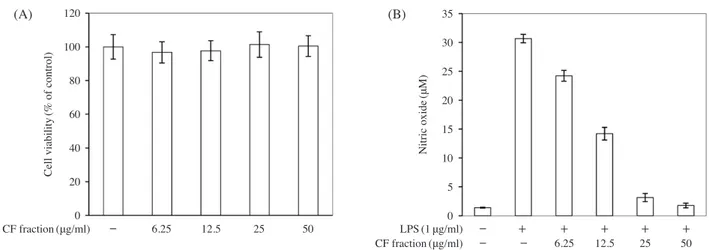
Effects of the chloroform fraction on cell viability and NO production in LPS-stimulated RAW264.7 cells
Cell viability was measured after administration of the
chloroform fraction at various concentrations(6.25, 12.5, 25, and 50μgml-1); no cytotoxicity was observed at any concentration(Fig. 4A). On the other hand, NO production decreased in a concentration-dependent manner following treatment with the chloroform fraction(Fig. 4B); the IC50 was 12.58μgml-1.
Effects of the chloroform fractions on iNOS expression levels in LPS-stimulated RAW264.7 cells
The mRNA and protein levels of iNOS increased in the LPS-treated group but iNOS expression was suppressed in a concentration-dependent manner following treatment with Fig. 2. Effects of various solvent fractions on cell viability
and nitric oxide(NO) production in lipopolysaccharide
(LPS)-stimulated RAW264.7 cells. (A) Cell viability was evaluated using EZ-Cytox cell viability assay and the rel-ative absorbance values were compared. (B) Cells were
treated with the solvent fractions(50μgml-1) for 2h and
then incubated in LPS(1μgml-1) for 18h. NO was
mea-sured with the culture media using the Griess Reagent.
Each value represents the mean±SD.(n=6). #p<0.05 vs.
control, *p<0.05 vs. LPS alone-treated group. M:
metha-nol, HX: hexane, C: chloroform, EA: ethyl acetate, B: buta-nol. 100 80 60 40 20 0 (A) (B) 30 25 20 15 10 5 0 Cell viability (% of control) Nitric oxide (μM) Fractions(50μg/ml) - M HX C EA B - - M HX C EA B - + + + + + + FractionsLPS(50(1μg/ml)μg/ml)
1000 900 800 700 600 500 400 300 200 100 0 180 160 140 120 100 80 60 40 20 0 800 700 600 500 400 300 200 100 0 700 600 500 400 300 200 100 0 (A) (B) (C) (D) IL-6 (pg/ml) MCP-1 (pg/ml) TNF-α (pg/ml) IFN-β (pg/ml) Fractions(50μg/ml) Fractions(50μg/ml) Fractions(50μg/ml) Fractions(50μg/ml) LPS(1μg/ml) LPS(1μg/ml) LPS(1μg/ml) LPS(1μg/ml) - - M HX C EA B - - M HX C EA B - - M HX C EA B - - M HX C EA B
Fig. 3. Inhibitory effects of the solvent fractions on interleukin(IL-6), tumor necrosis factor-α(TNF-α), monocyte chemoattractant protein-1
(MCP-1), and interferon-β(IFN-β) production levels in LPS stimulated RAW264.7 cells. The cells were pre-incubated with various
solvent fractions(50μgml-1) for 2h and then incubated with LPS(1μgml-1) for 4h(IL-6 and TNF-α) or 18h(MCP-1 and IFN-β).
Cytokines were measured with enzyme-linked immunosorbent assay(ELISA) assays. Each value represents the mean±SD.(n=3).
#p<0.05 vs. control, *p<0.05 vs. LPS alone-treated group.
- + + + + + +
- + + + + + +
- + + + + + +
- + + + + + +
Fig. 4. Effects of the chloroform fractions on cell viability and NO production in LPS-stimulated RAW264.7 cells. (A) Cell viability was
evaluated using EZ-Cytox cell viability assay and relative absorbance values were compared. (B) Cells were treated with different
concentrations of the chloroform fraction(6.25, 12.5, 25, and 50μgml-1) for 2h and then incubated in LPS(1μgml-1) for 18h. NO
was measured with the culture media using Griess reagent. Each value represents the mean±SD.(n=6).
120 100 80 60 40 20 0 (A) (B) 35 30 25 20 15 10 5 0 Cell viability (% of control) Nitric oxide (μM) CF fraction(μg/ml) - 6.25 12.5 25 50 - - 6.25 12.5 25 50 - + + + + + CF fractionLPS(1(μg/ml)μg/ml)
the various chloroform factions(Fig. 5). These findings in-dicate that NO production was decreased by the inhibition of iNOS expression. Therefore, it appears that the anti- inflammatory effects of P. frutescens are the greatest in the chloroform fraction.
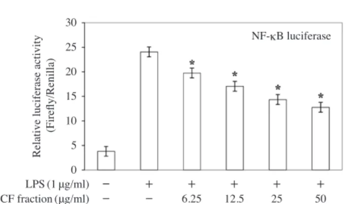
Effects of the chloroform fractions on NF-κB activation in LPS-stimulated RAW264.7 cells
To determine whether NF-κB activation was influenced by treatment with the chloroform fraction, its activation levels were measured using a luciferase assay. Compared with the non-LPS-treated group, NF-κB activation was sup-pressed by approximately six-fold in the LPS-treated group. Additionally, NF-κB was inhibited in a concentration-de-pendent manner when treated with the various concentra-tions of the chloroform faction(Fig. 6).
Ingredient isolation and structural analyses of the active components
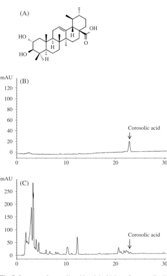
The chloroform fraction exhibited significant anti-inflam-matory effects relative to the other solvent fractions. This fraction was divided into 16 sub-fractions using Silica gel column chromatography with a chloroform:methanol ratio of 100:1. Sub-fraction 14 had inhibitory effects on NO production without cytotoxicity and, thus, was structurally analyzed using NMR. The structure of this isolated single ingredient was identified by comparing the present findings with the NMR data of previous papers(Woo et al. 2014); the ingredient contained in sub-fraction 14 determined to be corosolic acid(Fig. 7A). This result was confirmed by high-performance liquid chromatography(HPLC) using a corosolic acid analytical standard(Fig. 7B, C).
DISCUSSION
The anti-inflammatory effects of P. frutescens have been demonstrated using various extraction methods(Choi et al. 2004; Kim et al. 2007; Cho et al. 2011; Lee et al. 2012). In other study, after extraction with 80% ethanol, P. frutescens was fractionated in the order of hexane, ethyl acetate, and butanol. Among these fractions, ethyl acetate fraction has the highest reported inhibitory effects on NO and PGE2 production(Kim et al. 2006). In contrast, the present study found that the ethyl acetate fraction had a lower inhibitory effect on NO production than the chloroform fraction, which Fig. 5. Effects of the chloroform fraction on inducible nitric
ox-ide synthase(iNOS) expression levels in LPS-stimulated
RAW264.7 cells.(A) The expressions of the iNOS and
β-tubulin proteins were analyzed with Western blot anal-yses. Cells were treated with different concentrations of
the chloroform fraction(6.25, 12.5, and 25μgml-1) for 2
h and then incubated with LPS(1μgml-1) for 18h.(B)
iNOS mRNA levels were determined with an real-time
polymerase chain reaction(RT-PCR) assay. The mRNA
ex-pression of iNOS was determined in the cells stimulated by
LPS(1μgml-1) in the presence of different concentration of
the chloroform fraction(6.25, 12.5, and 25μgml-1) for 24h.
Each value represents the mean±SD.(n=3). *p<0.05 vs.
LPS alone-treated group. (A) (B) 700 600 500 400 300 200 100 0 iNOS β-Tubulin
Relative iNOS mRNA
level (iNOS/β-actin) - - 6.25 12.5 25 50 - - 6.25 12.5 25 - + + + + + - + + + + CF fraction(μg/ml) CF fraction(μg/ml) LPS(1μg/ml) LPS(1μg/ml)
Fig. 6. Effects of the chloroform fraction on nuclear factor-κB
(NF-κB) activation in LPS-stimulated RAW264.7 cells.
The cells were transfected with 5μg of pNF-κB
lucifer-ase and 1μg of pRL-TK control reporter vector. The cells
were treated with the chloroform fraction prior to treatment
with LPS(1μgml-1) and then incubated for 24h. Each
value represents the mean±SD.(n=3). *p<0.05 vs. LPS
alone-treated group. 30 25 20 15 10 5 0
Relative luciferase activity
(Firefly/Renilla)
NF-κB luciferase
- - 6.25 12.5 25 50
- + + + + +
may indicate that the chloroform fraction has a greater anti- inflammatory effect than the ethyl acetate fraction. Further-more, in a previous study that employed the same extraction method as the present study, the chloroform fraction of P. frutescens showed anti-carcinogenic effects(Han et al. 1994). Thus, the chloroform fraction of P. frutescens could be used for anti-cancer research as well as anti-inflammatory re-search.
Once activated by LPS, macrophages secrete inflammato-ry cytokines to mediate the inflammatoinflammato-ry response. IL-6 is not only involved in inflammation and infection responses, but also in the regulation of metabolic, regenerative, and neural processes(Kim et al. 2004). TNF-α is a cytotoxic molecule that induces tumor necrosis and its production is enhanced at site of inflammation(Park et al. 2012). MCP-1
plays an important role in the initiation of the inflammato-ry response and is secreted by a number of cells related to inflammation including fibroblasts, endothelial cells, vas-cular smooth muscle cells, mononuclear cells, and T cells (Melarejo et al. 2009). IFN-β is a lymphokine produced by T cells that regulates antiviral activity, macrophage activa-tion, and apoptosis(Boehm et al. 1997). IFN-β is also asso-ciated with the expression of iNOS through the JAK/STAT-1 pathways(Jin et al. 2010).
In LPS-stimulated RAW264.7 cells, NO is produced via iNOS expression and the activation of NF-κB(Kwai et al. 2006). In the present study, RT-PCR and Western blot anal-yses were carried out to determine whether the inhibition of NO production by the chloroform fraction in LPS-stimu-lated RAW264.7 cells was due to decrease in the expression of iNOS. The chloroform fraction induces the inhibition of NO production by suppressing iNOS expression in murine microbial BV-2 cells(Kim et al. 2007). NF-κB is an inflam-matory transcription factor that regulates the expression of genes involved in the mediation of the inflammatory re-sponse, apoptosis, and cell proliferation through the TLR4-MyD88 signal transduction pathways(Kwai et al. 2006).
Corosolic acid is a compound that is widely known as a triterpenoid in the leaves of the banabá plant(Lagerstroemia speciosa L.; Jayakuma et al. 2014). More specifically, this plant is called the “insulin plant” due to its excellent ability to lower blood glucose levels; it has also been reported to have anti-diabetic(Toshihiro et al. 2006), anti-carcinogenic (Yangeng et al. 2009), and anti-inflammatory(Hong et al. 2012) effects. Corosolic acid inhibited the activation of NF- κB during the LPS-stimulated inflammatory response, which suggests that it was responsible for the inhibition of NO ac-tivity induced by the chloroform fraction of radiation-bred P. frutescens. The content of corosolic acid from chloroform- soluble fraction was 0.3%. Even if the content of that was very small, that had highest anti-inflammatory activity in chloroform-soluble fraction. In the present study, the coro-solic acid isolated from the chloroform fraction had a sim-ilar inhibitory effect on NO production to that observed in other studies(Hong et al. 2012) and many studies have demonstrated the anti-inflammatory properties of P. frute-scens using various extraction methods(Kim et al. 2007; Lee et al. 2012). However, no studies have assessed the an-ti-inflammatory effects of a single ingredient isolated from the chloroform fraction.
Fig. 7. Structure of corosolic acid and the high-performance liquid
chromatography(HPLC) chromatogram of corosolic acid at
210nm. (A) Molecular structure of the corosolic acid. (B)
HPLC chromatogram of the corosolic acid standard. (C) Chloroform fraction of P. frutescens.
(A) (B) (C) mAU 120 100 80 60 40 20 0 mAU 250 200 150 100 50 0 HO HO OH O H H H 0 10 20 30 0 10 20 30 Corosolic acid Corosolic acid
CONCLUSION
The present study demonstrated that the chloroform frac-tion from the radiafrac-tion-bred mutant P. frutescens had strong anti-inflammatory effects. The present findings may serve as a basis for future studies investigating the anti-inflamma-tory and anti-carcinogenic effects of P. frutescens.
ACKNOWLEDGMENT
This work was supported by the R&D program of the Korea Atomic Energy Research Institute and the National Research Foundation of Korea(NRF) grant funded by the Korea government(MSIP)(No. 2012M2A2A6010575).
REFERENCES
Boehm U, Klamp T, Groot M and Howard JC. 1997. Cellular responses to interferon-γ. Ann. Rev. Immunol. 15:749-795. Cho BO, Park HY, Ryu HW, Jin CH, Choi DS, Kim DS, Lim
ST, Seo KI, Byun MW and Jeong IY. 2011. Protective ef-fect of Perilla frutescens cv. Chookyoupjaso mutant water extract against oxidative injury in vitro and in vivo. Korean J. Food Sci. Technol. 20:1705-1711.
Cho BO, Ryu HW, So YK, Lee CW, Jin CH, Yook HS, Jeong YW, Park JC and Jeong IY. 2014. Anti-inflammatory effect of mangostenone F in lipopolysaccharide-stimulated RAW 264.7 macrophages by suppressing NF-κB and MAPK ac-tivation. Biomol. Ther.(Seoul) 22:299-294.
Cho W, Nam JW, Kang HJ, Windono T, Seo EK and Lee KT. 2009. Zedoarondiol isolated from the rhizoma of Curcuma heyneana is involved in the inhibition of iNOS, COX-2 and pro-inflammatory cytokines via the downregulation of NF-κB pathway in LPS-stimulated murine macrophages. Int. Immunopharmacol. 9:1049-1057.
Choi WH, Um MY, Ahn JY, Kim SR, Kang MH and Ha TY. 2004. Acetylcholinesterase inhibitory activity and protec-tive effect against cytotoxicity of perilla seed methanol extract. Korean J. Food Sci. Technol. 36(6):1026-1031. Coussens LM and Werb Z. 2002. Inflammation and cancer.
Na-ture 420:860-867.
Han DS, Chung BH, Yoo HG, Kim YO and Baek SH. 1994. Studies on the cytotoxicity and antitumor activity of Peril-la frutescens. Kor. J. Pharm. 25(3):249-257.
Han HS, Park JH, Choe HJ, Son JH, Kim YH, Kim S and Choe C. 2004. Biochemical analysis and physiological activity of perilla leaves. J. Korean Soc. Food Cult. 19:94-105.
Hong C, Jie Y, Qin Z, Li HC and Qiang W. 2012. Corosolic acid ameliorates atherosclerosis in apolipoprotein E-De-ficient mice by regulating the nuclear factor-κB signaling pathway and inhibiting monocyte chemoattractant protein- 1 expression. Cir. J. 76:995-1003.
Jayakuma KS, Sajan JS, Aswati Nair R, Padmesh Pillai P, Deepu S and Padmaja R. 2014. Corosolic acid content and SSR markers in Lagerstroemia specios(L.) pers.: a com-parative analysis among populations across the southern western Ghats of India. Phytochemistry 106:94-103. Jin CH, Lee HJ, Park YD, Choi DS, Kim DS, Kang SY, Seo
KI and Jeong IY. 2010. Isoegomaketone inhibits lipopoly-saccharide-induced nitric oxide production in RAW 264.7 macrophages through the heme oxygenase-1 induction and inhibition of the interferon-β-STAT-1 pathway. J. Agric. Food Chem. 58:860-867.
Kim JY, Jung KS and Jeong HG. 2004. Suppressive effects of the kahweol and cafestol on cyclooxygenase-2 expression in macrophages. FEBS Lett. 569:321-326.
Kim JY, Kim JS, Jung CS, Jin CB and Ryu JH. 2007. Inhib-itory activity of nitric oxide synthase and peroxynitrite scavenging activity of extracts of Perilla frutescens. Kor. J. Pharm. 38(2):1-24.
Kim MH, Lee MH, Lee NH, Kwon DJ and Choi UK. 2007. Antimicrobial activity of aqueous ethanol extracts of Pe-rilla frutescens var. acuta leaf. J. Korean Soc. Food Cult.
22:182-189.
Kim SN, Lee EJ, Lee HJ, Nam GS, Kim HS, Hwang SW and Hwang SY. 2006. Effect on inflammatory-cytokines produc-tion inhibiproduc-tion and analgestic activity of Perilla frutescens extracts. J. Physiol. & Pathol. Korean Med. 20(2):414-419. Kwai T and Akira S. 2006. TLR signaling. Cell Death Differ.
13:816-825.
Lee CB. Coloured flora of Korea. 1st ed. 1982. Hyang Mun Sa, Seoul, Korea. p. 659.
Lee HA and Han JS. 2012. Anti-inflammatory effect of Perilla frutescens(L.) britton var. frutescens extract in LPS-stim-ulated RAW264.7 macrophages. Prev. Nutr. Food Sci. 17: 109-115.
Libby P. 2006. Inflammation and cardiovascular disease mech-anisms. Am. J. Clin. Nutr. 83:456S-460S.
Madoka H, Michino I, Toru Y, Robert PA and Gisho H. 2004. cDNA isolation and functional expression of myrcene syn-thase from Perilla frutescens. Biol. Pharm. Bull. 27:1979-1985.
Melarejo E, Medina MA, Sanchez JF and Uradiales JL. 2009. Monocyte chemoattractant protein-1α key mediator in inflammatory processes. Int. J. Biochem. Cell Biol. 41(5): 998-1001.
Okuda T, Hatano Y, Agata I and Nishibe S. 1986. The compo-nents of tannic plants in Japan. Yakugaku zasshi.
106:1108-1111.
Park HY, Kim GY, Hyun JW, Hwang HJ, Kim ND, Kim BW and Choi YH. 2012. 7,8-Dihydroxyflavone exhibits anti-in-flammatory properties by down regulating the NF-κB and MAPK signaling pathway in lipopolysaccharide-treated RAW264.7 cells. Int. J. Mol. Med. 29:1146-1152.
Park PH, Kim HS, Jin XY, Jin F, Hur F, Ko G and Sohn DH. 2009. KB-34, a newly synthesized chalcone derivative, inhibits lipopolysaccharide-stimulated nitric oxide produc-tion in RAW264.7 macrophages via heme oxygenase-1 induction and blockade of activator protein-1. Eur. J. Phar-macol. 606(1-3):215-224.
Park YD, Kang MA, Lee HJ, Jin CH, Choi DS, Kim DS, Kang SY, Byun MW and Jeong IY. 2009. Inhibition of an induc-ible nitric oxide synthase expression by a hexane extract from Perilla frutescens cv. Chookyoupjaso mutant induced by mutagenesis with gamma-ray. J. Radiat. Ind. 3(1):13-18. So YK, JO YH, Nam BM, Lee SY, Kim JB, Kang SY, Jeong
HG and Jin CH. 2015. Anti-obesity effect of isoegomake-tone isolated from Perilla frutescens(L.) britt. cv. leaves. Kor. J. Pharm. 46(4):283-288.
Stuehr D, Cho HJ, Kwon NS, Weise M and Nathan CF. 1991. Purification and characterization of the cytokine-induced macrophage nitric oxide synthase: an FAD- and FMN-con-taining flavoprotein. Proc. Natl. Sci. 88:7773-7777. Toshihiro M, Naoya U, Koutaro Y, Mitsuo F, Torao I, Tetsuo
K, Futoshi M and Yutaka S. 2006. Antidiabetic effects of corosolic acid in KK-Ay diabetic mice. Biol. Pharm. Bull. 29(3):585-587.
Woo KW, Han JY, Choi SU, Kim KH and Lee KR. 2014. Tri-terpenes from Perilla frutescens var. acuta and their cyto-toxic activitiy. Nat. Prod. Sci. 20(2):71-75.
Yanfeng X, Ruiliang G, Juan D, Hailiang X, Tingjiao Y and Jiayu S. 2009. Corosolic acid induces apoptosis through mitochondrial pathway and caspases activation in human cervix adenocarcinoma HeLa cells. Cancer Lett. 284:229-237.
Received: 31 July 2016 Revised: 23 August 2016 Revision accepted: 6 September 2016