The Anti-cancer Effects of Bigihwan, Daechilgithang, and Mokwhyangbinranghwan Ethanol Extracts in Human Hepatocellular Carcinoma Cells
So Young Kim
1,2, Su Hyun Hong
1,3, Sung Hyun Choi
4, JaeHun Cheong
2and Yung Hyun Choi
1,3*
1Anti-Aging Research Center, Dong-eui University, Busan 47340, Korea
2Department of Molecular Biology, Pusan National University, Busan 46241, Korea
3Department of Biochemistry, Dong-eui University College of Korean Medicine, Busan 47227, Korea
4Department of System Management, Korea Lift College, Geochang 50141, Korea
Received January 14, 2020 /Revised January 28, 2020 /Accepted January 28, 2020
Hepatocellular carcinoma (HCC) is one of the most commonly diagnosed cancers in the word.
Although radiation and chemotherapy are generally effective, there are various side effects that great- ly limit the effectiveness of these treatments. Therefore, traditional herbs may have potential as im- portant resources for the discovery of liver cancer therapeutics. In this study, we selected three Korean herbal medicine formulas from the Donguibogam, namely Bigihwan (BGH), Daechilgithang (DCGT), and Mokwhyangbinranghwan (MHBRH), and evaluated their anti-cancer effects on HCC cells. According to our results of three ethanol extracts, BGH was more effective at suppressing HCC growth than DCGT or MHBRH. Furthermore, flow cytometry analysis showed that inhibition of HCC proliferation by the three extracts was associated with the induction of apoptosis and autophagy. In particular, BGH significantly increased mitochondrial impairment and showed the possibility of inducing mi- tophagy in comparison with the other two extracts. BGH prominently upregulated the levels of micro- tubule-associated protein light chain-3 which was accompanied by a decrease in the expression of an- ti-apoptotic Bcl-2 without altering the expression of pro-apoptotic Bax. In addition, the levels of PTEN-induced kinase 1 were also markedly increased in BGH-treated HCC cells. Moreover, autoph- agy blocking improved cell viability and reduced apoptosis after the three treatments, indicating that autophagy by these extracts enhances HCC cells against cytotoxicity. In conclusion, our findings show that BGH demonstrates the highest anti-cancer activity among the three formulas and inhibits the pro- liferation of HCC cells through autophagy induction.
Key words : Apoptosis, autophagy, HCC cells, herbal medicine formulas
*Corresponding author
*Tel : +82-51-890-3319, Fax : +82-51-853-4036
*E-mail : [email protected]
This is an Open-Access article distributed under the terms of the Creative Commons Attribution Non-Commercial License (http://creativecommons.org/licenses/by-nc/3.0) which permits unrestricted non-commercial use, distribution, and reproduction in any medium, provided the original work is properly cited.
Journal of Life Science 2020 Vol. 30. No. 5. 460~467 DOI : https://doi.org/10.5352/JLS.2020.30.5.460
서 론
간세포 암종(肝癌, hepatocellular carcinoma)은 전 세계적 으로 가장 일반적으로 진단되는 암 중 하나이며, 대부분 만성 적인 간 질환에서 발생된다. 그 원인으로는 간염바이러스, 알 코올, 지방간 등을 포함한다. 간 절제술, 간 이식 및 방사선 치료는 간암 발병 초기에 유용하게 사용될 수 있지만, 초기에 는 증상이 거의 없기에 진단이 어려워 매우 제한적인 치료법 이며, 임상에서 간암의 치료에 광범위하게 사용되고 있는 multikinase inhibitor인 regorafenib이나 sorafenib 역시 제한 적일 뿐 아니라 다양한 부작용이 보고되고 있다[2, 22, 24]. 따 라서 항암제의 부작용을 경감시키면서 독성이 낮은 천연물
유래 간암 치료제의 발굴이 시급한 상황이며, 이를 위하여 전 통적으로 사용되어온 약제들은 간암 선택적 치료제의 발굴을 위한 중요한 자원이 될 수 있다. 이러한 측면에서 간암과 유사 하거나 간과 연관된 질병의 치료에 사용되어 온 몇 가지 처방 들의 효능에 대한 재검증이 요구된다.
비록 한의학에서 간암이라는 표현을 사용하지는 않았지만, 흉복강 내에 덩이가 있는 병증인 오적[五積: 간적(肝積), 심적 (心積), 비적(脾積), 폐적(肺積) 및 신적(腎積)]에는 간비종대 (肝脾腫大), 흉복부의 종괴(腫塊) 및 적액(積液)이 포함된다.
따라서 이러한 오적의 치료에 사용된 처방전들은 암의 치료를
위한 적용이 가능할 것이다. 특히 간(肝)과 관련되어 생긴 적
(積)을 의미하는 간적(肝積, 또는 肥氣)은 간기(肝氣)가 잘 통
하지 못하거나 간(肝)에 어혈이 몰려서 생기는 간암과 연계성
을 가지는 간 질환의 일종으로 이를 위한 대표적인 처방전이
비기환(肥氣丸, Bigihwan)이다. 동의보감(東醫寶鑑)에 의하면
비기환은 시호(柴胡, 40 g), 황련(黃連, 28 g), 후박(厚朴, 20 g),
화초(花椒, 16 g), 감초(甘草, 12 g), 봉아출(蓬莪朮, 10 g), 인삼
(人參, 10 g), 곤포(昆布, 10 g), 조협(皁莢, 6 g), 백복령(白茯苓,
6 g), 건강(乾薑, 2 g), 파두상(巴豆霜, 2 g) 및 천오두(川烏頭,
0.8 g)로 구성되어 있다. 현재까지 연구된 바로는 백혈병, 임파 종 및 혈구암 세포에서의 항암효능 및 신생혈관을 억제하는 효과가 보고된 바 있다[9, 12]. 그리고 오적(五積)과 장기의 기 능에 장애가 발생하는 병증인 적병(積病)의 초기 및 후기에 사용되는 대표적인 처방전으로 대칠기탕(大七氣湯, Daechil- githang)과 목향빈랑환(木香檳榔丸, Mokwhyangbinranghwan) 을 예로 들 수 있다. 대칠기탕은 삼릉(三稜), 봉아출, 청피(靑 皮), 진피(陳皮), 길경(桔梗), 곽향(藿香), 익지인(益智仁), 향부 자(香附子), 육계(肉桂), 감초 각 4 g과 생강(生薑) 3쪽 및 대조 (大棗) 2개로 구성된다. 대칠기탕은 따뜻한 성질을 가짐으로써 한증(寒症)인 적병의 초기에 사용하며, C6 신경 교질 세포에서 글루타민산염에 의해 유도된 apoptosis를 억제하는 효과가 보 고되어 있다[13]. 그리고 목향빈랑환은 찬 성질을 가지며 열증 (熱症)인 적병(積病)의 후기에 사용하며, 기체복통(氣滯腹痛) 및 식적복통(食積腹痛)에 대표적인 처방으로 대황(大黃, 160 g), 흑축(黑丑: 맏물가루, 80 g), 황금(黃芩, 80 g), 목향(木香, 40 g), 빈랑(檳榔, 40 g), 황련(40 g), 당귀(當歸, 40 g), 지각(枳 殼, 40 g), 청피(40 g), 진피(40 g), 향부자(40 g), 봉아출(40 g) 및 황백(黃柏, 40 g)으로 구성되어 있고, 위장관질환 및 궤양성 보호에 관한 효능이 밝혀진 바 있다[1]. 이러한 세 가지 처방 중 비기환이 유일한 간적의 처방으로 보고되어 있지만, 오적 과 적병의 초기 및 후기에 사용되는 대칠기탕과 목향빈랑환이 간암의 치료에 기여할 가능성이 있을 것으로 예상된다. 따라 서 본 연구에서는 한의학 처방전에서 간암치료를 위한 치료 소재 발굴의 과정으로 이들 3가지 처방전의 항암 활성을 인체 간암세포를 대상으로 평가하였다.
재료 및 방법
세포 배양
본 연구에서 사용된 Hep3B와 HepG2 간암세포는 American Tissue Culture Collection (Manassas, VA, USA)에서 구입하 였고, Dulbecco's Modified Eagle's Medium (DMEM, Wel GENE Inc., Daegu, Republic of Korea)에 10% fetal bovine serum (FBS), 1% penicillin 및 streptomycin (WelGENE Inc.) 을 첨가하여 37℃, 5% CO
2하에서 배양하였다.
시료의 에탄올 추출물 준비
본 연구에 사용된 비기환, 대칠기탕, 목향빈랑환은 동의대 학교 한방병원에서 구입하였으며, 비기환 162 g, 대칠기탕 40 g 및 목향빈랑환 72 g에 2 l의 70% 에탄올을 첨가하고 40 KHZ 초음파를 가하여 5시간 동안 반복 추출하였다. 각 추출물은 여과한 후 Rotary evaporator (Tokyo Rikakikai Co., Tokyo, Japan)를 이용하여 농축한 후 동결 건조하였다. 건조 분말 (ethanol extracts of Bigihwan; BGH, ethanol extracts of Daechilgithang; DCGT, ethanol extracts of Mokwhyangbin-
ranghwan; MHBLH)은 dimethyl sulfoxide (DMSO, Amresco Inc., Solon, OH, USA)를 이용하여 100 mg/ml로 stock sol- ution으로 만들어 세포 배양용 배지에 적절히 희석하여 사용 하였다.
세포 생존율 측정
비기환, 대칠기탕 및 목향빈랑환 에탄올 추출물에 의한 세 포독성을 비교하기 위하여 3-(4,5-Dimethylthiazol-2-yl)-2,5- diphenyltetra-zolium bromide (MTT) assay를 실시하였다. 이 를 위하여 6 well plate에 간암세포를 분주하여 24시간 동안 안정화시킨 후에 각 추출물을 적정 농도로 처리하였다. 24시 간 처리 후 0.5 mg/ml MTT 용액(Amresco Inc.)를 첨가하여 30분 동안 반응시킨 후, MTT가 포함된 배지를 제거하고 DMSO 를 첨가하여 생성된 formazan을 모두 녹이고 96 well plate에 200 μl씩 옮겨서 ELISA reader (Molecular Devices, Sunny- vale, CA, USA)를 이용해 540 nm에서 흡광도 값을 측정하였 다.
세포의 형태 변화 관찰
Hep3B 세포에서 각 에탄올 추출물의 처리에 따른 세포의 형태 변화는 도립현미경(Carl Zeiss, Oberkochen, Germany) 을 이용하여 관찰하였다. 또한 apoptosis 유발 여부를 확인하 기 위한 핵의 형태적 변화를 관찰하기 위하여 세포를 3.7%
formaldehyde 용액을 처리하여 상온에서 10분 동안 고정하였 다. 이들 세포를 phosphate buffered saline (PBS)로 수세 후 4',6-diamidino-2-phenylindole (DAPI, Sigma-Aldrich Chem- ical Co. St. Louis, MO, USA) 용액(1 mg/ml)으로 염색하였다.
PBS 및 증류수로 다시 세척한 다음 형광 현미경(Carl Zeiss)을 이용하여 400배의 배율로 핵의 형태 변화를 관찰하였다.
Apoptosis 유도의 정량화
각 추출물에 의한 apoptosis 유발의 정도를 비교하기 위하 여 annexin V-fluorescein isothiocyanate (FITC) apoptosis de- tection kit (BD Biosciences, Can Diego, CA, USA)를 사용하였 다. 이를 위하여 각 추출물을 24시간 동안 처리 후 PBS로 세척 하고 FITC-annexin V와 propidium iodide (PI)로 암실에서 20분 동안 염색한 후 flow cytometry (Becton Dickinson, San Jose, CA, USA) 분석을 실시하였다.
Western blot analysis
각 추출물이 24시간 동안 처리된 Hep3B 세포를 PBS로 수세
후, lysis buffer [25 mM Tris-Cl (pH 7.5), 250 mM NaCl, 5
mM ethylenediaminetetraacetic acid, 1% NP-40, 1 mM phe-
nymethyl sulfonyl fluoride, 5 mM dithiothreitol]를 첨가하여
4℃에서 30분간 반응시킨 후 14,000 rpm, 4℃, 30분간 원심분
리 후 상층액을 얻었다. 단백질 농도는 Bio-Rad 단백질 정량
A
B
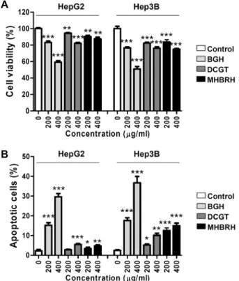
Fig. 1. Effects of ethanol extracts of Bigihwan (BGH), Daechilgi- tang (DCGT) and Mokwhyangbinranghwan (MHBRH) on the proliferation and apoptotic cell death in HepG2 and Hep3B human HCC cells. HepG2 and Hep3B cells were treated with the indicated concentrations of BGH, DCGT and MHBRH for 24 hr. (A) The cell viability was measured by an MTT assay. The data are expressed as the mean ± SD of three independent experiments (**p<
0.01 and *** p<0.0001 compared to the control). (B) The cells were fixed and stained with annexin V-FITC and PI for flow cytometry analysis. The percentages of apop- totic cells were determined by expressing the numbers of Annexin V+ cells as percentages of all the present cells.
The results are presented as the mean ± SD of three in- dependent experiments (*p<0.005, **p<0.001 and ***p<
0.0001 compared to the control).
시약(Bio-Rad Lab., Hercules, CA, USA)을 사용하여 정량하였 고, Laemilni sample buffer (Bio-Rad Lab.)를 섞어서 loading sample을 만들었다. 동량의 단백질을 sodium dodecyl sul- phate (SDS)-polyacrylamide gel을 이용하여 전기영동을 한 후 polyvinylidene difluoride (PVDF) membrane (Schleicher and Schuell, Keene, NH, USA)으로 전이시켰고, 5% skim milk를 30분 간 처리하고 적정 1차 항체(Santa Cruz Biotech- nology, Inc. 및 Cell Signaling Technology, Danvers, MA, USA)를 처리하여 4℃에서 over night 시킨 다음 PBS-T (PBS with Tween 20)를 사용하여 10분간 3번 세척하였다. 그 후 2차 항체를 상온에서 1시간 30분 동안 반응시킨 후 PBS-T로 10분간 3번 세척하고 암실에서 enhanced chemiluminoesence (ECL) solution (Amersham Corp., Arlington Heights, IL, USA)을 적용시킨 후 Chemi-smart (Vilber Lourmat, France) 를 이용하여 단백질들의 발현 변화를 분석하였다.
Mitochondrial membrane potential (MMP, Δψm) 측정 각 추출물 처리에 따른 미토콘드리아 기능 손상의 정도를 확인하기 위하여 5,5‘,6,6’-tetrachloro-1,1‘,3,3--tetraethyl-imi- dacarbocyanine iodide (JC-1) 염색을 실시하였다. 이를 위하 여 각 추출물이 24시간 동안 처리된 세포에 10 μM의 JC-1 (Sigma-Aldrich Chemical Co.) 용액을 처리하고 37℃에서 20 분 동안 염색시킨 후 PBS로 세포를 수세 후 flow cytometry 분석을 실시하였다.
Autophagy 유도의 확인
각 추출물에 의한 간암세포의 증식 억제가 autophagy 유도 와 연관성이 있는지를 확인하기 위하여 CYTO-ID
®autophagy detection kit (Enzo Life Sciences, Inc., Farmingdale, NY, USA)를 사용하였다. 이를 위하여 각 추출물이 처리된 세포에 assay buffer와 CYTO-ID green stain solution을 처리하여 37
℃에서 30분 동안 염색을 하였다. 그리고 assay buffer로 세척 한 후 PBS에 세포를 부유시켜 flow cytometry로 autophagy 유발 정도를 측정하였다. 또한 동일한 조건에서 배양된 세포 들을 대상으로 CYTO-ID 및 형광현미경을 이용하여 형광 강도 를 관찰하였다.
통계처리
GraphPad Prism® version 5.0 (Graphpad Inc., San Diego, CA, USA) one-way ANOVA를 사용하여 통계분석 하였으며, Turkey’s test로 사후 검정하여 p<0.01 값을 유의한 값으로 처 리하였다.
결과 및 고찰
비기환, 대칠기탕 및 목향빈랑환 에탄올 추출물이 간암세포 의 증식 및 apoptosis 유발에 미치는 영향
비기환, 대칠기탕 및 목향빈랑환 에탄올 추출물 처리에 따 른 간암세포(Hep3B 및 HepG2)의 생존율을 확인하기 위하여 각 추출물을 200 및 400 μg/ml의 농도로 24시간 동안 처리한 후 MTT assay를 수행하였다. Fig. 1A의 결과와 같이, 각 추출 물의 처리 농도 의존적으로 세포 생존율이 유의적으로 감소되 었으나, 비기환 추출물이 처리된 세포에서 다른 두 추출물에 비해서 생존율 감소가 크게 나타났고, 대칠기탕 및 목향빈랑 환 추출물 처리에 의한 생존율 억제 현상은 유사하였으며, HepG2 세포에 비해 Hep3B 세포에서 감수성이 다소 높았다.
각 추출물 처리에 의한 간암세포 생존율의 감소가 apopto-
sis 유도와 연관이 있는지 확인하기 위해서 Annexin V/PI 염
색을 한 후 flow cytometry 분석을 수행하였다. Fig. 1B의 결과
A
B
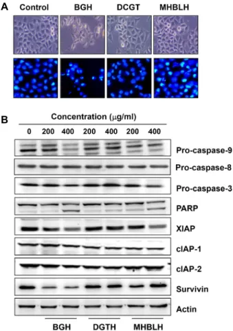
Fig. 2. Effects of BGH, DCGT and MHBRH on the cell morphol- ogy and the levels of apoptosis regulatory proteins in Hep3B cells. (A) Cells were treated with 400 μg/ml of BGH, DCGT and MHBRH for 24 hr. The morphological changes of Hep3B cells were observed using phase- con- trast microscopy. The DAPI-stained nuclei were pictured under fluorescence microscopy. Representative photo- graphs of the morphological changes are presented. (B) After treatment with the indicated concentrations of BGH, DCGT and MHBRH for 24 hr, total cell lysates were prepared. Equal amounts of cellular proteins were separated in SDS-polyacrylamide gels, and transferred to PVDF membranes. The membranes were probed with the indicated antibodies, and the proteins were visual- ized using an ECL detection system. Actin was used as an internal control for Western blot assays.
와 같이, 각 추출물의 처리 농도 의존적으로 apoptosis 유도가 증가되었고 세 가지 추출물 중에서 비기환 추출물에 의한 apoptosis 유발 효과가 가장 높게 나타났다. 아울러 apoptosis 유발 효과에서도 HepG2 세포보다 Hep3B 세포에서 다소 높게 나타났다.
각 추출물 처리에 따른 간암세포의 형태 변화의 예는 Hep3B 세포를 대상으로 제시하였으며, 비기환 추출물이 처리된 간암 세포에서 세포의 분지 형성, 밀도 감소와 부착력 상실과 같은 형태적 변화가 대칠기탕 및 목향빈랑환 처리군에 비하여 현저 하게 관찰되었다. 또한 전형적인 apoptosis가 유발된 세포에 서 관찰되는 염색질의 응축(chromatin condensation)에 의한 apoptotic body의 형성이 증가되었다(Fig. 2A). 따라서 3가지 에탄올 추출물 처리에 따른 간암세포의 증식억제는 apoptosis 유도와 연관이 있으며, 그중 비기환 추출물이 가장 항암 활성 이 높게 나타났다.
Hep3B 세포에서 apoptosis 관련 유전자들의 발현에 미 치는 비기환, 대칠기탕 및 목향빈랑환 에탄올 추출물의 영향
진핵세포에서의 apoptosis 유도 경로는 extrinsic (내재적) 및 intrinsic (외재적) 경로로 대별되며, caspase cascade가 핵 심적인 역할을 한다. Extrinsic 경로의 경우, 세포막에 존재하 는 death receptor에 death ligand의 결합에 따른 caspase-8의 활성화에 의하여 개시되며[7, 18], 이는 caspase-3 및 -7과 같은 effect caspase의 활성을 통하여 poly (ADP-ribose) polymer- ase (PARP)을 포함한 다양한 세포 내 기질 단백질의 분해를 유도한다[6, 8]. 반면 미토콘드리아를 중심으로 시작되는 in- trinsic 경로는 미토콘드리아에서 세포질로 유출된 cytochrome
c가 pro-caspase-9 및 apoptotic protease activating factor-1등과 apoptosome을 형성하여 caspase-9와 caspase-3/-7의 활 성을 촉진시켜 apoptosis를 종결하는 공동 경로로 유입된다[6, 18]. 그리고 caspase의 활성은 inhibitor of apoptosis protein (IAP) family에 속하는 단백질들에 의하여 그들의 활성이 억 제되어 apoptosis 유도를 억제하는 것으로 알려져 있다[3, 17].
따라서 3가지 추출물에 대한 증식억제 및 apoptosis 유도에 대한 감수성이 높았던 Hep3B 세포를 대상으로 각 추출물의 처리에 따른 apoptosis 유도 기전 연구를 수행하였다. Fig. 2B 의 결과에 의하면, caspase-9와 -3의 비활성형 발현이 비기환 추출물 처리 농도 증가에 따라 감소되었다. 반면, caspase-8의 발현에는 큰 변화가 없었으며, 활성화된 caspase-3의 기질 단 백질에 해당되는 PARP의 단편화가 대칠기탕 및 목향빈랑환 추출물 처리군에서 일부 증가되었지만, 비기환 추출물 처리군 에서 현저한 증가가 관찰되었다. 또한 비기환 추출물이 처리 된 세포에서 IAP family 단백질들 중에서는 survivin의 발현 이 현저하게 감소되었으며, XIAP의 발현도 대조군에 비하여 다소 억제되었다. 처방전에 따른 약간의 차이는 있었지만, 대 칠기탕 및 목향빈랑환 추출물 처리군에서는 큰 변화가 없었
다. 따라서 비기환 추출물 처리에 의한 apoptosis의 유발에는 extrinsic 경로 보다는 intrinsic 경로의 활성화가 더 관여를 함 을 알 수 있다. 비록 대칠기탕 및 목향빈랑환 추출물 처리군에 서는 apoptosis 유발의 정도는 낮지만, 처리 농도를 증가시킬 경우 비기환 추출물 처리군에서와 유사한 경로의 활성을 통하 여 apoptosis를 유발시킬 것으로 추정된다.
Hep3B 세포에서 Bcl-2 family의 발현 및 미토콘드리아의
기능에 미치는 비기환, 대칠기탕 및 목향빈랑환 추출물의 영향
A B
Fig. 3. Effects of BGH, DCGT and MHBRH on the levels of Bcl-2 family proteins and the MMP values in Hep3B cells. (A) After treatment with different concentrations of BGH, DCGT and MHBRH for 24 hr, total cell lysates were prepared and Western blotting was then performed using the indicated antibodies and an ECL detection system. Actin was used as an internal control. (B) The cells were collected and stained with JC-1 dye, and were then analyzed by a flow cytometer to evaluate the changes in MMP. Each bar represents the percentage of cells with JC-1 aggregates (mean SD of triplicate determinations,
**p<0.001 and ***p<0.0001, when compared to control)
Bcl-2 family에 속하는 인자들은 미토콘드리아 기능 손상과 연계된 intrinsic apoptosis 경로의 중요한 조절 인자로서 작용 한다[6, 10]. Bcl-2 family에 속하는 단백질은 apoptosis 유도를 억제하는 anti-apoptotic 단백질들과 반대 작용을 하는 pro- apoptotic 단백질들로 구성되어 있다. Bax를 포함한 pro-apop- totic 단백질이 Bcl-2와 같은 anti-apoptotic 단백질과 비교하여 상대적으로 발현이 증가하면 Bax가 미토콘드리아로 이동하여 MMP의 소실과 세포질로의 cytochrome c 방출을 유도하여 intrinsic apoptosis 경로가 활성화된다[7, 10]. 따라서 이상에 서 관찰된 Hep3B 세포의 apoptosis 유도와 미토콘드리아 기 능 손상과의 연관성을 조사하기 위하여 먼저 Bcl-2 family에 속하는 주요 단백질의 발현 변화를 확인하였다. Fig. 3A의 결 과와 같이, 비록 Bax의 발현은 각 추출물의 처리에 따라 큰 변화가 없었지만, Bcl-2의 발현은 비기환 추출물 처리에 따라 현저히 감소되었다. 그리고 미토콘드리아의 기능 손상의 지표 인 MMP 소실의 값이 비기환 추출물이 처리된 세포에서 처리 농도 의존적으로 현저히 증가하였다(Fig. 3B).
한편 손상된 미토콘드리아를 autophagy (자가포식)에 의하 여 제거되는 과정인 mitophagy에 중심적으로 관여하는 조절 자는 serine-threonine kinase인 PTEN-induced kinase 1 (PINK1)과 E3 ubiquitin ligase인 Parkin이다[16, 26]. 특히 MMP가 감소되면 단백질 분해효소(mitochondrial inner membrane protease)인 presenilin-associated rhomboid-like protease (PARL)가 PINK1을 분해하지 못하기 때문에 미토콘 드리아의 PINK1 단백질이 안정화되게 된다[11, 20]. 안정화된 PINK1은 Parkin을 미토콘드리아 막으로 이동시키게 되고 E3 ubiquitin ligase 효소작용을 활성화하여 미토콘드리아의 단백 질들의 ubiquitination을 일으켜 mitophagy 과정이 시작된다 [14, 21]. 따라서 비기환 추출물 처리군에서 관찰된 Bcl-2의 발 현 감소 및 MMP의 소실이 mitophagy 유도와 연관성이 있는
지를 확인하기 위하여 PINK1의 발현을 조사한 결과, 비기환 추출물이 처리된 Hep3B 세포에서 PINK1의 발현이 현저하게 증가된 반면, 대칠기탕 및 목향빈랑환 추출물 처리군에서는 유의적인 증가가 관찰되지 않았다(Fig. 3A). 이는 비기환 추출 물에 의한 apoptosis의 유도는 Bcl-2 family 단백질의 발현 변 화에 따른 미토콘드리아 손상이 관여하고 있음을 의미하며, 이를 통하여 intrinsic apoptosis 경로가 활성화되었음을 유추 할 수 있다.
Hep3B 세포의 autophagy 유도에 미치는 비기환, 대칠기 탕 및 목향빈랑환 추출물의 영향
다음은 세 가지 추출물 처리에 의한 apoptosis 유도 과정에 autophagy 현상이 동반되는지 확인하였다. Autophagy는 세 포 내에서 불필요하거나 기능이 손상된 세포 성분을 제거하는 과정으로 세포 구성 요소의 파괴와 재활용에 관여한다[15, 25].
이 과정은 분해 대상 성분들이 세포 내의 다른 성분들과는
격리되어 이중막에 둘러싸여 autophagosome (자가소화포)이
형성되고 lysosome과 융합되어 내용물들은 분해되어 재활용
되는 단계를 포함한다[5, 19]. Autophagy 동안 microtubule-
associated protein light chain-3 (LC3)-I는 LC3-II로 가공되어
autophagosome 막으로 전위되고, ubiquitin-like protein con-
jugation이라는 Atg (autophagy related gene) 계열 단백질들
이 막에 부착되는 현상이 동반된다. 즉 Atg 및 LC3-I 뿐만 아니
라 LC3-II 발현의 증가는 autophagy 유도의 지표로 활용된다
[23, 25]. 따라서 비기환, 대칠기탕 및 목향빈랑환 추출물이
Hep3B 세포에서 autophagy를 유도할 수 있는지를 확인하기
위하여 Atg 단백질들 중에서 단백질 및 소기관에 대한 lyso-
some이화 경로의 주요 인자인 Atg5 [4, 23] 및 LC3의 발현
변화를 조사하였다. 본 연구의 결과에 의하면, 비록 Atg5의 발
현에는 큰 변화가 없었지만, 비기환 추출물이 처리된 Hep3B
A B
C
Fig. 4. Effects of BGH, DCGT and MHBRH on the autophagy induction in Hep3B cells. (A) Cells were treated with the indicated concentrations of BGH, DCGT and MHBRH for 24 hr. Cell lysates were prepared, and Western blotting was then performed using the indicated antibodies. Actin was used as an internal control. (B) Relative accumulation of autophagosomes after treatment with BGH, DCGT and MHBRH for 24 hr measured by staining with Cyto-ID. Values represent mean ± SD of three independent experiments (*p<0.005, **p<0.001 and ***p<0.0001 compared to the control). (C) Cells were collected and then stained with Cyto-ID (green) and Hoechst 33342 (blue) for visualization of autophagosomes and nuclei, respectively, and analyzed by fluorescence microscopy.
세포에서 LC3-I 및 LC3-II의 발현이 모두 증가되었다(Fig. 4A).
이러한 autophagy 유도의 정량적인 평가를 위하여 CYTO-ID 염색에 의한 flow cytometry 분석을 실시한 결과, 대칠기탕 및 목향빈랑환 추출물 처리군에서도 다소 증가하였으나 비기 환 추출물이 처리된 세포에서 처리 농도 의존적으로 autoph- agy의 유도가 현저하게 증가하였다(Fig. 4B). 이러한 현상은 Cyto-ID green dye를 이용한 형광현미경적 분석에서도 유사 하게 관찰되었다(Fig. 4C).
다음은 이상에서 관찰된 세 가지 추출물 처리에 의한 au- tophagy의 유도가 간암세포의 증식억제에 어떠한 역할을 하 는지를 조사하기 위하여 autophagy 과정에서 autolysosome 형성을 선택적으로 억제하는 bafilomycin A1 (BA1)를 사용하 였다. Fig. 5A 및 Fig. 5B의 결과와 같이 BA1을 전처리하였을 때 비기환 추출물 처리에 의하여 증가된 apoptosis가 유의적 으로 감소되었으며, 이에 따라 억제된 세포생존율도 회복이 되었다. 대칠기탕과 목향빈랑환의 처리군의 경우에도 비기환 추출물 처리군에 비하여 감소의 폭은 낮았으나, apoptosis 유 도의 억제 및 생존율 회복의 현상이 관찰되었다. 이러한 결과
는 세 가지 추출물에 의한 생존율 억제와 연관된 apoptosis 유도에 동반된 autophagy는 apoptosis 유도에 관여하고 있음 을 유추할 수 있었다.
본 연구의 결과에 의하면 조사된 3가지 처방전의 에탄올
추출물 중에서 비기환이 대칠기탕과 목향빈랑환에 비하여 간
암세포에서 항암활성이 높게 나타났다. 이는 오적과 적병에
사용되어 온 대칠기탕과 목향빈랑환 보다 비기환이 간적에
사용되어온 처방이란 점과도 연계된다. 이들의 항암활성은
apoptosis 유도와 밀접한 연관성이 있었으며, 미토콘드리아
손상을 매개로 이루어짐을 알 수 있었고, 이 과정에서 autoph-
agy가 동반되었음을 확인하였다. 현재까지 항암활성을 지니
는 약물에 의한 암세포의 죽음에서 apoptosis와 autophagy
유도 사이에서의 연계성은 여전히 논란의 여지가 있다. 그러
나 세포의 죽음이나 생존의 조절 측면에서 autophagy가
apoptosis를 촉진시키거나 억제할 수 있다는 측면에서 항암제
발굴의 중요한 표적임은 확실하다. 따라서 이들 처방전의 간
암세포 증식 억제에 관한 세포 신호 전달계 및 에너지 대사
연계성 등을 포함한 추가적인 기전 연구의 수행과 각 처방전
A
B
Fig. 5. Effects of an autophagy inhibitor, bafilomycin A1, on BGH, DCGT and MHBRH-induced apoptosis and growth inhibition in Hep3B cells. (A) Cells were preincubated with 5 nM bafilomycin A1 (BA1) for 2 hr and then treat- ed with the indicated concentrations of BGH, DCGT and MHBRH for 24 hr. The cells were stained with annexin V-FITC and PI for flow cytometry analysis. The percen- tages of apoptotic cells were determined by expressing the numbers of Annexin V+ cells as percentages of all the present cells. (B) The cells grown under the same conditions as (A) were collected. Then the cell viability was evaluated with an MTT assay. The results are pre- sented as the mean ± SD of three independent experi- ments (**p<0.001 and ***p<0.0001 vs. untreated control;;
##p<0.001 and ###p<0.0001 present vs. absent BA1).
의 활성 성분에 대한 분석이 이루어져야 할 것이다. 본 연구의 결과는 이러한 추가 연구를 통하여 간암과 연관된 전통 처방 전의 효능과 기전을 이해하기 위한 기초 자료로서 활용될 것 이다.
감사의 글
본 연구는 한국연구재단(2018R1A2B2005705)의 지원을 받 아 수행되었음.
The Conflict of Interest Statement The authors declare that they have no conflicts of interest
with the contents of this article.
References
1. Baik, T. H. and Lee, I. 1997. An experimental study on the effects of mkwhyangbinrang-whan. J. Int. Kor. Med. 18, 373- 390.
2. Brown, Z. J., Heinrich, B. and Greten, T. F. 2018. Mouse models of hepatocellular carcinoma: an overview and high- lights for immunotherapy research. Nat. Rev. Gastroenterol.
Hepatol. 15, 536-554.
3. Chaudhary, A. K., Yadav, N., Bhat, T. A., O'Malley, J., Kumar, S. and Chandra, D. 2016. A potential role of X-linked in- hibitor of apoptosis protein in mitochondrial membrane per- meabilization and its implication in cancer therapy. Drug Discov. Today 21, 38-47.
4. Codogno, P. and Meijer, A. J. 2006. Atg5: more than an au- tophagy factor. Nat. Cell Biol. 8, 1045-1047.
5. Condello, M., Pellegrini, E., Caraglia, M. and Meschini, S.
2019. Targeting autophagy to overcome human diseases. Int.
J. Mol. Sci. 20, E725.
6. Dorstyn, L., Akey, C. W. and Kumar, S. 2018. New insights into apoptosome structure and function. Cell Death Differ.
25, 1194-1208.
7. Fulda, S. and Debatin, K. M. 2006. Extrinsic versus intrinsic apoptosis pathways in anticancer chemotherapy. Oncogene 25, 4798-4811.
8. Hajra, K. M. and Liu, J. R. 2004. Apoptosome dysfunction in human cancer. Apoptosis 9, 691-704.
9. Han, S. I. and Kang, B. K. 1991. Antitumor effects of bi- gihwan on tumor cells derived from leukemia and lympho- ma patients. J. Int. Kor. Med. 12, 1-15.
10. Hata, A. N., Engelman, J. A. and Faber, A. C. 2015. The BCL2 family: Key mediators of the apoptotic response to targeted anticancer therapeutics. Cancer Discov. 5, 475-487.
11. Jin, S. M., Lazarou, M., Wang, C., Kane, L. A., Narendra, D. P. and Youle, R. J. 2010. Mitochondrial membrane poten- tial regulates PINK1 import and proteolytic destabilization by PARL. J. Cell Biol. 191, 933-942.
12. Kang, D. G. and Kang, B. K. 1991. Antitumor effects of sig- bunhwan and bigihwan on tumor cells derived from leuke- mia and lymphoma patients. J. Int. Kor. Med. 12, 96-112.
13. Kim, H. Y., Ko, S. J., Bang, C. H., Shin, S. H., Lee, D. Y.
and Lee, I. 2010. Effects of daechilgi-tang on glutamate-in- duced apoptosis in C6 glial cells. Kor. J. Orient. Int. Med.
31, 693-705.
14. Kim, Y., Park, J., Kim, S., Song, S., Kwon, S. K., Lee, S. H., Kitada, T., Kim, J. M. and Chung, J. 2008. PINK1 controls mitochondrial localization of Parkin through direct phos- phorylation. Biochem. Biophys. Res. Commun. 377, 975-980.
15. Lin, L. and Baehrecke, E. H. 2015. Autophagy, cell death, and cancer. Mol. Cell. Oncol. 2, e985913.
16. Miller, S. and Muqit, M. M. K. 2019. Therapeutic approaches to enhance PINK1/Parkin mediated mitophagy for the treatment of Parkinson's disease. Neurosci. Lett. 705, 7-13.
초록:인체 간암세포에서 비기환(肥氣丸), 대칠기탕(大七氣湯) 및 목향빈랑환(木香檳榔丸)의 항암 활성 비교
김소영
1,2․홍수현
1,3․최성현
4․정재훈
2․최영현
1,3*
(1동의대학교 항노화연구소, 2부산대학교 분자생물학과, 3동의대학교 한의과대학 생화학교실, 4한국승강기대학교
승강기시스템관리과)
간암은 전 세계적으로 가장 높게 진단되는 암 중 하나이며, 방사선 및 화학 요법이 일반적으로 사용되는 치료법 이지만 다양한 부작용은 치료 효과를 크게 제한한다. 따라서 전통 의학에서 사용되어 온 처방법은 이를 극복할 수 있는 대안이 될 수 있다. 본 연구에서는 동의보감에 기술되어 있는 3가지 한약 처방전(비기환, 대칠기탕 및 목향빈랑환)을 선택하여 인체 간암세포에 대한 항암 효과를 평가하였다. 간암세포에서 3가지 처방전의 에탄올 추 출물의 세포 독성을 조사하기 위한 MTT 분석 결과, 비기환 추출물은 대칠기탕 및 목향빈랑환에 비하여 세포 생존 력을 현저하게 억제하였다. 그리고, flow cytometry 분석의 결과에서 3가지 처방전 추출물에 의한 간암세포의 증 식 억제가 apoptosis 및 autophagy 유도와 관련이 있었다. 특히, 비기환 추출물은 미토콘드리아의 기능을 크게 손상시켰으며 다른 두 처방전과 비교하여 mitophagy 유발 가능성을 보여주었다. 아울러 비기환 추출물은 LC3의 발현을 현저하게 증가시켰으며, 이는 Bcl-2의 발현 감소를 동반하는 반면, Bax의 발현에 영향을 미치지 않았다.
PINK1의 발현 또한 비기환 추출물이 처리된 세포에서 매우 증가하였다. 나아가, autophagy 억제제는 3가지 처방 전 추출물 처리에 의한 세포 생존율 감소와 apoptosis 유도를 억제하였으며, 이러한 결과는 이들 처방전 추출물 처리에 의한 autophagy가 apoptosis 개시에 관여하고 있음을 보여주는 것이다. 결론적으로, 본 연구 결과는 비기 환 추출물이 3가지 처방전 중에서 가장 높은 항암 활성을 보였으며, 비기환 추출물은 autophagy 유도제로서 간암 세포의 증식을 억제함을 의미한다.
17. Mohamed, M. S., Bishr, M. K., Almutairi, F. M. and Ali, A. G. 2017. Inhibitors of apoptosis: Clinical implications in cancer. Apoptosis 22, 1487-1509.
18. Reubold, T. F. and Eschenburg, S. 2012. A molecular view on signal transduction by the apoptosome. Cell. Signal. 24, 1420-1425.
19. Russo, M. and Russo, G. L. 2018. Autophagy inducers in cancer. Biochem. Pharmacol. 153, 51-61.
20. Sekine, S. and Youle, R. J. 2018. PINK1 import regulation;
a fine system to convey mitochondrial stress to the cytosol.
BMC Biol. 16, 2.
21. Sha, D., Chin, L. S. and Li, L. 2010. Phosphorylation of par- kin by Parkinson disease-linked kinase PINK1 activates par- kin E3 ligase function and NF-kappaB signaling. Hum. Mol.
Genet. 19, 352-363.
22. Soussi, T., Ishioka, C., Claustres, M. and Béroud, C. 2006.
Locus-specific mutation databases: pitfalls and good prac- tice based on the p53 experience. Nat. Rev. Cancer 6, 83-90.
23. Wesselborg, S. and Stork, B. 2015. Autophagy signal trans- duction by ATG proteins: from hierarchies to networks. Cell.
Mol. Life Sci. 72, 4721-4757.
24. Xu, W., Yu, J. and Wong, V. W. 2017. Mechanism and pre- diction of HCC development in HBV infection. Best Pract.
Res. Clin. Gastroenterol. 31, 291-298.
25. Yan, X., Zhou, R. and Ma, Z. 2019. Autophagy-cell survival and death. Adv. Exp. Med. Biol. 1206, 667-696.
26. Youle, R. J. and Narendra, D. P. 2011. Mechanisms of mitophagy. Nat. Rev. Mol. Cell Biol. 12, 9-14.