면역세포에서 Bioconversion 전후 제주 감귤 과피 추출물의 항염증 효과
서지은1․임희진1․장윤희1․박혜련1․한복경2․정중기2․최경숙2․박수범2․최혁준2․황진아1
1명지대학교 식품영양학과
2(주)비케이바이오
Effects of Jeju Citrus unshiu Peel Extracts Before and After Bioconversion with Cytolase on Anti-Inflammatory Activity in RAW264.7 Cells
Jieun Seo1, Heejin Lim1, Yun-Hee Chang1, Hye-Ryeon Park1, Bok-Kyung Han2, Jung-Ky Jeong2, Kyoung-Sook Choi2, Su-Beom Park2, Hyuk-Joon Choi2, and Jinah Hwang1
1Department of Food and Nutrition, College of Natural Sciences, Myongji University
2BK Bio Co., Ltd.
ABSTRACT Citrus and its peels, which are by-products from juice and/or jam processing, have long been used in Asian folk medicine. Citrus peels show an abundant variety of flavanones, and these flavanones have glycone and aglycone forms. Aglycones are more potent than glycones with a variety of physiological functions since aglycone absorption is more efficient than glycones. Bioconversion with cytolase converted narirutin and naringin into naringenin and hesperidin into hesperetin. Therefore, this study aimed to investigate the anti-oxidant and anti-inflammatory effects of bioconversion of Citrus unshiu (CU) peel extracts with cytolase (CU-C) in RAW264.7 cells. HPLC chromatograms showed that CU and CU-C had 23.42% and 29.39% total flavonoids, respectively. There was substantial bioconversion of narirutin to naringenin and of hesperidin to hesperetin. All citrus peel extracts showed DPPH scavenging activities in a dose-dependent manner, and CU-C was more potent than intact CU. RAW264.7 cells were pre-treated with 0∼500 μg/mL of citrus peel extracts for 4 h and then stimulated by 1 μg/mL of lipopolysaccharide (LPS) for 8 h. All citrus peel extracts showed decreased mRNA levels and protein expression of LPS-induced inducible nitric oxide synthase (iNOS) and cyclooxygenase-2 (COX-2) in a dose-dependent manner. Especially, CU-C markedly inhibited mRNA and protein expression of iNOS and COX-2 compared to intact citrus peel extracts. All citrus peel extracts showed decreased NO production by iNOS activity. This result suggests that bioconversion of citrus peel extracts with cytolase may provide potent functional food materials for prevention of chronic diseases attributable to oxidation and in- flammation by boosting the anti-inflammatory effects of citrus peels.
Key words: Citrus unshiu peel extract, bioconversion, anti-oxidant, anti-inflammation, RAW264.7 cells
Received 12 September 2014; Accepted 29 January 2015 Corresponding author: Jinah Hwang, Department of Food and Nutrition, College of Natural Sciences, Myongji University, Yongin, Gyeonggi 449-728, Korea
E-mail: [email protected], Phone: +82-31-330-6205
서 론
염증(inflammation)은 물리적 작용이나 화학적 물질, 바 이러스나 박테리아와 같은 외부의 자극원이나 다른 질병에 대한 생체방어 기작으로 당뇨, 심혈관질환, 관절염, 암, 신경 변성질환 등의 많은 만성질환에서 근본적인 병태생리학적 기전으로 알려져 있다(1-3). 면역력에 결함이 생겨 면역반 응이 충분하게 일어나지 않으면 감염을 유도할 수 있는 반면 면역을 위한 염증반응이 과도하게 일어나면 만성질환을 일 으키게 된다(4).
대식세포(macrophage)는 면역과 염증반응에서 주요 역
할을 하고 면역반응 시에는 감염 초기에 생체방어를 위해서 여러 사이토카인(cytokine)과 산화질소(nitric oxide, NO) 등을 생산한다. 그람양성균인 lipopolysaccharide(LPS)는 염증반응을 유도하는 강한 자극물질로 대식세포에 LPS와 같은 물질에 의해 자극이 가해지면 NO, 염증성 사이토카인, 키모카인(chemokine)뿐만 아니라 prostaglandin, leuko- triene 등의 mediator를 과도하게 생성한다(5,6).
감귤류(Citrus unshiu)의 과피는 동양의학에서 기관지와 천식 환자에게 사용되어 왔으며 최근에는 감염성질환이나 암 등을 예방한다고 보고되었다(7,8). 감귤류는 비타민 C, 엽산, 식이섬유, 카로티노이드(carotenoids), 플라보노이드 (flavonoids) 등이 함유되어 있고, 감귤류 플라보노이드는 항균, 항산화, 항암, 항염증 효과 등 많은 기능을 갖는다고 보고되었다(9,10). 감귤류에 존재하는 플라보노이드는 분자 구조에 따라 플라본(flavones), 플라바논(flavanones), 플라 보놀(flavonols), 이소플라본(isoflavones), 안토시아니딘
(anthocyanidins), 플라바놀(flavanols) 등의 6개 그룹으로 분류되며(9,11), 특히 과육보다는 과피와 씨에 플라바논이 풍부하다(11). 감귤류 플라바논은 narirutin, naringin, hesperidin 등의 배당체(glycone), naringenin과 hesper- etin 등의 비배당체(aglycone) 형태로 존재한다(9,12). 비 배당체는 배당체보다 좀 더 효과적으로 우리 몸에서 흡수되 기 때문에 플라바논의 rutinoside 형태의 물질이 가수분해 과정을 거쳐 비배당체의 형태로 전환되면 흡수가 촉진되어 혈장, 담즙, 소변 등에 더 높은 농도로 존재한다(9,13).
Naringin은 α-D-rhamnosidase와 β-D-glucosidase에 의 해서 naringenin으로 전환되고, hesperidin은 β-D-gluco- sidase나 α-D-rhamnosidase에 의해서 hesperetin으로 전 환된다. Cytolase PLC5는 Aspergillus niger의 배양물에 서 얻어진 효소제제로서 pectinases 외에 cellulase, hemi- cellulase, β-glucosidase 등의 다양한 활성을 가지고 있 다. 상업적으로는 산도가 높은 과실류 가공 시 침전 제거, 수율 및 여과력 향상에 주로 사용된다. 또한 당분해효소로서 배당체 형태의 플라보노이드를 비배당체 형태로 전환시켜 추출 효율을 높일 수 있다(14). 인삼 추출물을 β-glucosi- dase와 cytolase로 효소 반응시키면 ginsenosides의 형태 로 전환되었으며 대식세포에서 염증을 억제시키고 결장선암 세포에서 항암 효과를 나타내었다(15,16). 따라서 최근에 미생물이나 효소적 반응의 bioconversion(물질전환)을 통 한 비배당체 형태의 플라보노이드 개발이 활성화되고 있다.
제주도의 감귤류 생산량이 지속적으로 증가함에 따라 소 비보다 공급되는 양이 많아지고, 감귤이나 한라봉에 대한 가공제품 또한 계속 개발되고 있다. 가공제품을 생산하는 과정에서 생기는 가공부산물인 과피는 비용 및 환경문제를 유발하며 대부분 폐기물 처리되고 있다. 감귤류는 과육보다 과피에 많은 생리활성 성분이 있으므로 본 연구에서는 상업 화된 효소를 이용하여 감귤 과피 내의 생리활성 물질 중 배 당체를 비배당체의 형태로 물질전환 하였을 때 면역세포에 서 항산화와 항염증 효과가 있는지를 확인하고자 하였다.
재료 및 방법
감귤 과피 추출물의 조제
감귤 과피 추출물(Citrus unshiu, CU)과 cytolase의 가 수분해로 효소 처리한 감귤 과피 추출물(CU-C)은 (주)BK Bio(Seongnam, Korea)로부터 제공받았다. 감귤 과피는 제 주연구소에서 공급받아 과피분말 100 g을 0.5%의 cyto- lase PCL5(DSM Food Specialties, Heerlen, Nether- lands)를 첨가하여 물을 용매로 pH 4.0, 60°C에서 14시간 동안 효소 처리 후 여과시켜 잔사를 제거하였다. 여기에 4배 부피(v/v)의 80% ethanol을 가하여 60°C에서 8시간 동안 교반추출 후 여과시켜 분리된 상층액만 취해 농축 후 동결건 조 하여 분말화하였다. CU와 CU-C의 플라보노이드 구성성 분의 분석을 위해 고속 액체 크로마토그래피(high per-
formance liquid chromatography, HPLC; YL9100HPLC, Younglin, Anyang, Korea)와 Eclipse plux C18 column (4.6 mm I.D.×250 mm, Agilent Co., Santa Clara, CA, USA)을 이용하였고, 시료 주입량은 10 μL, column oven 온도는 35°C, 유속 1 mL/min, 검출 파장은 280 nm에서 60분 동안 분석하였다. 이동상은 0.5%(v/v) acetic acid/
acetonitrile과 0.5%(v/v) acetic acid/distilled water를 gradient 조건에 따라 사용하였다. 시료의 플라보노이드 함 량을 정량 분석하기 위해서 internal control로 naringin, narirutin, hesperidin, naringenin, hesperetin(Sigma- Aldrich Co., St. Louis, MO, USA)을 이용하여 비교 분석하 였으며 이를 위해서 YLClarity chromatography station system을 이용하였다.
세포의 배양과 분화
세포주는 면역세포주인 RAW264.7 cell line(KCLB No 40071, Korea Cell Line Bank, Seoul, Korea)을 이용하였 고, Dulbecco's modified Eagle's medium(DMEM, Gibco, Grand Island, NY, USA) 배지에 10% fetal bovine serum (FBS, Gibco)과 1% penicilin/streptomycin(P/S, Gibco) 을 첨가하여 37°C, 5% CO2의 환경에서 배양하였다. 배지는 2일 간격으로 교환하여 주었고 80% 정도 배양되면 cell scraper로 세포를 조심스럽게 떼어내서 1:6~10의 비율로 계대배양 하였다.
1% P/S를 첨가한 DMEM 배지에 감귤 과피 bioconver- sion 전후 시료 200, 500 μg/mL의 농도로 제조하여 0.2 μm syringe filter(Pall Corporation, Port Washington, NY, USA)를 이용하였다. 시료를 각 농도별로 4시간 동안 처리한 뒤 1 μg/mL의 lipopolysaccharide(LPS, Sigma- Aldrich Co.)를 처리하고 8시간 배양한 뒤 실험을 진행하였 다. Sinetrol(Fytexia, Vendres, France)은 양성대조군으 로 사용하였으며 시료와 동일하게 처리하였다(17).
MTT assay를 이용한 세포생존율 측정
96-well plate에 약 2×10⁵ cells/mL 농도의 세포를 200 μL씩(약 4×10⁴ cells/well) 분주하여 배양한 뒤, 0~750 μg/
mL 농도의 Sinetrol, 감귤 과피 추출물을 24시간 처리한 배지에 10 μL의 thiazolyl blue tetrazolium bromide(5 mg/mL MTT, Amresco, Solon, OH, USA)를 넣고 1분간 잘 섞어주었다. 3시간 동안 배양한 후 well의 배지를 제거하 고 isopropanol에 용해시킨 0.4 N HCl 100 μL를 넣어서 생성된 보라색의 formazan crystal을 잘 녹여내었다. 분광 광도계(Gen5.2, BioTek, Winooski, VT, USA)를 이용하여 570 nm의 파장에서 흡광도를 측정하였다.
DPPH radical assay를 이용한 항산화 능력 측정 산화적 스트레스를 유발하는 radicals의 소거 능력을 통하 여 항산화 효과를 측정하기 위해 Brand-Williams 등(18)의
Table 1. HPLC chromatograms of Citrus unshiu peel extracts1) CU2) (%) CU-C3) (%) Glycone
Naringin Narirutin Hesperidin
0.01 3.38 19.97
0.00 1.59 15.20 Aglycone
Naringenin Hesperetin
0.02 0.05
6.10 6.49
Total 23.42 29.39
1)Provided by BK Bio Co., Ltd.
2)CU: Citrus unshiu peel extract.
3)CU-C: bioconversion of Citrus unshiu peel extract with cytolase.
방법을 수정하여 이용하였다. 먼저 메탄올에 용해시킨 0.1 mM 2,2-diphenyl-1-picrylhydrazyl(DPPH, Sigma-Aldrich Co.) 용액 120 μL를 Sinetrol과 감귤 과피 추출물을 메탄올 에 용해시켜 200과 500 μg/mL 농도로 만든 80 μL에 섞어 주고 차광 상태를 유지하며 실온에서 30분간 반응시킨 후 분광광도계(Epoch, BioTek)를 이용하여 515 nm의 파장에 서 흡광도를 측정하였다. 양성대조군으로는 대표적인 항산 화 물질인 quercetin(50 μg/mL, Sigma-Aldrich Co.)뿐 아 니라(19) 현재 시판되고 있는 감귤류의 폴리페놀을 이용한 건강기능식품인 프랑스의 Sinetrol(Fytexia)을 사용하여 시 료와 동일하게 처리하였다(17). 항산화력의 수치화는 다음 과 같은 식을 이용하였다.
DPPH 라디칼 소거능(%)=
(
1- Blank 흡광도시료 흡광도)
×100Real time-polymerase chain reaction을 이용한 mRNA levels 측정
RAW264.7 세포의 mRNA는 Trizol reagent(Invitro- gen, Grand Island, NY, USA)를 이용하여 추출하였고 re- verse transcription master premix(ELPIS, Daejeon, Korea)로 역전사를 진행시켜 cDNA를 합성시켰다. 유전자 증폭을 위한 primers(Bioneer, Daejeon, Korea)는 induc- ible nitric oxide synthase(iNOS) forward 5'-ACGCTT- CACTTCCAATGC-3', backward 5'-GGGCTCTGTTGAG- GTCTA-3'; cyclooxygenase-2(COX-2) forward 5'-CT- CTCTGAACTATGGTGTGAAC-3', backward 5'-GTC- TGCTTTATGCGTAAATTCC-3'; β-actin forward 5'-GG- GAAGGTGACAGCATTG-3', backward 5'-ATGAAGTA- TTAAGGCGGAAGATT-3'로 제작하여 실험하였다. 0.25 μg의 cDNA와 10 pM primers 1 μL, 2X SYBR green su- permix(Biorad, Hercules, CA, USA) 10 μL, RNase-free water 8 μL를 섞어 CFX96 Touch(Biorad)를 이용하여 유 전자 증폭 및 발현을 확인하였다.
Western blotting을 이용한 단백질 검출
시료를 처리한 RAW264.7 세포를 분리하여 whole cell lysates를 얻었다. 이것을 10% SDS-PAGE에 전기영동을 통해 단백질을 분리하고 polyvinylidene difluoride mem- brane(Roche, Nutley, NJ, USA)으로 transfer 과정을 거친 후에 iNOS, COX-2, β-actin(Santa Cruz Biotechnology, Inc., Dallas, TX, USA)의 항체를 붙이고, 그 후 화학발광반 응법을 이용하여 가시화시켰다.
Griess assay를 이용한 NO 생성 측정
NOS에 의하여 생성되는 NO의 생성에 미치는 영향을 확 인하기 위하여 시료 처리가 끝난 RAW264.7 세포의 배지만 모아서 NO level을 측정하였다. 40 μL의 배지시료와 표준 물질인 sodium nitrite(0~35 μM, Sigma-Aldrich Co.)에
5 μL의 nitrate reductase cofactor와 5 μL의 nitrate re- ductase enzyme을 잘 섞어준 후 실온에서 2시간 동안 반응 시켰다. 반응이 끝난 well에 5% phosphoric acid(Sigma- Aldrich Co.)에 용해시킨 1% sulfanilamide 25 μL와 0.1
% N-(1-naphthyl)-ethylenediamine dihydrochloride(Sig- ma-Aldrich Co.) 25 μL를 넣었다. 빛을 차단하며 10분 동안 실온에서 반응시켜준 후 분광광도계(Epoch, BioTek)를 이 용하여 540 nm의 파장에서 흡광도를 측정하였다.
통계처리
각 실험은 3회 이상 반복실험을 실시하였고 결과는 평균
±표준편차로 표시하였다. 또한 모든 실험 결과는 IBM SPSS (Ver. 20.0, IBM, Armonk, NY, USA)를 이용하여 Stu- dent's t-test와 one-way ANOVA로 분석하였다. 통계적 유의성은 Tukey test를 이용하여 검증하였고 5% 이내에서 통계적 유의성을 제시하였다(P<0.05).
결과 및 고찰
감귤 과피 추출물의 효소 처리에 의한 플라보노이드 형태 전환
감귤 과피 추출물의 플라보노이드의 형태가 cytolase를 통해 효과적으로 전환되었는지를 확인하기 위해 배당체 형 태의 narirutin, naringin, hesperidin과 비배당체 형태의 naringenin과 hesperetin을 HPLC로 분석하였다(Table 1).
Naringin과 narirutin은 α-L-rhamnosidase에 의해 prunin 으로 전환된 후 β-glucosidase에 의해 최종적으로 agly- cone인 naringenin으로 전환되며, hesperidin은 α-L- rhamnosidase에 의해 hesperetin glucoside로 전환된 후 β-glucosidase에 의해 aglycone인 hesperetin으로 전환 된다(9,11,13). CU와 CU-C의 총 플라보노이드 양은 각각 23.42%, 29.39%이며 배당체와 비배당체의 형태에 따른 수 치가 다르게 나타남을 확인하였다. CU에서 CU-C로 전환시 켰을 때 glycone 중 naringin과 narirutin의 수치가 감소하 였고, aglycone 형태인 naringenin은 0.02%에서 6.10%로 증가하였다. 또 glycone 형태의 hesperidin은 효소 처리 전
ab
a a a a a
c
0 20 40 60 80 100 120 140
Sinetrol CU CU-C
Cell viability (% Control) .
0 100 250
400 500 600
750 μg/mL
Fig. 1. Effects of CU and CU-C on cell viability. RAW264.7 cells were treated with different concentrations (0∼750 μg/mL) of Sinetrol, CU or CU-C for 24 h. Cell viability was measured by MTT assay. The viability of untreated control cells was de- fined as 100%. Each bar represents the mean±SEM (n=3).
Means with different letters (a-c) above the bars are significantly different according to ANOVA, Tukey test (P<0.05).
d d
d d
a
b bc
b
a
b a
0 20 40 60 80 100
Sinetrol CU CU-C Quercetin50
DPPH radical scavenging activity . (% Control) .
0 200 500 μg/mL
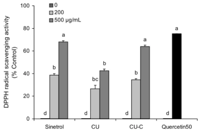
Fig. 2. Effects of CU and CU-C on DPPH radical scavenging activity. Quercetin (50 μg/mL) was used as a positive control.
Each bar represents the mean±SEM (n=3). Means with different letters (a-d) above the bars are significantly different according to ANOVA, Tukey test (P<0.05).
에는 19.97%였으나 처리 후에 15.20%로 감소한 대신 aglycone인 hesperetin이 효소 처리 전에는 0.05%로 미미 하다가 처리 후에 6.49%로 증가하였다(Table 1). 이것은 cytolase를 이용한 효소 처리로 naringin과 narirutin이 naringenin으로, 또는 hesperidin이 hesperetin으로 변환 된 것으로 사료된다. 이는 cytolase가 장관 내에서 D-glu- cosides의 비환원 말단에서 포도당을 가수분해 시키는 β- glucosidases와 유사한 역할을 하는 것으로 보인다. Agly- cone 형태는 glycone 형태보다 더 효과적으로 흡수되어 혈 청과 소변, 담즙에서 더 높은 농도의 aglycone류가 존재한 다(9,13,14). Naringin은 CU군에서 거의 검출되지 않았는 데 이것은 naringin이 감귤의 주요 플라보노이드가 아님을 확인할 수 있었다. Rutinose가 붙어있는 narirutin과 hes- peridin은 쓴맛이 거의 없으나 neohesperidose가 붙어있는 naringin은 쓴맛이 난다(9). 양성대조군으로 사용한 Sin- etrol은 감귤류의 폴리페놀 중 특히 플라보노이드의 nar- ingin, narirutin, hesperidin, neohesperidin으로 주로 구 성되어 있다(17). 그중 naringin의 양이 가장 많으며 이것은 Sinetrol이 강한 쓴맛을 내는 것과 연관이 있어 알약의 형태 를 제외하고는 산업적으로 이용하는 데 많은 제약이 있다고 보고되었다(17).
감귤 과피 추출물이 세포생존율에 미치는 영향
면역세포에서 감귤의 bioconversion 전후 추출물의 세포 독성과 차후 실험의 적정 농도를 결정하기 위해서 Sinetrol 군, CU군과 CU-C군을 0~750 μg/mL의 다양한 농도로 24 시간 처리하였다. 그 결과 세 군 모두에서 세포생존율에 유 의적인 변화가 없었으나 750 μg/mL 농도의 CU-C군에서 49% 이하로 유의적으로 감소하였다(Fig. 1). 즉 CU는 처리 된 농도에서는 세포독성을 나타내지 않았고, CU-C는 750 μg/mL 이상의 농도에서 세포독성을 나타내었으므로 두 시 료 모두에서 세포독성을 나타내지 않는 200과 500 μg/mL
농도에서 차후 실험을 진행하였다.
감귤 과피 추출물이 항산화 능력에 미치는 영향
감귤류 플라보노이드의 항산화 효과는 여러 연구를 통해 보고되었으므로 본 연구에서는 대표적인 항산화 지표인 DPPH radical 소거능을 측정하였다. 양성대조군인 Sin- etrol은 200 μg/mL에서 38.7%, 500 μg/mL에서 70%의 라 디칼 소거능을 나타내었다. CU군은 200 μg/mL에서 21.5
%, 500 μg/mL에서 42.6%의 라디칼 소거능을 나타내었고, CU-C군은 200 μg/mL에서 34.6%, 500 μg/mL에서 63.8%
의 라디칼 소거능을 나타내었다. 즉 CU보다 CU-C가 농도 의존적으로 DPPH radical 소거능이 더 뛰어난 것을 확인하 였다. 뛰어난 항산화 효과를 가진 quercetin이 50 μg/mL 농도에서 75.2%의 라디칼 소거능을 나타내었는데, 500 μg/
mL 농도의 Sinetrol과 CU-C군이 유사한 수준의 라디칼 소 거능을 나타내었다(Fig. 2). 이는 CU-C가 cytolase에 의해 배당체인 narirutin과 naringin, hesperidin이 비배당체인 naringenin, hesperetin으로 전환되어 항산화 능력이 더 강 한 플라보노이드로 함량이 변화한 것이 원인이라고 사료된 다. DPPH radical 소거능이 hesperetin, naringenin, hes- peridin, naringin 순서로 나타나며 비배당체 플라보노이드 에서 좀 더 강한 활성을 보인다고 보고되었으므로 감귤 과피 추출물의 비배당체의 함량이 DPPH radical 소거능에 영향 을 미친다는 것을 알 수 있다(20). Lee와 Kim(21)은 hes- peridin과 hesperetin이 radical 소거 능력이 있고, nar- ingin과 naringenin, hesperidin은 환원력을 가지고 있어 항 산화 효과도 나타내었다고 보고하였다. 양성대조군으로 사 용한 Sinetrol은 감귤류의 폴리페놀 중 특히 플라보노이드의 naringin, narirutin, hesperidin, neohesperidin으로 주로 구성되어 있으며 현재 항산화 및 다이어트에 효과를 가진 건강기능식품으로 판매되고 있다(17).
a
c
b bc
bc bc
b
c
0 0.2 0.4 0.6 0.8 1 1.2
LPS(-) LPS(+) Sinetrol CU CU-C
Relative mRNA levels of . iNOS/β-actin .
0 200 500 μg/mL
LPS (+)
A
c
a a
ab a a
b
b
0 0.2 0.4 0.6 0.8 1 1.2 1.4 1.6 1.8 2
LPS(-) LPS(+) Sinetrol CU CU-C
Relative mRNA levels . of COX-2/β-actin .
0 200 500 μg/mL
B
LPS (+)
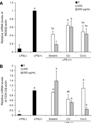
Fig. 3. Effects of CU and CU-C on mRNA levels of iNOS and COX-2 in RAW264.7 cells. Cells were pre-treated with 0, 200, or 500 μg/mL of Sinetrol, CU or CU-C for 4 h and then stimu- lated by 1 μg/mL of LPS for 8 h. Real time-PCR was performed using each cDNA and gene specific primers. Each bar represents the mean±SEM (n=3). Means with different letters (a-c) above the bars are significantly different according to ANOVA, Tukey test (P<0.05).
iNOS
COX-2
β-actin LPS(+)
Sinetrol CU CU-C LPS(-) PLS(+) 200 500 200 500 200 500
Fig. 4. Effects of CU and CU-C on protein expression of iNOS and COX-2 in RAW264.7 cells. Cells were pre-treated with 0, 200, or 500 μg/mL of Sinetrol, CU or CU-C for 4 h and then stimulated by 1 μg/mL of LPS for 8 h. Immunoblotting was performed with whole cell lysates and specific antibodies.
감귤 과피 추출물이 염증관련 mRNA 및 단백질 발현에 미치 는 영향
염증반응에서 과다하게 생성된 NO는 세포독성, 혈관확 장, 조직손상과 같은 작용으로 염증을 심화시키는 염증성 물질로 NOS 중에 염증반응에 관여하는 iNOS에 의해 생성 된다. 그리고 아라키돈산(arachidonic acid)으로부터 pro- staglandin, thromboxane A2, prostacyclin의 생성에 관여 하는 효소인 COX-2 역시 염증반응 시 급증하는 염증관련 효소이다. 그 밖에 tumor necrosis factor-alpha(TNF-α) 는 염증성 사이토카인으로 tumor promoter로서 작용하여 여러 암 조직에서 발현된다. 이와 같이 염증반응이 일어날 때는 여러 염증조절인자가 발현되어 작용한다(22).
염증반응을 유도하는 강한 자극물질인 LPS를 처리하였 을 때 iNOS와 COX-2의 mRNA level은 증가하였으나 Sinetrol과 감귤 과피 추출물이 이를 감소시켰다. 이러한 효 과는 500 μg/mL를 처리한 Sinetrol군만 제외하고는 시료 처리 농도에 따라 억제 효과가 더 증가되었으며 물질전환 전보다 후 추출물인 CU-C군이 더 유의적으로 iNOS와 COX- 2의 mRNA 발현을 억제시켰다(Fig. 3).
단백질의 경우에는 LPS에 의해 증가된 iNOS를 감귤 과
피 추출물이 억제시켰으며, 특히 CU군보다 CU-C군에서 더 억제 효과가 뛰어났다. 또 다른 염증관련 효소인 COX-2의 경우에도 LPS가 처리에 의해서 발현량이 증가하였다(Fig.
4). Sinetrol에서는 오히려 COX-2의 발현이 훨씬 더 강하게 증가하였고, CU와 CU-C군에서 LPS에 의해 증가된 COX- 2의 발현이 억제되었다. 저농도보다는 고농도의 CU-C군이 더 효과적으로 COX-2의 발현을 억제하였으며 이러한 결과 들은 mRNA level과 단백질 발현의 결과가 일치하는 것을 보여주었다(Fig. 4).
LPS에 의해 유도된 iNOS는 naringenin과 hesperetin, COX-2의 경우에는 naringenin에 의해서 유의적으로 억제 되므로(23,24) 이 결과는 물질전환으로 naringenin과 hes- peretin 함량이 높아진 CU-C군에서 iNOS 발현 억제 효과 가 더 증가한 것으로 사료된다. 또한 iNOS와 달리 COX-2는 hesperetin에 의해 억제되지 않으므로 iNOS보다 억제 효과 가 약한 것으로 사료된다(23,24).
감귤 과피 추출물이 NO 생성에 미치는 영향
저농도의 NO는 혈관확장이나 이물질에 대한 면역과정을 진행하는 데 쓰이는 유익한 면이 있지만 iNOS에 의해서 과 량 생성된 NO의 경우 superoxide와 반응하여 독성이 강한 peroxynitrite를 생성하고 많은 질환과 염증의 원인이 된다 (6).
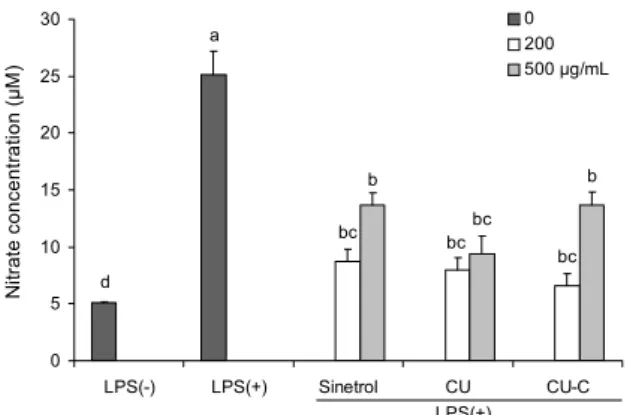
실험 결과 NO의 생성량은 LPS를 처리하지 않았을 때보 다 LPS를 처리하였을 때 급격히 증가하였고, 양성대조군인 Sinetrol군과 물질전환 여부와 관계없이 감귤 과피 추출물 이 LPS에 의해 과도하게 증가된 NO를 감소시켰다(Fig. 5).
L-Arginine이 NOS에 의해서 L-citrulline으로 전환되며 NO를 생성하므로 앞에서 진행한 실험에서 iNOS의 mRNA 와 단백질 발현이 LPS에 의해 급격히 증가되지만 감귤 과피 추출물을 처리한 군의 경우 이러한 염증성 효소들이 감소된 결과가 NO의 생성을 억제한 것으로 사료된다(25). 즉 iNOS 의 mRNA와 단백질 발현의 억제 효과가 과도한 NO의 생성
a
d
bc bc bc
b bc
b
0 5 10 15 20 25 30
LPS(-) LPS(+) Sinetrol CU CU-C
Nitrate concentration (μM) .
0 200 500 μg/mL
LPS(+)
Fig. 5. Effects of CU and CU-C on nitric oxide (NO) production in RAW264.7 cells. Cells were pre-treated with 0, 200, or 500 μg/mL of Sinetrol, CU or CU-C for 4 h and then stimulated by 1 μg/mL of LPS for 8 h. NO release in cell media was meas- ured by Griess method. Each bar represents the mean±SEM (n=3). Means with different letters (a-d) above the bars are sig- nificantly different according to ANOVA, Tukey test (P<0.05).
을 억제하므로 감귤 과피 추출물이 염증이 유도된 상태에서 iNOS의 발현을 억제하여 인체에 해로운 NO와 그 대사물을 억제한다고 사료된다.
이러한 감귤류의 항염증 또는 항암 효과도 여러 연구를 통해 보고되고 있다. 세포신호전달과정에서 필수적인 kin- ase와 phosphodiesterase를 억제할 수 있고(26,27), T림 프구와 B림프구의 활성에도 영향을 미치는 것으로 알려져 있으며(28,29), 감귤류의 nobiletin은 염증관련효소인 iNOS 와 COX-2의 발현을 억제시켰다(30). 최근의 보고들에 의하 면(31-34) 감귤류의 naringenin은 자외선 조사 모델에서 자외선으로 인한 DNA의 손상을 보호하는 효과가 있었고 (31), naringin은 과산화수소로 유도된 apoptosis 관련 유전 자나 단백질 발현에 영향을 미쳤다(32). 또한 감귤류 플라보 노이드는 protein kinase C의 활성과 피부암 발암촉진과정 등을 억제하였고, 구조상 A와 B 고리의 수산화 그룹의 위치 와 개수에 따라 암세포의 증식 억제에 효과를 보였다(29, 33). 이와 같이 감귤류 플라보노이드는 발암요소로부터 DNA 손상을 보호하고 발암촉진과정을 억제하며, 암세포의 증식을 억제하는 항암 효과가 나타내었다(29,33).
요 약
감귤류와 감귤류의 과피는 오랜 기간 동양의학의 약재로 사 용되어 왔고 최근에는 생과와 주스로 많이 소비되고 있다.
하지만 감귤류 가공 공정 시 많은 과피 부산물이 발생하므로 활용방안이 필요하다. 감귤류의 과피에는 플라바논(flav- anone)이 풍부하며 이 플라바논의 형태 중 배당체보다 비배 당체의 체내 흡수가 더 효과적이므로 비배당체가 생리적 효 과가 더 뛰어나다. Bioconversion(물질전환)은 cytolase에 의해 narirutin과 naringin은 naringenin으로, hesperidin 은 hesperetin으로 각각 전환되며, 본 연구는 감귤의 과피에
다량 존재하는 플라바논을 물질전환 한 뒤 면역세포에서 항 산화 및 항염증 효과를 확인하였다. 물질전환 후에 감귤 과 피 추출물의 전자공여능이 농도 의존적으로 높아지는 것을 확인하였고, 면역세포인 RAW264.7에 200, 500 μg/mL 감 귤 bioconversion 전(CU)・후(CU-C) 과피 추출물을 4시간 동안 처리한 후 LPS(1 μg/mL, 8시간)를 처리하였다. 염증 관련 효소인 iNOS와 COX-2의 mRNA와 단백질 발현을 확 인한 결과 물질전환과 관계없이 감귤 과피 추출물이 LPS에 의해 유도된 iNOS와 COX-2의 mRNA와 단백질 발현을 농 도 의존적으로 감소시켰지만 물질전환 전보다 후 추출물의 억제 효과가 더 높았다. iNOS에 의해 생성되는 NO 역시 감귤 과피 추출물이 LPS에 의해 유도된 NO 생성을 억제시 켰다. 본 연구 결과는 감귤 과피 추출물이 항산화와 항염증 생리활성을 보였지만 cytolase를 이용하여 물질전환 한 감 귤 과피 추출물이 물질전환 전보다 이러한 생리활성이 강화 됨을 보임으로써 향후 감귤류 부산물로 폐기되어온 감귤류 과피가 산화적 스트레스와 염증반응에 기인하는 만성질환 을 위한 기능성 소재로 활용이 가능할 것으로 기대된다. 감 귤 과피 추출물의 in vitro 상에서 검증된 항염증 효능이 향 후에 in vivo 상에서 여러 염증 조직에 미치는 영향에 대한 추가 검증이 필요하다고 사료된다.
감사의 글
본 연구는 2011~2013년에 농림수산식품기술기획평가원 공동연구사업과 2014년 명지대학교 교책중점연구소 지원 사업에 의해서 수행한 연구 결과의 일부이며 이에 감사드립 니다.
REFERENCES
1. Himaya SW, Ryu B, Qian ZJ, Kim SK. 2012. Paeonol from Hippocampus kuda Bleeler suppressed the neuro-inflam- matory responses in vitro via NF-κB and MAPK signaling pathways. Toxicol in Vitro 26: 878-887.
2. Trowbridge HO, Emling RC, Fornatora M. 1997. Inflamma- tion. A review of the process. Implant Dent 6: 238-278.
3. Zamora R, Vodovotz Y, Billiar TR. 2000. Inducible nitric oxide synthase and inflammatory diseases. Mol Med 6: 347- 373.
4. Tracey KJ. 2002. The inflammatory reflex. Nature 420: 853- 859.
5. Fan GW, Zhang Y, Jiang X, Zhu Y, Wang B, Su L, Cao W, Zhang H, Gao X. 2013. Anti-inflammatory activity of baicalein in LPS-stimulated RAW264.7 macrophages via es- trogen receptor and NF-κB-dependent pathways. Inflamma- tion 36: 1584-1591.
6. Hu W, Yang X, Zhe C, Zhang Q, Sun L, Cao K. 2011.
Puerarin inhibits iNOS, COX-2 and CRP expression via suppression of NF-κB activation in LPS-induced RAW264.7 macrophage cells. Pharmacol Rep 63: 781-789.
7. Kang SI, Shin HS, Kim HM, Hong YS, Yoon SA, Kang SW, Kim JH, Kim MH, Ko HC, Kim SJ. 2012. Immature Citrus sunki peel extract exhibits antiobesity effects by β-
oxidation and lipolysis in high-fat diet-induced obese mice.
Biol Pharm Bull 35: 223-230.
8. Ejaz S, Ejaz A, Matsuda K, Lim CW. 2006. Limonoids as cancer chemopreventive agents. J Sci Food Agric 86: 339- 345.
9. Tripoli E, Guardia ML, Giammanco S, Majo DD, Giammanco M. 2007. Citrus flavonoids: Molecular structure, biological activity and nutritional properties: A review. Food Chem 104: 466-479.
10. Tsai SH, Lin-Shiau SY, Lin JK. 1999. Suppression of nitric oxide synthase and the down-regulation of the activation of NFkappaB in macrophages by resveratrol. Br J Pharma- col 126: 673-680.
11. Peterson J, Dwyer J. 1998. Flavonoids: Dietary occurrence and biochemical activity. Nutr Res 18: 1995-2018.
12. Choi SY, Ko HC, Ko SY, Hwang JH, Park JG, Kang SH, Han SH, Yun SH, Kim SJ. 2007. Correlation between fla- vonoid content and the NO production inhibitory activity of peel extracts from various citrus fruits. Biol Pharm Bull 30: 772-778.
13. Fuhr U, Kummert AL. 1995. The fate of naringin in humans:
a key to grapefruit juice-drug interactions? Clin Pharmacol Ther 58: 365-373.
14. Santamaría RI, Reyes-Duarte MD, Bárzana E, Fernando D, Gama FM, Mota M, López-Munguía A. 2000. Selective en- zyme-mediated extraction of capsaicinoids and caratenoids from chili guajillo puya (Capsicum annum L.) using ethanol as solvent. J Agric Food Chem 48: 3063-3067.
15. Seo JY, Lee JH, Kim NW, Her E, Chang SH, Ko NY, Yoo YH, Kim JW, Seo DW, Han JW, Kim YM, Choi WS. 2005.
Effect of a fermented ginseng extract, BST204, on the ex- pression of cyclooxygenase-2 in murine macrophages. Int Immunopharmacol 5: 929-936.
16. Yang G, Park D, Lee J, Song BS, Jeon TH, Kang SJ, Jeon JH, Shin S, Jeong HS, Lee HJ, Kim YB. 2011. Suppressive effects of red ginseng preparations on SW480 colon cancer xenografts in mice. Food Sci Biotechnol 20: 1649-1653.
17. Dallas C, Gerbi A, Tenca G, Juchaux F, Bernard FX. 2008.
Lipolytic effect of a polyphenolic citrus dry extract of red orange, grapefruit, orange (SINETROL) in human body fat adipocytes. Mechanism of action by inhibition of cAMP- phosphodiesterase (PDE). Phytomedicine 15: 783-792.
18. Brand-Williams W, Cuvelier ME, Berset C. 1995. Use of free radical method to evaluate antioxidant activity. Lebensm Wiss u-Technol 28: 25-30.
19. Clarke G, Ting KN, Wiart C, Fry J. 2013. High correlation of 2,2-diphenyl-1-picrylhydrazyl (DPPH) radical scaveng- ing, ferric reducing activity potential and total phenolics
content indicates redundancy in use of all three assays to screen for antioxidant activity of extracts of plants from the Malaysian rainforest. Antioxidants 2: 1-10.
20. Cha JY. 2001. Biofunctional activities of citrus flavonoids.
J Korean Soc Agric Chem Biotech 44: 122-128.
21. Lee JH, Kim GH. 2010. Evaluation of antioxidant and in- hibitory activities for different subclasses flavonoids on en- zymes for rheumatoid arthritis. J Food Sci 75: H212-H217.
22. Mekhora C, Muangnoi C, Chingsuwanrote P, Dawilai S, Svasti S, Chasri K, Tuntipopipat S. 2012. Eryngium foetidum suppresses inflammatory mediators produced by macro- phages. Asian Pac J Cancer Prev 13: 653-664.
23. Yang HL, Chen SC, Senthil Kumar KJ, Yu KN, Lee Chao PD, Tsai SY, Hou YC, Hseu YC. 2011. Antioxidant and anti- inflammatory potential of hesperetin metabolites obtained from hesperetin-administered rat serum: an ex vivo approach.
J Agric Food Chem 60: 522-532.
24. Chao CL, Weng CS, Chang NC, Lin JS, Kao ST, Ho FM.
2010. Naringenin more effectively inhibits inducible nitric oxide synthase and cyclooxygenase-2 expression in macro- phages than in microglia. Nutr Res 30: 858-864.
25. Wang L, Tu YC, Lian TW, Hung JT, Yen JH, Wu MJ. 2006.
Distinctive antioxidant and antiinflammatory effects of fla- vonols. J Agric Food Chem 54: 9798-9804.
26. Gamet-Payrastre L, Manenti S, Gratacap MP, Tulliez J, Chap H, Payrastre B. 1999. Flavonoids and the inhibition of PKC and PI 3-kinase. Gen Pharmacol 32: 279-286.
27. Kusano A, Nikaido T, Kuge T, Ohmoto T, Delle Monache G, Botta B, Botta M, Saitoh T. 1991. Inhibition of adenosine 3',5'-cyclic monophosphate phosphodiesterase by flavonoids from licorice roots and 4-arylcoumarins. Chem Pharm Bull (Tokyo) 39: 930-933.
28. Middleton E Jr, Kandaswami C. 1992. Effects of flavonoids on immune and inflammatory cell functions. Biochem Phar- macol 43: 1167-1179.
29. Manthey JA, Grohmann K, Guthrie N. 2001. Biological prop- erties of citrus flavonoids pertaining to cancer and inflam- mation. Curr Med Chem 8: 135-153.
30. Li S, Sang S, Pan MH, Lai CS, Lo CY, Yang CS, Ho CT.
2007. Anti-inflammatory property of the urinary metabolites of nobiletin in mouse. Bioorg Med Chem Lett 17: 5177-5181.
31. Kootstra A. 1994. Protection from UV-B-induced DNA dam- age by flavonoids. Plant Mol Biol 26: 771-774.
32. Kanno S, Shouji A, Asou K, Ishikawa M. 2003. Effects of naringin on hydrogen peroxide-induced cytotoxicity and apoptosis in P388 cells. J Pharmacol Sci 92: 166-170.
33. Hwang SL, Shih PH, Yen GC. 2012. Neuroprotective effects of citrus flavonoids. J Agric Food Chem 60: 877-885.