40(1) : 6 12 (2009)
6
암세포 특이적 세포 사멸을 유도하는 자생식물 추출물의 항암 효과
윤이관1·이승은2·이동진1·노문철3·성정숙4·박충범2·장영주
*
단국대학교
,
나노바이오의과학과,
세포주기신호전달연구실&
조직재생공학연구소,
1단국대학교,
생명자원과학대학,
식량생명공학과
,
2농촌진흥청 인삼특작부,
3한국생명공학연구원,
생물산업기술연구센터,
4농촌진흥청농업유전자원센터Anti-cancer Activity of Korean Local Plant Extracts Inducing Apoptosis in Various Carcinoma Cells
Yi Kwan Yoon
1, Seung Eun Lee
2, Dong Jin Lee
1, Mun Chual Rho
3, Jung-Suk Sung
4, Chung Berm Park
2and Young Joo Jang
*Cell cycle & Signal Transduction Laboratory, Department of Nanobiomedical Science & Institute of Tissue Regeneration Engineering, Dankook University, Cheonan 330-714,
1
The College of Bio-Resources Science, Dankook University, Cheonan 330-714,
2
Department of Herbal Crop Research, RDA, Chung-Buk 369-873,
3
Bioindustry Research Center, Korea Research Institute of Bioscience and Biotechnology, Jeonbuk 580-185,
4
Department of National Agrobiodiversity Center, RDA, Suwon 441-853
Abstract −
Thirty five methanol extracts from 19 natural local plants, which have been used as traditional anti-cancer medicine, were prepared. They were analyzed the cytotoxic effects on primary fibroblast cells and carcinoma cells. The root extract of Solanum nigrum were highly toxic in both cell lines with IC
50values of less than 0.01
µg/
µl, and 26 of 35 extracts were toxic in all cells with IC
50values of 0.1~2
µg/
µl. Three extracts including the fruit extracts of Solanum nigrum and Morus alba had no cytotoxic activity in both cell lines. Five of 35 extracts were highly toxic in cancer cells than in primary cells. Because pri- mary cells were more resistant on these extracts, the five extracts were selected for anti-cancer agent candidates. Apoptosis or programmed cell death has an essential role in chemotherapy-induced tumor cell killing. Recently, inducers of apoptosis have been used in cancer therapy. When two of 5 cancer cell-specific cytotoxic extracts ( Ulmus parvifolia and Zelkova serrata ) were treated in concentration of 0.02~0.1
µg/
µl, apoptosis were increased at 3-5 times in cancer cell lines. Finally, the apoptotic effects of these extracts were confirmed by cleavages of both poly-(ADP-ribose)-polymerase and caspase-3 as apoptotic mark- ers. In this report, we suggested that two of 35 medicinal herb extracts can be useful anti-cancer drug candidates inducing apo- ptosis in several carcinoma cell lines.
Key words
−Ulmus parvifolia , Zelkova serrata , Cytotoxicity, Apoptosis, Anti-cancer
서 론
최근세계각국에서자생자원식물들에대한관심이높 아져인간에게유용한식물에대한기본연구가활발히진 행되고있다
.
특히약용자생식물은그이용가치가매우높으며
,
항암,
항염제를비롯한신약개발,
건강식품의소재및신기능성소재의자원으로써응용되고있을만큼응용
분야도다양하다
.
1)식물의종류뿐아니라식물의각부위별쓰임새도매우다양한데
,
대표적인예로인삼,
당귀,
작약등은다양한기능으로수요가많은중요한약초로써대 부분뿌리만사용되어왔지만
,
최근작약의경우지상부인꽃잎
,
줄기,
그리고잎으로부터도생리활성물질을분리동정하였다는보고가있다
.
2) 자생식물들은생리활성물질뿐아니라항암물질의원료로이용되어왔다
.
예로부터까마중
( Solanum nigrum )
과개미취( Aster tartaricus )
는부인병및암에쓰였던약재이며
,
느티나무( Zelkova serrata ),
꾸지뽕나무
( Cudrania tricuspidata ),
시무나무( Hemiptelea
*교신저자(E-mail):[email protected] (Tel):82-41-550-1936
nigrum )
도위암및대장암을비롯한암의치료를위한항암약재로쓰여왔다는보고가있다
.
3)이와함께왕느릅나무( Ulmus macrocarpa )
의수피,
느릅나무( Ulmus parvifolia )
의근피와삼백초
( Saururus chinensis )
에서추출한물질이항암활성이있다는실험결과가발표된바가있다
.
4-7)이들약용식물의항암효과가어떤기전을통하여일어나는지는 명확하게알려진바는없지만
,
일반적으로세포독성을유발할것이라는것을예측할수있다
.
그러나단순히세포독성을유발하여세포를파괴하는약물은투여시심한부작 용때문에좋은약물이될수는없다
.
세포사멸현상
(apoptosis
또는programmed cell death)
은정상적인발생단계및생리작용에서세포의수를일정하 게유지하기위한필수적인과정이다
.
인간에발생하는다양한암에서세포사멸이제대로일어나지않는경우가많 아세포사멸기전의파괴는정상세포가암세포로변화되는 주요한원인이라할수있다
.
8)최근항암제로써개발하기위한물질을스크리닝할 때강한독성으로세포의 파괴
(necrosis)
를유도하는 것보다는암세포특이적인세포사멸을 유도하는기전을이용하여하고있다
.
8-11) 따라서위와같이 세포사멸현상은 암세포를죽이기위한화학항 암치료법의 주요한원리가될수도있다
.
암세포에세포사멸을유도하기위해서는사멸과정의주요단백질인
Bcl2
계열의 단백질
, Caspase
효소, Tumor necrosis factor (TNF)-related apoptosis-inducing ligand (TRAIL)
단백질들의활성을직접또는간접으로증가시키면된다
.
12-14)이와더불어사멸 기전관련단백질인산화효소
,
탈인산화효소
,
전사인자및세포표면의수용체, proteosome
등을표적으로하여세포사멸을유도할수도있다
.
15-16)최근세포사멸유도를위한다양한약물의스크리닝실험들이진행 되고있지만
,
대부분의후보약물들이 낮은항암효율과약물에 대한저항성 유발때문에 전임상단계에서개발 이제한되고있다
.
따라서암세포특이적인세포사멸을유도하여항암효과를가지는약물을개발하기위해서는
1
차로자생식물과 같은다양한 천연물로부터다량의후보물 질을이용하여스크리닝한후많은 종류의후보물질을 확보하여진행하는것이바람직할것이다
.
이논문에서저자는한국의전통적인약용식물로 알려진다양한자생식 물의각부위별추출물을이용하여암세포특이적인세포 사멸유도능이있는항암후보물질을 도출하기위한스 크리닝결과를제시하였다
.
재료 및 방법
식물 재료 − 실험에이용한
35
종의시료는2001
년부터2005
년까지전국산야에서자생하는식물또는수원농촌진흥청약용작물시험포장에서증식한자생식물중전통적으
로항암효과가있다고알려진
19
종의식물을채취하여지상부및뿌리부위를나누어음건
,
쇄절후메탄올로환류추출장치를이용하거나
ASE (accelerated solvent extractor,
DIONEX, USA)
를사용하여얻은물질을감압농축하여사용하였다
.
각시료들에메탄올을넣어50 mg/ml
의농도로녹인후스크리닝실험에적용하였다
.
각시료의원료가된식물명과부위별추출조건은
Table 1
에정리하였다.
세포 배양 −
3
종의구강암세포주(KB, YD15, YD38), 3
종의간암세포주(SNU387, SNU739, SK-Hep1),
위암세포주인
SNU216
및SNU668
들은모두인간에서유래된세포들이며
,
한국세포주은행(Korea cell line bank, KCLB, http://cellbank.snu.ac.kr)
로부터분양받아본실험에이용하였다
.
17)암세포주들은10% fetal bovine serum (FBS)
이함유된
Dulbecco’s Modified Eagle Medium (DMEM)
을배지로이용하여
, 37
oC, 5% CO
2가공급되는항온세포배양기에 서배양하였다.
치은섬유아세포(human gingival fibroblast,
HGF)
는사랑니의발치후환자의동의를얻어발치치아의주변조직으로부터일차배양되었고
,
암세포실험에대한대조군으로이용하였다
.
Cell cytotoxicity test − 식물의추출물이세포성장의억
제효과가있는지의여부를확인하기위해세포들을
96 well
plate
에10
4~10
5 개/ml
의농도로접종하고37
oC, 5% CO
2가 공급되는항온세포배양기에서8
시간이상전배양하였다.
추출물을세포배양액
100
µl
에0.001, 0.01, 0.1, 2, 10, 50, 200 mg
씩을각각처리한후18
시간동안배양하였다.
세포의독성은
Cell Counting Kit-8
TM(CCK-8, Dojindo, Japan)
시약을이용하여분석하였다
.
각배양액에CCK-8
용액10
µl
씩넣은후
2~4
시간반응하고,
수용액에녹아있는tetrazolium salt (WST-8, [2-(2-methoxy-4-nitrophenyl)-3-(4-nitrophenyl)- 5-(2,4-disulfophenyl)-2H-tetrazolium, monosodium salt])
의양을
microplate reader
기를사용하여450 nm
에서의흡광도를측정하였다
.
Caspases assay − 각추출물들의세포사멸유발효과 를다량으로분석하기위해
Homogeneous Caspases Assay Kit
TM(Roche Applied Science, Germany)
을이용하였다.
세포들을
96 well plate
에10
5~10
6개/ml
의 농도로접종하고37
oC, 5% CO
2가공급되는항온세포배양기에서8
시간이상전배양한후각추출물들을적절한농도로처리하여
18
시간이상배양하였다
.
세포사멸이일어난세포안에서활성이증가되어있는
caspases
의양을측정하기위해kit
에제공된
caspase
의기질단백질(DEVD-R110)
을세포에첨가하여
1
시간동안효소반응한후기질단백질이caspases
에 의해 분해된 정도를
microplate reader
기를 사용하여560 nm
에서의흡광도로써측정하였다.
Western analysis − 세포사멸이진행되면서발생하는몇 몇단백질의생화학적변화를
Western analysis
을통해증명하였다
.
세포배양액에식물추출물을적당한농도로첨가하여
18
시간처리한후모든세포를모아lysis
용액(20 mM Tris-HCl, 100 mM NaCl, 0.5% NP-40, protease inhibitors)
을넣고세포를용해하였다
. 4
oC
의원심분리기에서12000rpm
의속도로원심분리한후
,
상층액에있는단백질들을SDS-
PAGE
상에서 분리하였고,
분리된 단백질을PVDF
membrane
으로옮긴후,
세포사멸과정의표지단백질중대표적인단백질인
poly-(ADP-ribose)-polymerase
에대한항체
(Roche Applied Science, Germany)
와caspase-3
에대한항체
(Santa Cruz Biotechnology Inc., California, USA)
를이용하여
Western analysis
를진행하였다. 결과 및 고찰
자생식물
19
종의각부위로부터만들어진35
종의메탄올추출물을암세포및정상세포에적용하여항암효과가있는 물질을스크리닝하고자하였다
.
먼저35
종의추출물을정상치은조직으로부터
1
차배양된치은섬유아세포와암세Table I.
Used medicinal plants and their methanol extracts
Drug no Korean name Scientific name Family name Used part Place of origin IC
50in HeLa cells (mg/ml) 1
가시오갈피Acanthopanax senticosus Araliaceae Root
약용작물시험장1.0 2
가시오갈피Acanthopanax senticosus Araliaceae Stem
약용작물시험장0.3
3
강활Ostericum koreanum Apiaceae Root
정선0.75
4
강활Ostericum koreanum Apiaceae Aerial part
약용작물시험장NA
5
강활Ostericum koreanum Apiaceae Root
약용작물시험장0.75
6
개미취Aster tartaricus Asteraceae Flower
약용작물시험장0.25
7
개미취Aster tartaricus Asteraceae Aerial part
약용작물시험장0.75
8
개미취Aster tartaricus Asteraceae Root
약용작물시험장0.55
9
까마중Solanum nigrum Solanaceae Aerial part
약용작물시험장0.075
10
까마중Solanum nigrum Solanaceae Root
약용작물시험장<0.01
11
까마중Solanum nigrum Solanaceae Fruit
약용작물시험장NA
12
꾸지뽕나무Cudrania tricuspidata Moraceae Leaf
약용작물시험장0.2
13
느릅나무Ulmus parvifolia Ulmaceae Root bark
제천0.07
14
느릅나무Ulmus parvifolia Ulmaceae Stem
제천0.3*
15
느릅나무Ulmus parvifolia Ulmaceae Bark
단양0.02*
16
느티나무Zelkova serrata Ulmaceae Leaf
약용작물시험장0.5
17
느티나무Zelkova serrata Ulmaceae Whole stem
약용작물시험장0.25*
18
느티나무Zelkova serrata Ulmaceae twigs
약용작물시험장0.25*
19
닥나무Broussounetia kazinoki Moraceae Leaf
약용작물시험장1.5
20
두릅나무Aralia elata Araliaceae Bark
금산1.0
21
둥굴레Polygonatum odoratum Liliaceae Aerial part
약용작물시험장>2
22
둥굴레Polygonatum odoratum Liliaceae Root
약용작물시험장1.5
23
백화사설초Hedyotis corymbosa Rubiaceae Whole
약용작물시험장>2
24
뱀딸기Duchesnea chrysantha Rosaceae Aerial part
약용작물시험장0.3*
25
뽕나무Morus alba Moraceae Leaf
약용작물시험장0.75
26
뽕나무Morus alba Moraceae Fruit
약용작물시험장NA
27
뽕나무Morus alba Moraceae Root bark
강진0.1
28
섬오갈피Acanthopanax koreanum Araliaceae Leaf
약용작물시험장0.3
29
시무나무Hemiptelea davidii Ulmaceae Root bark
제천1.5
30
시무나무Hemiptelea davidii Ulmaceae Bark
제천1.5
31
컴프리Symphytum officinale Boraginaceae Aerial part
약용작물시험장1.0
32
혹느릅나무Vulmus davidiana Ulmaceae Leaf
약용작물시험장1.5
33
화살나무Euonymus alatus Celastraceae Stem
상주2.0
34
활나물Crotalaria sessiliflora Fabaceae Root
약용작물시험장0.3
35
활나물Crotalaria sessiliflora Fabaceae Aerial part
약용작물시험장0.75
포에각각처리한후
,
나타나는세포독성정도를측정하였다
.
분석실험에이용한암세포의종류는구강암,
간암,
위암,
자궁암등모두
9
종이었으며,
이들의doubling time
은18~20
시간으로분석되었다
(
재료및방법참고). 35
종의추출물을
0.00001
µg/
µl~2
µg/
µl
의농도로세포에처리하여18
시간후에살아있는세포의양을측정하였다
.
각9
종의암세포에서측정된값을평균하여
IC
50값을결정하였다(Table 1). 35
종의추출물중강활( Ostericum koreanum )
의지상부
(Table 1
의4
번추출물),
까마중( Aster tartaricus )
의열매
(Table 1
의11
번추출물),
뽕나무( Morus alba .)
의열매(Table 1
의26
번추출물)
로부터얻은추출물들은치은섬유아세포와암세포모두에
2
µg/
µl
이상의고농도를처리하여도 세포에는 독성이 나타나지 않았다
.
까마중( Aster
tartaricus )
의열매추출물(11
번)
의독성분석결과는Fig. 1
의그래프
b
에나타내었다.
반면, 27
종의추출물들은정상세포와암세포모두에서비슷한
IC
50값으로독성을가지고 있는것으로분석되었다.
이중까마중( Aster tartaricus )
의뿌리에서추출한시료
(Table 1
의10
번추출물)
는매우독성이강하여
0.01
µg/
µl
이하의IC
50값을가지며,
매우저농도에서도세포들을죽이는것으로관찰되었다
(Fig. 1, a).
이결과에서동일식물의경우추출부위에따라독성정도 가확연히다르다는사실을알수있다
.
이번실험에서흥미로운것은정상세포에서는독성이없
거나약하지만암세포에는비교적강한독성을가지는식 물의추출물이있다는점이다
.
지금까지항암활성이있는것으로보고된추출물들은대부분항암제의투여시정상 조직에심각한부작용을일으키는문제가있으므로
,
항암제가암세포만을특이적으로파괴한다는점은항암제의효율 적인측면에서중요한의미가있다
.
본실험에서5
종의추출물이 이와 같은 특징을 보였다
.
느릅나무( Ulmus
parvifolia )
의줄기및수피추출물들(Table 1
의14
와15
번추출물
),
느티나무( Zelkova serrata )
의나무줄기의전체부분과잔가지부분
(Table 1
의17
과18
번추출물),
그리고뱀딸기
( Duchesnea chrysanta )
의지상부추출물(Table 1
의24
번추출물
)
이정상세포에는독성이약하지만암세포특이적인독성을보였다
.
이들의암세포에서의IC
50값은0.5
µg/
µl
이하였다
.
이들의세포독성분석곡선은Fig. 1
에나타내었다
(Fig. 1, c-f & h).
정상세포및암세포에모두독성이있는 비특이적인 독성을 보이는 추출물의 예로 둥굴레
( Polygonatum odoratum )
뿌리에서얻은시료의독성곡선을
Fig. 1
의그래프g
에나타내었다.
최근항암제의개발을위한전략으로천연물들을스크리 닝할때단순한세포의파괴
(necrosis)
를유도하는것보다는암세포특이적인생리현상을응용하여스크리닝하는전 략이고려되고있다
.
특히암세포에는정상세포에서발생하는세포사멸기전이억제되어있는경우가빈번하므로
,
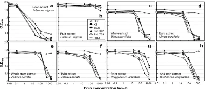
8-11)Fig. 1.
Cytotoxic effects of methanol extracts on primary and cancer cells. Indicated concentrations of 8 methanol extracts were treated in normal and cancer cell cultures. After 18 hours, the amounts of soluble tetrazolium salt were measured in 450 nm.
a,cytotoxicity by treatment of root extract of Solanum nigrum ;
b,cytotoxicity by fruit extract of Solanum nigrum ;
c,cytotoxicity by
whole extract of Ulmus parvifolia ;
d,cytotoxicity by bark extract of Ulmus parvifolia ;
e,cytotoxicity by whole stem extract of
Zelkova serrata ;
f,cytotoxicity by twig extract of Zelkova serrata ;
g,cytotoxicity by treatment of root extract of Polygonatum
odoratum ;
h,cytotoxicity by treatment of aerial part extract of Duchesnea chrysantha . The symbols in graphs indicated cell lines
used in this experiment.
세포사멸을유도하는기전을항암제스크리닝에응용할수 있다
. 5
종의추출물(Table 1
의14, 15, 17, 18, 24
번-
순서대로느릅나무줄기및수피추출물
,
느티나무줄기전체와잔가지부분
,
그리고뱀딸기의지상부추출물)
을이용하여
9
종의암세포에서세포사멸을유도할수있는지스크리닝하였다
.
세포사멸의과정에서활성이일어나는다양한종류의
caspase
들의활성을한꺼번에분석할수있는kit
를이용하여
,
여러종의암세포에서의caspases
활성을그래프로나타내었다
(Fig. 2).
까마중( Aster tartaricus )
의뿌리에서추출한시료
(10
번추출물)
는매우독성이강하여0.01
mg/ml
이하의IC
50값을가짐에도불구하고독성이없는과실추출물
(11
번시료)
과마찬가지로caspases
의활성은관찰되지않았다
(Fig. 2
의3 & 4
번막대들).
이결과에서까마중뿌리에서추출한물질이세포의괴사는유도하지만
,
세포사멸을유도하지는않는다는사실을알수있다
. Fig. 2
의막대그래프에서기존에세포사멸을유발한다고알려진
doxorubicin
의caspases
활성정도(Fig. 2
의2
번막대들)
와같거나증가된정도를보이는추출물은느릅나무수피추 출물
(Table 1
의15
번추출물, Fig. 2
의6
번막대들)
과느티나무의잔가지부분에서얻어진추출물
(Table 1
의18
번, Fig. 2
의8
번막대들)
추출물이었다.
이결과는매우흥미로운데
, Fig. 1
의독성결과에서보는바와같이세포독성이비슷한수준으로일어난다고하여도세포의독성정도가세 포사멸과일치하지는않는다는것이다
.
느릅나무수피추출물과느티나무의잔가지추출물이세포사멸을유도하는것 으로보이므로이를생화학적으로증명하기위해세포사멸 과정을예측하는데유용한표지단백질인
poly-(ADP-ribose)- polymerase
와caspase-3
의활성분할을단백질수준에서분석하였다
.
구강세포3
종과간암세포3
종, HeLa
세포에두추출물을각각처리하고
18
시간후에세포를모아Western
분석을하였다
.
추출물을처리하지않은세포에서는두단백질은모두활성분할되지않아작은크기의단백질조각
(
그림3
의세모표시)
이관찰되지않았다(
그림3, A).
느릅나무수피추출물을처리하였을경우에는구강암세포
2
종과 간암세포인
SNU387, HeLa
세포에서poly-(ADP- ribose)-polymerase
와caspase-3
모두활성이증가하는것을관찰할수있었다
(
그림3, B
의lane 1, 3, 5 & 6).
이와는달리느티나무의잔가지추출물은구강암세포주인
KB
와SNU387
세포에만세포사멸을유발하였고(Fig. 3, C
의lane 1 & 3),
느릅나무수피추출물과는달리HeLa
및YD15
에는전혀세포사멸을유발하지않았다
(Fig. 3, C
의lane 5
& 6). SNU387
세포와는달리또다른종류의간암세포주인
SNU739
와SK-Hep-1
은두 추출물에 의하여PARP
와caspase-3
의활성이일어나지않았다(
그림3, B & C, lane 4 & 7).
결론으로
,
전통적으로항암활성이있다고알려진약용식물인 느릅나무
( Ulmus parvifolia )
의 수피와 느티나무( Zelkova serrata )
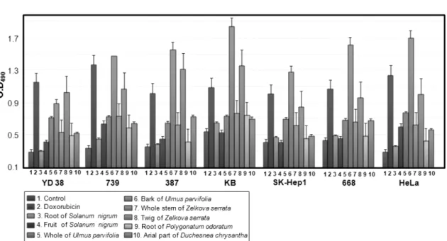
의잔가지로부터암세포특이적인세포독Fig. 2.
Caspases activity on various cancer cells by treatment of methanol extracts. 0.5
µg/
µl of extracts were treated in cell culture
for 18 hours and the caspases activity was measured at 490 nm (see Materials & Methods). Each bar indicated the used extract (see
Table 1).
1,negative control;
2,0.1
µg/
µl of doxorubicin;
3-10,methanol extracts. The used cancer cell lines were indicated as
YD38, 739 (SNU739), 387 (SNU387), KB, SK-Hep1, 668 (SNU668), and HeLa cell. Each value of means +/- S.D. was calculated
from three separated experiments.
성을가질뿐만아니라
,
나아가세포사멸을유도하는좀더효과적인항암후보물질을얻을수있었으며
,
이추출물들을암조직특이적인항암제로써응용할수있는가능성 도제시하였다
.
이후위의두가지추출물을화학적으로분석
,
분리정제하여각순수화합물의세포사멸능을검증하게될것이며
,
세포사멸과정중에두약물의표적이되는인자를연구하게될것이다
.
사 사
이논문은농촌진흥청국책기술개발사업
(200802A01030005)
의연구비를지원받아수행된연구입니다
.
인용문헌
1. Lee, C. H., Lee, H. K., Park, K. K., Park, C. H., Lee, B. Y., Sung, R. S. and Lee, S. H. (2002) Use of Resources a Plant for Biotechnology Industry development. Research Center for Bioresource and Health at Chungbuk National University.
2. Kim, S. J., Park, J. H., Choi, S. Y., Son, K. H. and Kim, K.
U. (2007) Isolated and identification of biological activity compounds from leaves and stem of Paeonia lactiflora Pallas.
Korean Journal of Medicinal Crop Science.
15: 6-11.
3. Lee, S. R., Lee, J. I. and Jung, K. J. (2004) Anticancer Med- ical Herbs of Korea. Jinsol Publishing company.
4. Lee, K. H., Cho, C. H. and Yoon, W. H. (2004) In vivo anti- tumor activity of mansonone E isolated from Ulmus davidiana
var. japonica NAKAI. Korean Journal of Pharmacognosy .
35: 199-202.
5. Lim, S. Y. (2007) Effect of extracts from root bark of Ulmus parvifolia on inhibition of growth and DNA synthesis of human cancer cells. Journal of Life Science.
17: 1232-1236.
6. Park, S. J., Yoo, H. J., Choi, H. S., Seo, B. Y., Yang, S. H., Kim, Y. H., Jeong, J. Y. and Lee, K. N. (2004) Effects of Sau- rurus chinensis Bail extracts on anticancer activity and cad- mium induced cytotoxicity. The Korean Journal of Oriental Preventive Medicine.
8: 81-98.
7. Kwon, Y. M., Lee, J. H. and Lee, M. W. (2002) Phenolic compounds from barks of Ulmus macrocarpa and its anti- toxidative activities. Korean Journal of Pharmacognosy.
33: 404-410.
8. Hu, W. and Kavanagh, J. J. (2003) Anticancer therapy tar- geting the apoptotic pathway. The Lancet Oncologyol.
4: 721- 9. Kaufmann, S. H. and Vaux, D. L. (2003) Alterations in the 729.
apoptotic machinery and their potential role in anticancer drug resistance. Oncogene .
22: 7414-7430.
10. Fulda, S. and Debatin, K. (2004) Exploiting death receptor signaling pathways for tumor therapy. Biochimica et Bio- physica Acta, CR. Reviews on Cancer .
1705: 27-41.
11. Pisano, M., Pagnan, G., Loi, M., Mura, M. E, Tilocca, M. G., Palmieri, G., Fabbri, D., Dettori, M. A., Delogu, G., Ponzoni, M. and Rozzo, C. (2007) Antiproliferative and pro-apoptotic activity of eugenol-related biphenyls on malignant melanoma cells. Molecular Cancer.
6: 8-19.
12. LeBlanc, H. N. and Ashkaenazi, A. (2003) Apo2L/TRAIL
Fig. 3.
Activated cleavages of caspase-3 and poly-(ADP-ribose)-polymerase in various cancer cells. 0.5
µg/
µl of bark extract of
Ulmus parvifolia (B) and stem extract of Zelkova serrata (C) were treated in 7 cancer cell cultures for 18 hours. Cells were
harvested and lysed, and the cell extracts were separated on SDS-PAGE. Proteins were electro-transferred to PVDF membrane, and
caspase-3 and poly-(ADP-ribose)-polymerase were detected by using proper antibodies. Upper panels, immunoblots of caspase-3 (a-
casp3); lower panels, immunoblots of poly-(ADP-ribose)-polymerase (
α-PARP). Lane 1, KB oral cancer cells; lane 2, YD38 oral
cancer cells; lane 3, SNU387 liver cancer cells; lane 4, SNU739 liver cancer cells; lane 5, HeLa cervical cancer cells; lane 6, YD15
oral cancer cells; lane 7, SK-Hep1 liver cancer cells. Filled triangles and star indicated the cleaved fragments of proteins and
nonspecific protein bands, respectively.
and its death and decoy receptors. Cell Death and Differ- entiation .
10: 66-75.
13. Baetu, T. M. and Hiscott, J. (2002) On the TRAIL to apo- ptosis. Cytokine Growth Factor Rev .
13: 199-207.
14. Trauzold, A., Wermann, H., Arlt, A., Schutze, S., Schafer, H., Oestern, S., Roder, C., Ungefroren, H., Lampe, E., Heinrich, M., Walczak, H. and Kalthoff, H. (2001) CD95 and TRAIL receptor-mediated activation of protein kinase C and NF-kap- paB contributes to apoptosis resistance in ductal pancreatic
adenocarcinoma cells. Oncogene.
20: 4258-4269.
15. Jaattela, M. (2002) Programmed cell death: many ways for cells to die decently. Annals of Medicine.
34: 480-488.
16. Reed, J. C. (2002) Apoptosis-based therapies. Nature Reviews. Drug Discovery .
1: 111-121.
17. Ku, J. L. and Park, J. G. (2005). Biology of SNU cell lines.
Cancer Research and Treatment.
37: 1-19.
(2008년 12월 22일 접수)