• 검색 결과가 없습니다.

Effects of Recombinant Aquaporin 3 and Seawater Acclimation on the Expression of Aquaporin 3 and 8 mRNAs in the Parr and Smolt Stages of Rainbow Trout, Oncorhynchus mykiss

N/A
N/A
Protected

Academic year: 2021

Share "Effects of Recombinant Aquaporin 3 and Seawater Acclimation on the Expression of Aquaporin 3 and 8 mRNAs in the Parr and Smolt Stages of Rainbow Trout, Oncorhynchus mykiss"

Copied!
11
0
0

로드 중.... (전체 텍스트 보기)

전체 글

(1)

Article

http://dx.doi.org/10.4217/OPR.2016.38.2.103 Ocean and Polar Research June 2016

Effects of Recombinant Aquaporin 3 and Seawater Acclimation on the Expression of Aquaporin 3 and 8 mRNAs in the Parr and Smolt Stages of

Rainbow Trout, Oncorhynchus mykiss

Na Na Kim

1

, Young Jae Choi

2

, Sang-Gu Lim

3

, Bong-Seok Kim

3

, and Cheol Young Choi

2*

1

Marine Ecosystem and Biological Research Center, KIOST Ansan 15627, Korea

2

Division of Marine BioScience, College of Ocean Science and Technology Korea Maritime and Ocean University, Busan 49112, Korea

3

Jeju Fisheries Research Institute, National Institute of Fisheries Science Jeju 63068, Korea

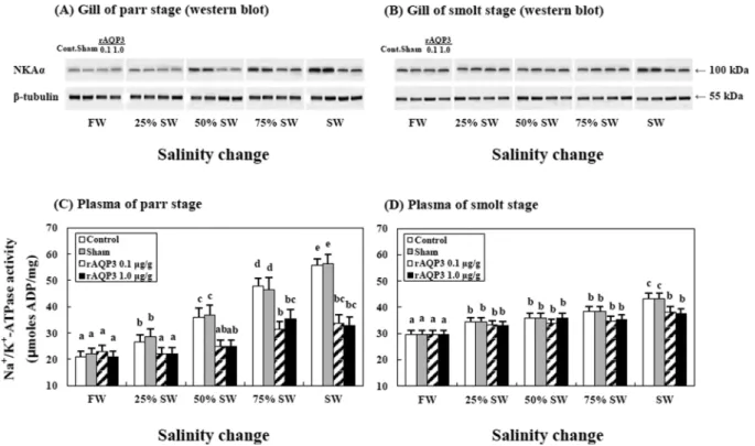
Abstract : This study aimed to examine the role of two aquaporin isoforms (AQP3 and AQP8) in response to the hyperosmotic challenge of transitioning from freshwater (FW) to seawater (SW) during parr and smoltification (smolt) using the rainbow trout, Oncorhynchus mykiss. We examined the changes in the expression of AQPs mRNAs in the gills and intestine of the parr and smolt stages of rainbow trout trans- ferred from FW to SW using quantitative real-time PCR in an osmotically changing environment [FW, SW, and recombinant AQP3 (rAQP3) injection at two dosage rates]. Correspondingly, AQPs were greater during smoltification than during parr stages in the rainbow trout. Plasma osmolality and gill Na

+

/K

+

- ATPase activity increased when the fish were exposed to SW, but these parameters decreased when the fish were exposed to SW following treatment with rAQP3 during the transition to seawater. Our results suggest that AQPs play an important role in water absorbing mechanisms associated with multiple AQP isoforms in a hyperosmotic environment.

Key words : osmoregulation, recombinant aquaporin 3, rainbow trout, parr, smolt

1. Introduction

Teleosts inhabit diverse aquatic environments, but environmental disturbances and changes in salinity can cause stress in these fish and affect their growth, reproduction, metabolism, osmolality, and immune function (Ackerman et al. 2000). Changes in the salinity and ion concentrations of a teleost’s aquatic environment can cause osmotic pressure on these fish. To cope with these changes in salinity, fish osmoregulate within their gills by regulating the movement of ions, such as Na

+

and Cl

, and water molecules (Evans 1993). Salmonids encounter a wide range of salinity changes in their lives, which causes

significant endocrine changes, as they are anadromous fish that hatch in freshwater rivers, migrate to the ocean, and return to freshwater rivers to breed (Ueda 2011).

Their organs that are involved in osmoregulation, such as the gills, kidneys, and liver, have proteins and hormones that regulate ions and water so these fish can adapt to the local environment. The hormones and proteins involved in osmoregulation include aquaporins (AQPs), cortisol, prolactin, growth hormone, Na

+

/K

+

-ATPase (NKA), and arginine vasotocin (Matsuzaki et al. 2002; Marshall et al.

2005; Park et al. 2012; Sakamoto and McCormick 2006).

AQPs are a group of membrane proteins that form water transfer channels and are important in maintaining water balance in the organs involved in osmoregulation and homeostasis of the body’s fluids (Borgnia et al. 1999;

*Corresponding author. E-mail : [email protected]

(2)

Matsuzaki et al. 2002). Several studies have shown that AQPs are important in the movement of water in euryhaline teleosts (Choi et al. 2013; Kim et al. 2014;

Martinez et al. 2005).

Thus far, 13 types of AQPs (AQP0 −AQP12) have been cloned from a variety of mammals (Ishibashi et al. 2011;

Zardoya 2005). These AQPs are divided into 3 subunits, on the basis of their genomic structure and amino acid homology. The 3 subunits are: the aquaporins group, which selectively moves water (AQPs 0, 1, 2, 4, 5, 6 and 8); the aquaglyceroporins group, which regulates the movement of water, glycerol, and urea (AQPs 3, 7, 9 and 10); and the superaquaporins group, which displays low amino acid homologies and has indistinct characteristics (AQP 11, 12) (Finn and Cerdà 2011; Ishibashi et al. 2009;

Tingaud-Sequeira et al. 2010). AQP3 is a member of the aquaglyceroporins group, and is permeable to water, glycerol, urea, and ammonia/ammonium. In the gills of the sockeye salmon, Oncorhynchus nerka, AQP3 mRNA expression is higher in seawater (SW)-adapted fish than that in fresh-water-adapted (FW)-adapted fish (Choi et al.

2013). In the gills, intestine, and kidneys of Atlantic salmon, Salmo salar, AQP3 mRNA expression is significantly increased when the fish migrate from FW to SW (Tipsmark et al. 2010). AQP8 is a member of the aquaporins group, which is almost exclusively expressed in the mucosal layer of the intestines. This reflects the significant role this pore plays in water uptake (Tipsmark et al. 2010). The mRNA expression of AQP8 peaks during smoltification in the sockeye salmon and Atlantic salmon (Choi et al.

2013; Tipsmark et al. 2010).

In fish, the hormones and proteins that maintain homeostasis, including osmoregulation, varies between fish. NKA is one of the most important hormones associated with ion regulation and is adjusted by directly controlling the discharge of Na

+

and Cl

ions from the gills of marine fish (Marshall et al. 2003; Marshall and Grosell 2006; McCormick 2001).

Salmonids migrate to the ocean following complex morphological, physiological, and behavioral changes.

This transition is known as the parr-smolt transformation, smoltification, or smolting (McCormick and Saunders 1987). As an example, parr salmon discharges water from their intestine, but during the smoltification period, the fish develops the ability to absorb water and ions in a similar way to other SW-adapted fish (McCormick and Saunders 1987). McCormick and Saunders (1987) found that the increase in salinity tolerance during smoltification is at least in part due to increased Na

+

/N

+

-ATPase activity

in the gill epithelium, as well as increased plasma glucose (McCormick and Saunders 1987). However, research into the role of AQP3 and AQP8 in the osmoregulatory organs of fish is still lacking.

Recently, the aquaculture of rainbow trout in seawater has become more prevalent. Therefore, this study was designed to determine the physiological changes that occur in juvenile rainbow trout during the parr-smolt transformation and seawater adaptation. We investigated the effect of recombinant AQP3 (rAQP3) on the physiological changes of rainbow trout, Oncorhynchus mykiss, that were acclimated to FW and were transferred to SW. We injected rainbow trout with rAQP3, then determined the physiological responses of fish to the injection of rAQP3 by measuring AQP3 and AQP8 mRNA and protein expression (in vivo and in vitro), as well as changes in NKA activity and expression (protein), and plasma osmolality.

2. Materials and Methods

Experimental fish

Rainbow trout (O. mykiss; parr: 2.4 ± 1.0 g; smolt:

105.6 ± 5.2 g) were purchased from the Ewhajung Trout Aquarium (Gyeongsangbuk-do, Korea), and housed in eight 40-L tanks fitted with circulation pumps prior to the start of experiments.

The transfer of the rainbow trout from FW (0 psu) to SW (35 psu) followed a strict protocol. Briefly, ground water was poured into square 40-L tanks, with SW added in stages to convert all the water in the tanks to SW. The fish were maintained at 25% SW, 50% SW, and 75% SW over subsequent 24-h periods. The temperature was maintained at 12 ± 0.5

o

C, and the photoperiod was maintained at a 12:12-h light:dark cycle.

Recombinant AQP3 treatment

To investigate the role of AQP3 in adapting to salinity changes, rainbow trout were treated with rAQP3 (TP301856;

OriGene Technologies, Inc., MD, USA), and the expression of AQPs mRNA was measured. The fish were allowed to acclimate in 40-L tanks. They were then anesthetized with 0.005% eugenol (4-ally-2-methoxyphenol) prior to injection.

Two experimental groups received an intraperitoneal

injection of rAQP3 dissolved in saline; one group

received the treatment at a dosage rate of 0.1 μg/g body

mass and the other at 1.0 μg/g body mass. Fish in the

sham group were injected with an equal volume of saline

(10 μL/g body mass). After the intraperitoneal injection,

(3)

fish were transferred from FW to 25% SW.

Gill tissue cultures

We prepared the gill filaments for organ culture following the methods of McCormick and Bern (1989).

The second gill arch was excised from the gill basket and washed in Dulbecco’s modified Ca

2+

/Mg

2+

-free phosphate buffer saline (PBS [1 mM]: 2.6 KCl, 1.5 KH

2

PO

4

, 137 NaCl, 8 Na

2

HPO

4

·2H

2

O). The gill filaments were separated from the gill arch just above the septum and were gently dispersed by passage through a pipette. The gill filaments were washed 3 times in the PBS, which contained 200 IU/

mL penicillin, 200 pg/mL streptomycin, and 5 mg/mL amphotericin B. Then, two gill filaments were placed in 1 mL of sterile Leibovitz-15 (L-l5) media that was supplemented with

L

-glutamine (Gibco Ltd., NY, USA).

This solution contained 100 IU penicillin, 100 mg streptomycin, and 2.5 pg amphotericin B. We placed these in sterile 24-well culture dishes, and then incubated the dishes at 20 ± 1

o

C. The cultured gills were sampled at 24- h intervals when the water was changing from 25% SW to 100% SW. Each sample was centrifuged (20

o

C, 10,000 × g, 15 s), and the supernatant was removed and stored at

−80

o

C until RNA extraction was performed.

We added recombinant AQP3 that was dissolved in 0.9% physiological saline to the culture medium at a ratio:

of 1/1,000 (v/v). The specified concentrations of rAQP3 of either 0.1 μg/mL or 1.0 μg/mL were added. Each sample was centrifuged (20

o

C, 10,000 × g, 15 s), and the supernatant was removed and stored at −80

o

C until RNA extraction was performed.

Intestine tissue culture

Each intestine was cut into pieces of approximately 1 × 1.5 mm and placed (in duplicate) in a 24-well culture plate that contained a pre-incubation medium [minimum essential medium (MEM) with Hanks' salts, 5 mg/mL bovine serum albumin (BSA), 250 U/mL penicillin G, and 250 μg/mL streptomycin sulfate, and with an adjusted pH of 7.8]. After 1 h, the medium was replaced with MEM that contained Earle’s salts (pH 7.8), 4 mg/mL BSA, 292 μg/mL

L

-glutamine, 50 U/mL penicillin G, and 50 μg/

mL streptomycin sulfate in a sterile 24-well culture dish.

Although the explants occasionally adhered to the bottom of the wells, they typically remained unattached during culturing. The cultured intestine was sampled at 24-h intervals during the period where the fish transitioned from FW to 100% SW. Each sample was centrifuged (20

o

C, 10,000 × g, 15 s) and the supernatant was removed

and stored at −80

o

C until it was required for RNA extraction.

Recombinant AQP3 that was dissolved in 0.9%

physiological saline was added to the culture medium at a ratio of 1/1,000 (v/v), with the specified concentrations of rAQP3 (0.1 and 1.0 μg/mL) added. Each sample was centrifuged (20

o

C, 10,000 × g, 15 s) and the supernatant removed and stored at −80

o

C until RNA extraction.

Sampling

Five fish from each group (100% FW, 25% SW, 50%

SW, 75% SW, 100% SW, and the rAQP3 injection group) were randomly selected for blood and tissue sampling.

Immediately after collecting the tissue specimens, the samples were frozen in liquid nitrogen and stored at −80

o

C until total RNA extraction was performed. Additionally, blood was taken from the caudal vein using a 1-mL heparinized syringe. After centrifugation (10,000 × g, 4

o

C, 5 min) the plasma was stored at −80

o

C until analysis was performed.

Quantitative PCR (QPCR)

QPCR was performed to determine the relative

expression of AQP3 (KJ737372) and AQP8 (KJ872772)

mRNA using the total RNA that was extracted from the

rainbow trout. Primers for the QPCR were designed with

reference to the known sequences of rainbow trout as

follows: AQP3 forward primer (5'-TGA TGC GTT GTG

GGA CTA-3'), AQP3 reverse primer (5'-GCT GCT GTG

CCT ATA ATC TG-3'), AQP8 forward primer (5'-CCA

TTG CTA TCT ACC TGT GT-3'), AQP8 reverse primer

(5'-TAG TTC TCT GTT GTG GTC ATC-3'), β-actin

forward primer (5'-ATC TGG CAT CAC ACC TTC TA-

3'), and β-actin reverse primer (5'-CTT CTC CCT GTT

GGC TTT G-3'). PCR amplification was conducted using

a Bio-Rad CFX96™ Real-Time PCR Detection System

(Bio-Rad, Hercules, CA, USA) and iQ™ SYBR Green

Supermix (Bio-Rad) according to the manufacturer's

instructions. QPCR was carried out as follows: 1 cycle of

denaturation at 95

o

C for 5 min, 35 cycles each consisting

of denaturation at 95

o

C for 20 s, and annealing and extension

at 55

o

C for 20 s. Each sample was run in triplicate to

ensure accuracy. As an internal control, β-actin was also

amplified for each sample, and all data were expressed as

the change with respect to the corresponding β-actin

calculated threshold cycle (Ct) levels. All analyses were

based on the Ct values of the PCR products. Based on two

QPCR assays, the intra- and inter-assay coefficients of

variation for the mRNA expression of AQP3 and AQP8

(4)

were less than 8%. The calibrated ΔCt value (ΔΔCt) for each sample and internal control (β-actin) was calculated [ΔΔCt = 2

^

− (ΔCt

sample

− ΔCt

internal control

)].

Western blot analysis

We extracted the total protein isolated from the brains of rainbow trout during changes in salinity using a protein extraction buffer (5.6 mM Tris, 0.55 mM EDTA, 0.55 mM EGTA, 0.1% SDS, 0.15 mg/mL PMSF, and 0.15 mg/mL leupeptin), followed by sonication, and quantification using the Bradford method (Bio-Rad). The total protein (30 μg) was loaded into a lane on a 4% acrylamide stacking gel and a 12% acrylamide resolving gel. A protein ladder (Fermentas, Glen Burnie, MD, USA) was used as a reference. Samples were electrophoresed through the stacking gel at 80 V and through the resolving gel at 150 V until the bromophenol blue dye front had run off the gel. The gels were immediately transferred to a 0.2- μm polyvinylidene difluoride (PVDF) membrane (Bio- Rad) at 85 V for 1.5 h at 4

o

C. Thereafter, the membranes were blocked with 5% milk in Tris-buffered saline (TBS;

pH 7.4) for 45 min, followed by washing in TBS. The membranes were incubated with polyclonal rabbit anti- AQPs (AQP3, 1:1,000 dilution; AQP8, 1:4,000 dilution) (Choi et al. 2013) and NKAα (dilution 1:2,000;

monoclonal mouse antibody to α-subunits of chicken, α5) (Development Studies Hybridoma Bank, USA), followed by horseradish peroxidase-conjugated anti-rabbit IgG secondary antibody (dilution 1:4,000) (Bio-Rad, Hercules, CA, USA) for 60 min. The internal control was β-tubulin (dilution 1:2,000; ab6046) (Abcam, Cambridge, UK), followed by horseradish peroxidase-conjugated anti-rabbit IgG secondary antibody (1:4,000) (Bio-Rad) for 60 min.

Bands were detected using WesternBright

TM

ECL (Advansta, Menlo Park, CA, USA) and 30 s exposure with a Molecular Imager

®

ChemiDoc

TM

XRS

+

System (Bio-Rad). The membrane images were scanned with a high resolution scanner and the band density was estimated using a computer program (Image Lab

TM

Software, version 3.0, Bio-Rad). The ratio of the internal control (β-tubulin) and the AQP3/AQP8 for each concentration was calculated and plotted against the concentration of the internal control.

Na

+

/K

+

-ATPase activity

NKA activity was measured following the methods described by Uchida et al. (1996) with some modifications.

Gills (approximately 4 −6 primary gill filaments from just above the septum) were collected from anesthetized fish.

The gills were then immersed in 100 μL of ice-cold SEI buffer (150 mM sucrose, 10 mM EDTA, 50 mM imidazole, and pH 7.3) and frozen at -80ºC. The filaments were thawed, homogenized in SEI buffer that contained 0.1%

deoxycholic acid, and centrifuged at 5,000 × g for 30 s to remove insoluble materials. The supernatant was assayed for NKA activity and protein content. Homogenate samples (10 μL) were added to a 200-μL assay mixture in 96-well microplates at 25ºC with and without 0.5 mM ouabain and read at 340 nm for 10 min with intermittent mixing.

The protein content of samples was determined using a Protein Assay Standard II (Bio-Rad, USA). The NKA activity was calculated as the difference in ATP hydrolysis between the presence and absence of ouabain, and expressed as μmoles of ADP per milligram protein per hour.

Plasma parameter analysis

Plasma osmolality was examined using a vapor pressure osmometer (Vapro 5600, Wescor Co., Logan, UT).

Statistical analysis

All data were analyzed using the SPSS statistical package (version 10.0; SPSS Inc., USA). Two-way ANOVA followed by Tukey’s post hoc test was used to compare differences in the data (P < 0.05). Values are expressed as mean ± standard error (SE).

3. Results

Expression of AQPs mRNA in parr/smolt rainbow trout ( in vivo)

QPCR analyses were used to show the relative expression levels of AQP3 and AQP8 mRNA in the gill and intestinal tissues of parr and smolt rainbow trout when they are transitioning from FW to SW.

The expression of both AQPs mRNA was higher in all

tissues when the fish were in SW than in FW (Figs. 1 and

2). In the gills of parr stage fish, AQP3 and AQP8 mRNA

significantly increased along with increasing levels of

salinity (Figs. 1A and 2A). In the gills of smolt stage fish,

AQP mRNAs peaked when the fish were in 25% SW for

AQP3 and 50 % SW for AQP8, and then decreased as the

fish were exposed to 100% SW (Figs. 1B and 2B). In the

intestinal tissue of parr stage fish, AQP mRNAs significantly

increased along with the salinity (Figs. 1C and 2C). For

both the AQPs studied, mRNA expression peaked when

the fish were exposed to 50% SW, and then decreased as

they were exposed to greater levels of salinity (Figs. 1D

and 2D).

(5)

Furthermore, the expression of AQPs in the rAQP3- injected group was significantly greater after beginning the transition to seawater compared with the other experimental groups (Figs. 1 and 2).

Using western blot analysis, AQP proteins were detected at a size corresponding to that predicted for rainbow trout (AQP3: approximately 33 kDa and AQP8: approximately 27 kDa). The expression of these respective proteins correlated with the AQP3 and AQP8 mRNA levels in the parr and smolt rainbow trout gill and intestinal tissues.

More particularly, AQP3 protein levels in the gills and AQP8 protein levels in the intestines were higher in SW- than FW-adapted fish. Furthermore, after fish in the rAQP3-injected group were exposed to seawater, the expression of AQPs measured in these fish was greater than in the other experimental groups.

Expression of AQP mRNAs in cultured the gill and intestinal tissue of rainbow trout ( in vitro)

QPCR analyses showed the relative levels of AQP3 and Fig. 1. Western blot and expression levels of AQP3 mRNAs in the gill (A, B) and intestine (C, D) of parr (A, C) and

smolt (B, D) rainbow trout using QPCR ( in vivo), after transitioning from freshwater to salt water (FW →

SW) and recombinant AQP3 (rAQP3) injection. β-tubulin (55 kDa) was used as the internal control during

western blot analysis. We reverse transcribed 3 μg of the total RNA prepared from gill tissue and amplified the

samples using gene-specific primers. Results are expressed as normalized fold-expression (relative to control)

with respect to β-actin levels for the same sample. Values are given as mean ± SE (n = 10). Values with dissim-

ilar letters indicate significant differences ( P < 0.05)

(6)

AQP8 mRNA expression in the cultured gill and intestinal tissues of parr and smolt rainbow trout during exposure to changing salinity levels (Figs. 3 and 4). The expression of AQP3 and AQP8 mRNAs was significantly greater in the cultured gill and intestinal tissues of fish exposed to SW, than in fish exposed to FW. Moreover, the expression of AQP mRNAs in the rAQP3-injected groups exposed to 100% SW was significantly greater than either the sham or the control group that were exposed to SW.

Gill NKA protein expression and activity

Using western blot analysis, NKAα proteins were

detected in the gills at a size corresponding to that predicted for rainbow trout NKAα (approximately 100 kDa, Figs.

5A and 5B). The expression of these proteins patterns resembled the pattern of plasma NKA levels in parr and smolt rainbow trout. In the gills, NKAα protein levels were higher in SW- than FW-adapted fish. Furthermore, in the rAQP3-injected groups the expression of NKAα was great after fish were exposed to seawater than in any other experimental group.

In the gills, NKA activity was 21.0 ± 1.8 μmoles ADP/

mg protein/h in parr fish, and 30.1 ± 1.6 μmoles ADP/mg protein/h in smolt fish at the beginning of the experiments.

Fig. 2. Western blot and expression levels of AQP8 mRNAs in the gill (A, B) and intestine (C, D) of parr (A, C) and

smolt (B, D) rainbow trout using QPCR ( in vivo), after transitioning from freshwater to salt water (FW →

SW) and recombinant AQP3 (rAQP3) injection. β-tubulin (55 kDa) was used as the internal control during

western blot analysis. Values with dissimilar letters indicate significant differences ( P < 0.05). Values are given

as mean ± SE (n = 10)

(7)

This reached a peak of 56.4 ± 2.3 μmoles ADP/mg protein/h in parr fish and 43.2 ± 2.1 μmoles ADP/mg protein/h in smolt fish during the transition to 100% SW (Figs. 5C and 5D). Additionally, in the gill tissue of parr and smolt rainbow trout that were given rAQP3 treatment, the NKA levels of SW-adapted fish decreased after being exposed to SW to 33.2 ± 2.0 μmoles ADP/mg protein/h in parr fish (Fig. 5C) and 37.6 ± 1.9 μmoles ADP/mg protein/h in smolt (Fig. 5D).

Plasma osmolality

The plasma osmolality of the rainbow trout was 227.5 ± 5.0 mOsm/kg in parr fish, and 244.6 ± 10.2 mOsm/kg in smolt fish at the beginning of the experiments. Plasma osmolality peaked at 516.4 ± 13.3 mOsm/kg in parr fish and 406.3 ± 15.6 mOsm/kg in smolt fish after being exposed to SW. In the groups treated with rAQP3, plasma

osmolality in the SW-adapted parr and smolt rainbow trout decreased to 330.6 ± 15.4 mOsm/kg in parr fish and 302.2 ± 17.2 mOsm/kg in smolt fish after exposure to SW (Fig. 6).

4. Discussion

In this study, we used freshwater rainbow trout in parr and smolt stages. Fish were exposed to SW at increasing levels of salinity following the injection of one of two concentrations of recombinant AQP3 (0.1 μg/g body mass or 1.0 μg/g body mass). We determined the AQP genes activity and the osmoregulatory response of the fish to the injection of rAQP3. We exposed the fish to increasing levels of salinity and measured AQP3 and AQP8 mRNA and protein expression (both in vivo and in vitro), as well as changes in NKA activity and expression (protein), and Fig. 3. Expression levels of AQP3 mRNAs in the cultured gill (A, B) and intestine (C, D) of parr (A, C) and smolt (B,

D) rainbow trout using QPCR ( in vitro), after transitioning from freshwater to salt water (FW → SW) and

recombinant AQP3 (rAQP3) injection. Values with letters indicate significant differences for the FW → SW

transition, within the period of the salinity changes. Values with dissimilar letters indicate significant differ-

ences ( P < 0.05). Values are given as mean ± SE (n = 10)

(8)

plasma osmolality.

We observed changes in the AQPs mRNA and protein expression in the gills and intestinal tissue of rainbow trout in parr and smolt stages (in vivo and in vitro), which were associated with changes in salinity. We observed that the expression of AQP3 and AQP8 mRNA and protein expression in the gill and intestinal tissue of parr stage fish increased with increasing levels of salinity. A similar pattern was observed in the cultured gill and intestinal tissues of parr stage fish in in vitro experiments. However, the expression of AQP3 and AQP8 mRNA and protein in the gill and intestinal tissue of smolt stage fish peaked at 50% SW, and then decreased. A similar pattern was observed in the cultured gills and intestine of smolt stages (in vitro). We observed that in the rAQP3-treated groups, the expression of AQP3 and AQP8 mRNA and protein was greater in both the gill and intestinal tissue than in the

control group, regardless of whether fish were in the parr or smolt stage.

Previous studies of European eels, Anguilla anguilla, and Atlantic salmon similarly found that the mRNA expression of AQP3 and AQP8 significantly increased during increasing exposure to SW (Martinez et al. 2005;

Tipsmark et al. 2010). Choi et al. (2013) found that parr and smolt stage sockeye salmon exposed to SW had an increased expression of AQP3 and AQP8 mRNA and protein in the osmoregulatory organs, such as the gills and the intestine.

This study found that in the gills and the intestine of rainbow trout, AQP3 and AQP8 play an important role in maintaining water balance during changes in water salinity. AQP3 and AQP8 mRNA expression differs between the parr and smolt stages of the control group, but not the rAQP3-treated groups, during increasing Fig. 4. Expression levels of AQP8 mRNAs in the cultured gill (A, B) and intestine (C, D) of parr (A, C) and smolt (B,

D) rainbow trout using QPCR ( in vitro), after transitioning from freshwater to salt water (FW → SW) and recombinant AQP3 (rAQP3) injection. Values with dissimilar letters indicate significant differences ( P < 0.05).

Values are given as mean ± SE (n = 10)

(9)

exposure to salinity. The expression of AQPs decreases after smolt stage rainbow trout are exposed to 50% SW, therefore we suggest that rainbow trout have already adapted their osmoregulatory abilities at that point.

Ion transport factors related to the discharge of ions from an increasingly saline environment are controlled by

NKA, an essential enzyme (McCormick 2001). It is known that in the gills, the expression of NKA’s mRNA and proteins increased to control plasma osmolality when fish were exposed to increasing salinity (McCormick 1995; Uchida et al. 2000). We investigated the effect of rAQP3 on the physiological activity when rainbow trout Fig. 5. Effects of recombinant AQP3 (rAQP3) treatment on NKA protein expression (A, B) and plasma NKA levels

(C, D) after transitioning from freshwater to salt water (FW → SW), and after rAQP3 injection in parr (A, C) and smolt (B, D) rainbow trout. Values are mean ± SE (n = 10). A western blot of NKA protein expression in the gills of parr and smolt rainbow trout after transitioning from freshwater to salt water has also been shown.

β-tubulin (55 kDa) was used as the internal control. Values with dissimilar letters indicate significant differ- ences ( P < 0.05)

Fig. 6. Effects of recombinant AQP3 (rAQP3) treatment on plasma osmolality levels after transitioning from freshwa-

ter to salt water (FW → SW), and after rAQP3 injection in parr (A) and smolt (B) rainbow trout. Values are

mean ± SE (n = 10). Values with dissimilar letters indicate significant differences (P < 0.05)

(10)

that were acclimated to FW were transferred to SW. We injected fish with rAQP3 at a dosage rate of 0.1 µg/g body mass or 1.0 µg/g body mass, and then measured any changes in NKA activity and protein expression, and plasma osmolality. We observed that the protein expression and activity of NKA in the gills and the plasma osmolality increased along with salinity in fish exposed to SW in both parr and smolt stage fish, but this effect was decreased by treatment with rAQP3. The activity and protein expression of NKA and the plasma osmolality of parr stage fish were significantly greater than those of smolt stage fish.

In a similar study, Singer et al. (2003) found that when FW-adapted Atlantic salmon were exposed to a SW environment, NKA activity in the gills increased significantly.

Furthermore, Choi et al. (2013) found that when FW- adapted sockeye salmon were exposed to a SW environment, plasma osmolality increased along with the increase in salinity. Choi et al. (2015) also found that the plasma levels of Na

+

, Cl

, and K

+

ions and cortisol, (a SW- adapted hormone) in parr stage fish increased more gradually than in smolt stage fish as salinity increased.

Previous studies have suggested that smolt stage rainbow trout have greater hypo-osmoregulatory abilities, a SW environment adaptation.

Our study found similar results to previous studies (Choi et al. 2015; Singer et al. 2003). The, activity and protein expression of NKA in the gills and plasma osmolality both increased significantly during increasing exposure to salinity. We suggest that the increased activity and protein expression of NKA in the gills and the increased plasma osmolality rapidly suppress the influx of ions from the increase in salinity, through an interaction with AQP3 following rAQP3 treatment. We determined that these levels (NKA and osmolality) were different between parr and smolt stage fish, which is a reflection of the differing abilities of these fish to osmoregulate in saline environments (parr → smolt).

In summary: 1) rAQP directly increased the expression of AQPs mRNA and proteins that play an important role in maintaining water homeostasis and plasma osmolality in rainbow trout. We suggested that rAQP3 have a positive function fish adapting to the SW environment by increasing the activity of the gills. 2) rAQP3 effectively increased the osmoregulation ability of parr stage rainbow trout, which have less osmoregulation ability compared to the that of smolt stage, to tolerate salinity. We conclude that rAQP3 enhance the ability of parr stage rainbow trout to adapt to saline environments. Thus, this study provides

basic data for understanding the function of AQPs with relation to osmotic control and maintaining water balance in a euryhaline teleost.

Acknowledgements

This study was supported by Korea Institute of Ocean Science and Technology (PO01285) and by a grant from the National Institute of Fisheries Science (R2106038).

References

Ackerman PA, Forsyth RB, Mazur CF, Iwama GK (2000) Stress hormones and the cellular stress response in salmonids. Fish Physiol Biochem 23:327 −336

Borgnia M, Nielsen S, Engel A, Agre P (1999) Cellular and molecular biology of the aquaporin water channels.

Annu Rev Biochem 68:425−458

Choi YJ, Shin HS, Kim NN, Shin SH, Yamamoto Y, Ueda H, Lee J, Choi CY (2013) Expression of aquaporin-3 and -8 mRNAs in the parr and smolt stages of sockeye salmon, Oncorhynchus nerka: Effects of cortisol treatment and seawater acclimation. Comp Biochem Physiol A 165:228 −236

Choi YJ, Kim NN, Choi CY (2015) Profiles of hypothalamus- pituitary-interrenal axis gene expression in the parr and smolt stages of rainbow trout, Oncorhynchus mykiss:

Effects of recombinant aquaporin 3 and seawater acclimation. Comp Biochem Phys A 182:14−21

Evans DH (1993) Osmotic and ionic regulation. In: Evans DH (ed) The Physiology of fishes. CRC Press, Boca Raton, Florida, pp 315−341

Finn RN, Cerdà J (2011) Aquaporin evolution in fishes.

Front Physiol 2:44

Ishibashi K, Hara S, Kondo S (2009) Aquaporin water channels in mammals. Clin Exp Nephrol 13:107 −117 Ishibashi K, Kondo S, Hara S, Morishita Y (2011) The

evolutionary aspects of aquaporin family. Am J Physiol 300:R566 −R576

Kim YK, Lee SY, Kim BS, Kim DS, Nam YK (2014) Isolation and mRNA expression analysis of aquaporin isoforms in marine medaka Oryzias dancena, a euryhaline teleost. Comp Biochem Phys A 171:1−8

Marshall W (2003) Rapid regulation of NaCl secretion by estuarine teleost fish: coping strategies for short-duration freshwater exposures. Biochim Biophys Acta 1618:95 − 105

Marshall WS, Cozzi RR, Pelis RM, McCormick SD (2005)

Cortisol receptor blockade and seawater adaptation in the

(11)

euryhaline teleost Fundulus heteroclitus. J Exp Zool Part A 303:132 −142

Marshall WS, Grosell M (2006) Ion transport, osmoregulation and acid-base balance. In: Evans DH, Claiborne JB (eds) The Physiology of Fishes, 3rd ed. CRC Press, Boca Raton, Florida, pp 177−230

Martinez A-S, Cutler CP, Wilson G, Phillips C, Hazon N, Cramb G (2005) Cloning and expression of three aquaporin homologues from the European eel (Anguilla anguilla): effects of seawater acclimation and cortisol treatment on renal expression. Biol Cell 97:615−627 Matsuzaki T, Tajika Y, Tserentsoodol N, Suzuki T, Aoki T,

Hagiwara H, Takata K (2002) Aquaporins: a water channel family. Anat Sci Int 77:85−93

McCormick SD (2001) Endocrine control of osmoregulation in teleost fish. Am Zool 41:781−794

McCormick SD, Bern HA (1989) In vitro stimulation of Na

+

-K

+

-ATPase activity and oubain binding by cortisol in coho salmon gill. Am J Physiol 256:R707−R715 McCormick SD, Saunders RL (1987) Preparatory physiological

adaptations for marine life of salmonids: osmoregulation, growth, and metabolism. Am Fish S S 1:211 −229 Park MS, Kim NN, Shin HS, Min BH, Kil G-S, Cho SH,

Choi CY (2012) Hypoosmotic shock adaptation by prolactin involves upregulation of arginine vasotocin and osmotic stress transcription factor 1 mRNA in the cinnamon clownfish Amphiprion melanopus. Anim Cells Syst 16:391 −399

Sakamoto T, McCormick SD (2006) Prolactin and growth hormone in fish osmoregulation. Gen Comp Endocr 147:24 −30

Singer TD, Finstad B, McCormick SD, Wiseman SD,

Schulte PM, McKinley RS (2003) Interactive effects of cortisol treatment and ambient seawater challenge on gill Na

+

, K

+

-ATPase and CFTR expression in two strains of Atlantic salmon smolts. Aquaculture 222:15−28

Tingaud-Sequeira A, Calusinska M, Finn RN, Chauvigné F, Lozano J, Cerdà J (2010) The zebrafish genome encodes the largest vertebrate repertoire of functional aquaporins with dual paralogy and substrate specificities similar to mammals. BMC Evol Biol 10:38

Tipsmark CK, Sørensen KJ, Madsen SS (2010) Aquaporin expression dynamics in osmoregulatory tissues of Atlantic salmon during smoltification and seawater acclimation. J Exp Biol 213:368 −379

Ueda H (2011) Physiological mechanism of homing migration in Pacific salmon from behavioral to molecular biological approaches. Gen Comp Endocr 170:222−232

Uchida K, Kaneko T, Yamauchi K, Hirano T (1996) Morphometrical analysis chloride cell activity in the gill filaments and lamellae and changes in Na

+

, K

+

-ATPase activity during seawater adaptation in chum salmon fry. J Exp Zool 276:193−200

Uchida K, Kaneko T, Miyazaki H, Hasegawa S, Hirano T (2000) Excellent salinity tolerance of mozambique tilapia (Oreochromis mossambicus): elevated chloride cell activity in the branchial and opercular epithelia of the fish adapted to concentrated seawater. Zool Sci 17:149−160 Zardoya R (2005) Phylogeny and evolution of the major

intrinsic protein family. Biol Cell 97:397 −414

Received Feb. 22, 2016

Revised Mar. 13, 2016

Accepted Mar. 17, 2016

수치

Fig. 2. Western blot and expression levels of AQP8 mRNAs in the gill (A, B) and intestine (C, D) of parr (A, C) and smolt (B, D) rainbow trout using QPCR ( in vivo), after transitioning from freshwater to salt water (FW → SW) and recombinant AQP3 (rAQP3) i
Fig. 6. Effects of recombinant AQP3 (rAQP3) treatment on plasma osmolality levels after transitioning from freshwa- freshwa-ter to salt wafreshwa-ter (FW → SW), and affreshwa-ter rAQP3 injection in parr (A) and smolt (B) rainbow trout

참조

관련 문서

4-8 Evaluation of rail corrugation for artificial specimen A (30 mm wavelength); (a) LVDT signal, (b) FFT analysis, (c) STFT analysis and (d) digitizing result

Frequency of conidial germination (A) and appressorium formation (B) of Colletotrichum orbiculare and callose formation of plant cells (C) at the

The matrix A show the cost per computer (in thousands of dollars) and B the production figures for the year 2005 (in multiples of 1000 units).. Find a matrix C that

The number of migrating individuals of the Oriental Honey Buzzard by hour (A: Hong island, B: Eocheong island, C: Socheong island, D: Busan and Geoje island) ···92

Effect of LRP6 on survivin expression in hypoxic cardiomyocytes (A) Expression of survivin in Ad-LRP6 and silencing LRP6 infected cardiomyocytes as determined by western

The combination treatment of PBA and 6-gingerol induced the cleavage of caspase-3, caspase-4, PARP, GRP78, p-eIF2α, and CHOP in DU145 cells and Western blot analysis for

GDP impact of COVID-19 spread, public health response, and economic policies. Virus spread and public

1 John Owen, Justification by Faith Alone, in The Works of John Owen, ed. John Bolt, trans. Scott Clark, &#34;Do This and Live: Christ's Active Obedience as the