184
종격동에 발생한 혼합생식세포종양 1예
을지대학교 의과대학 내과학교실1, 흉부외과학교실2, 병리학교실3 조 욱1, 한민수1, 김길동2, 김성호3, 김준형1, 이양덕1, 조용선1
A Case of Mixed Germ Cell Tumor of the Mediastinum
Wook Cho, M.D.1, Min Soo Han, M.D.1, Kil Dong Kim, M.D.2, Sung Ho Kim, M.D.3, Yang Deok Lee, M.D.1, Yong Seon Cho, M.D.1, Jun Hyoung Kim, M.D.1
Department of Internal Medicine1, Chest Surgery2, and Pathology3, Eulji University School of Medicine, Daejeon, Korea
The Mixed germ cell tumors of the mediastinum are very quite rare. The Prognosis is generally dominated by the most aggressive component, which is represented by a choriocarcinoma, an endodermal sinus tumor, an embryonal carcinoma, and a seminoma, in descending order of in the degree of malignancy. We experienced one a case of a mixed germ cell tumor at the anterior mediastinum. The patient was 27-year-old male, who complained of hemoptysis and cough. The Chest X-ray showed a well-defined lobulated mediastinal mass in the left upper lung field. The operation was done and The mass was excised surgically. A Biopsy showed elements of mature tissues, immature neuronal components, and seminoma components. (Tuberc Respir Dis 2005; 58:184-187)
Key words : Germ cell tumor, Mixed germ cell tumor, Mediastinum
Address for correspondence : Min Soo Han, M.D.
Department of Internal Medicine, Eulji University School of Medicine, 1036, Dunsan-Dong, Seo-Ku, Daejeon, 301-726, Korea
Phone : 042-611-3151 Fax : 042-611-3174 E-mail : [email protected]
Received : Dec. 9. 2004 Accepted : Jan. 25. 2005
서 론
종격동 생식세포종양은 종격동에서 발생하는 모든 종양의 약 20%정도를 차지하며 주로 전종격동에서 발생하고 대개 20-49세 남성에서 호발하며 예후는 고 환의 생식세포종양보다 좋지 않다1-5. Weidner N.의 보고에1 의하면종격동 생식세포종양의 44%는 성숙 낭성의 기형종이며 18%가 정상피종, 6%가 미성숙 기 형종, 5%가 순수한 배아성 암종, 2%가 순수한 내배엽 동 종양, 2%가 융모막암종, 23%가 악성의 혼합생식세 포종양이다1,5. 기형종은 하나 또는 그 이상의 다른 생 식세포종양성분들과 혼합되어 드물게 혼합생식세포 종양으로 발생하는데 이는 대개 악성으로 진단되며 종양의 정도, 크기에 따라 환자들은 흉통, 기침, 체중 감소, 호흡곤란, 상대정맥 증후군, 림프절병증, 골통증, 여성형 유방증, 객혈 등의 증상을 호소한다1,2,4,6. 증상 발현시 주위 조직에 침윤이 심하기 때문에 완전한 수
술적 절제가 어려워, 화학요법, 방사선요법 등 다양한 치료방법이 필요하다6-8. 국내에서는 종격동에 발생한 미성숙 기형종에 대한 증례 보고가6,7,9있었으나 미성 숙 기형종과 정상피종이 혼합된 생식세포종양에 대한 증례 보고는 없었다. 저자들은 객혈을 주소로 내원한 27세 남자 환자에서 수술로 확진한 미성숙 기형종과 정상피종이 혼합된 생식세포종양의 증례를 경험하였 기에 보고하는 바이다.
증 례
환 자 : 유 ○ ○, 27세, 남자 주 소 : 객혈
현병력 : 내원 15일전에 약 100mL 정도의 객혈이 발 생하여 인근 병원을 방문하여 촬영한 단순흉부사진 에서 이상소견이 있다는 말을 듣고 자세한 검사 및 치료를 위해 내원하였다.
과거력 : 특이 소견 없음.
흡연력 : 2 갑․년
진찰소견 : 내원 당시 혈압은 110/60 mmHg, 맥박은 72회/분, 호흡수는 20회/분, 체온은 37.2℃ 이었으며 고환 촉지 검사상 만져지는 종괴소견 없었음.
검사실 소견 : 내원시 말초혈액검사상 백혈구 8,470/
㎣, 혈색소 13.6 g/dL, 혈소판 224,000/㎣ 이었다. 대
Tuberculosis and Respiratory Diseases Vol. 58. No. 2, Feb, 2005
185 Figure 2. Chest CT shows a lobulated, smooth, well-
defined mass containing multiple calcifications and multiple low density area in the left anterior media
stinum. Ground-glass attenuation and consolidation is observed in the anterior segment of the contiguous left upper lobe.
Figure 4. Seminoma with a sheet-like arrangement of cells with clear cytoplasm, well-defined cell membranes, and nuclei with prominent, central nucleoli.(H&E, ×200)
Figure 3. Immature teratoma with immature neuroe
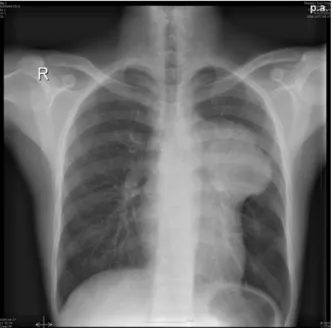
pithelial tubules. (H&E, ×200) Figure 1. Chest PA shows a well-defined lobulated
mediastinal mass in the left upper lung field.
기중에서 시행한 동맥혈 가스분석 검사는 pH 7.43, PaCO2 43.9 mmHg, PaO2 125.5 mmHg, HCO3-
29.0 mmol/L, 동맥혈 산소포화도는 98.6% 이었다. 일반 생화학 검사는 AST 18 IU/L, ALT 18 IU/L, 총단백
7.6 g/dL, 알부민 4.1 g/dL, 총빌리루빈 0.6 mg/dL, LDH 309 IU/L이었다. 종양 표지자 검사는 Alpha fetoprotein(αFP) 600ng/mL이상, human chorionic gonadotropin(βHCG) 10.1 mIU/mL이었다.
방사선 소견 : 단순흉부사진에서 종격동 좌측에 경 계가 분명한 종괴가 관찰되었으며(Fig. 1), 흉부전산 화단층촬영에서 전종격동 좌측에 8.6×8×10.5 cm 크 기의 경계가 분명한 종괴가 관찰되었고 다발성의 석회화와 저밀도 부분을 함유하고 있었다. 인접한 좌상엽의 앞부분에는 간유리음영과 경화가 동반된 소견이 관찰되었다(Fig. 2). 복부초음파검사는 정상 소견이었다.
수술 및 병리 소견 : 개흉 수술 소견상 전종격동 좌
W Cho, et al.: A case of mixed germ cell tumor of the mediastinum
186
측에 심장막과 심하게 유착된 종양이 관찰되었으며 조직 생검 및 절제를 시행하였다. 광학현미경 소견 에서 종양은 미성숙 신경상피세관을 포함하는 미성숙 기형종과(Fig. 3) 세포막이 분명하고 세포질은 투명 하며 중앙에 위치한 뚜렷한 핵을 포함하는 세포들 의 시트같은 배열을 보이는 정상피종의(Fig. 4) 소 견이 관찰되었다.
임상경과 : 환자는 수술 후 외래 추적 관찰 중단된 상태이다.
고 찰
생식선외에 발생하는 생식세포종양은 전체 생식세 포종양의 1-2%를 차지하는 드문 질환으로 주로 종격 동, 후복막강, 흉선, 송과선 등에서 발생하며 이중 종 격동의 빈도가 가장 높다1-3,7,8,10-12
. 종격동 생식세포종 양의 44%는 성숙 낭성의 기형종이며 18%가 정상피 종, 6%가 미성숙 기형종, 5%가 순수한 배아성 암종, 2%가 순수한 내배엽동 종양, 2%가 융모막암종, 23%
가 악성의 혼합생식세포종양이다1,5. 다른 생식세포종 양성분들과 혼합되어 드물게 발생하는 혼합생식세포 종양은 대개 악성으로 진단되며 예후는 혼합된 생식 세포종양성분의 악성정도에 따라 달라지는데 융모막 암종이 가장 나쁘고 정상피종이 가장 좋다1,2,4. 종격동 에 발생하는 생식세포종양은 모든 종격동의 낭종과 종양의 20%를 차지하며 주로 전종격동에서 나타나고 흉선종의 분포와 비슷하다1-5. 종격동 생식세포종양의 조직학적 형태는 고환 생식세포종양의 조직학적 구조 와 구별할 수 없기 때문에 종격동 생식세포종양이 발 견되었을 때에는 고환의 원발성 종양이 전종격동으로 전이를 하는 경우는 매우 드물지만 고환의 원발성 종 양의 가능성을 배제하는 것이 중요하다5. 종격동의 생식세포종양은 고환을 주의깊게 촉진하였을 때 종괴 가 없고, 후복막의 림프관 조영술에서 비정상적인 소 견이 발견되지 않을때 비로소 원발성으로 진단할 수 있다. 미성숙 기형종은 성숙 기형종과 배아성 암종의 중간에 위치하는 것으로 유배아체가 성숙한 조직으로 분화하는 중간 형태의 조직으로 구성된 종양이다. 성 숙 기형종과는 달리 세개 배아층에서 유래하는 조직
성분들의 분화가 불완전하고 기질화도 잘 이루어져 있지 않다. 미성숙 중간엽, 신경 로제트의 형태를 갖는 미성숙 신경외배엽조직들로 이루어진 특징을 보인다
2,3,9,12
. 세포학적으로 악성종양의 특징인 역형성 변화 가 없지만 악성종양의 임상 경과를 취한다2,4,12. 미성숙 기형종은 거의 모든 예가 20세 미만의 젊은 남자에 국 한되어 발생하며6 예후는 임상적인 병기결정과 나이 에 따라 결정되며 유아나 소아에서 발생하면 임상적 으로 양성 종양의 특성을 나타내고 10대나 젊은 성인 에서 발생하면 배아성암종과 유사한 악성 종양의 특 성을 나타낸다1,2,4-6,9. 양성 기형종은 대개 서서히 성장 하고, 경계가 명확하여 세가지 배엽을 포함하지만 외 배엽조직이 주된 성분이며, 반면에 미성숙기형종은 신속히 증식하며, 주위조직에 침범한다6. 따라서 미성 숙기형종의 경우 종양의 정도, 크기에 따라 환자들은 흉통, 기침, 체중감소, 호흡곤란, 상대정맥 증후군, 림 프절병증, 골통증, 여성형 유방증, 객혈등의 증상을 호
소한다1-6,8,9,13. αFP, βHCG는 임상적으로 악성 종격동
의 생식세포종양이 의심되는 경우에 양성기형종과의 감별을 위해 도움이 될 수 있으며5,6,12,15 미성숙기형종 의 치료에 대한 반응 및 조기 재발을 판정하는데에도 중요한 지표가 된다6. 종격동내에 발생하는 정상피종 은 종격동 생식세포종양의 과반수를 차지하며 주로 20-40대의 젊은 남자에게 호발하고 증상은 종양의 크 기, 인접장기의 침범정도 그리고 원격 전이등에 의하 여 나타나게 되며 주로 흉부압박감, 흉통, 기침, 체중 감소, 전신 무력감, 상대정맥 증후군등을 호소하게 된 다. 그러나 약 30-35% 정도에서는 전혀 증상없이 흉 부방사선 촬영에서 우연히 발견되고 있다4,8,10 . 종격동 정상피종은 흔히 양측폐나 주위 임파절, 골로 전이되 며5,14 방사선 치료에 잘 듣기 때문에 예후가 양호하여, 10년 생존율이 69%에 이른다5,8,14. 그러나 비정상피성 생식세포종들은 화학요법에 반응하지 않고, 재발이 잘 되며, 예후가 매우 불량하다1,5,8,14. 미성숙기형종을 포함한 모든 기형종의 치료는 근본적으로 외과적 절 제술이다6. 미성숙기형종은 신속히 증식하며 주위조 직으로 침윤이 심해 완전한 수술적 절제가 어려워서 화학요법, 방사선요법 등 다양한 치료방법이 필요할 뿐만 아니라 양성 기형종과는 현저한 예후의 차이를
Tuberculosis and Respiratory Diseases Vol. 58. No. 2, Feb, 2005
187 보이고 있다6,7.
요 약
종격동에 발생하는 생식세포종양에서 미성숙 기형 종과 정상피종이 혼합된 경우는 매우 드물며 예후는 좋지 않은 것으로 알려져 있다.
저자들은 객혈을 주소로 내원한 환자에서 수술로 확진한 미성숙 기형종과 장상피종이 혼합된 생식세포 종양의 1예를 경험하였기에 보고하는 바이다.
참 고 문 헌
1. Weidner N. Germ-cell tumors of the mediastinum.
Semin Diagn Pathol 1999;16:42-50.
2. Moran CA, Suster S. Germ-cell tumors of the mediastinum. Adv Anat Pathol 1998;5:1-15.
3. Moran CA, Suster S. Primary germ cell tumors of the mediastinum : I. Analysis of 322 cases with special emphasis on teratomatous lesions and a proposal for histopathologic classification and staging. Cancer 1997;80:681-90.
4. Dehner LP. Germ-cell tumors of the mediastinum.
Semin Diagn Pathol 1990;7:266-84.
5. Strollo DC, Rosado de Christenson ML, Jett JR.
Primary mediastinal tumors. part 1 : Tumors of the anterior mediastinum. Chest 1997;112:511-22.
6. Kang JK, Park JK, Wang YP, Kim SW, Lee HK.
Huge immature teratoma of mediastinum : A case report. Korean J Thorac Cardiovasc Surg 1989;22:
867-72.
7. Lee JP, Chung WS, Kim YH, Kang JH, Jee HO.
Immature teratoma at anterior mediastinum : Report of one case. Korean J Thorac Cardiovasc Surg 1992;25:435-7.
8. Ryu HM, Choi HJ, Shin GC, Chung JH, Lee KH, Lee HW, et al. A case of combined mixed germ cell tumor and angiosarcoma within the mediastinum.
Tuberc Respir Dis 1994;41:413-7.
9. Chung SH, Kang KM, Park SD, Park JH, Moon JH, Kang KH, et al. Clinical experience of mediastinal immature teratoma in a newborn. Korean J Thorac Cardiovasc Surg 1995;28:530-2.
10. Moran CA, Suster S. Primary germ cell tumors of the mediastinum :II.Mediastinal seminomas-A clini
copathologic and immunohistochemical study of 120 cases. Cancer 1997;80:691-8.
11. Sugitha I, Suwendra P, Semadi N, Danun M, Mulyadi K. Mediastinal teratoma. Jima 2002;1:307-12.
12. Ueno T, Tanaka TO, Nagata M, Tsunoda H, Anno I, Ishikawa S, et al. Spectrum of germ cell tumors : from head to toe. Radiolographics 2004;24:387-404.
13. Lee SY, Kang WT, Song HS, Lee YC, Lee YK. Clinical analysis of the primary mediastinal tumors. Tuberc Respir Dis 1991;38:128-34.
14. Moran CA, Suster S. Primary germ cell tumors of the mediastinum : III. Yolk sac tumor, embryonal carcinoma, choriocarcinoma, and combined nontera
tomatous germ cell tumors of mediastinum-A clinic
opathologic and immunohistochemical study of 64 cases. Cancer 1997;80:699-707.
15. Song YB, Kim KR, Choi IJ. Clinical and pathological analysis of germ cell tumors. Korean. J. Pathol 1986;20:295-304.