사염화탄소 유도 급성 간독성 모델에서 치자의 간 보호 효과
신전규·김효연*·이선미*,#
경희대학교 생명과학부, *성균관대학교 약학대학
(Received December 28, 2009; Revised January 5, 2010; Accepted January 5, 2010)
Protective Effect of Gardenia jasminoides Against Carbon Tetrachloride-Induced Acute Hepatotoxicity
Jun-Kyu Shin, Hyo-Yeon Kim* and Sun-Mee Lee*,#
College of Life Science, Kyung Hee University, Yongin 446-701, Korea
*School of Pharmacy, Sungkyunkwan University, Suwon 440-746, Korea
Abstract
— Gardenia jasminoides is one of the most widely used herbal preparations for the treatment of liver disorders.
This study evaluated the potential beneficial effect of G. jasminoides in a mouse model of carbon tetrachloride (CCl
4)- induced liver injury. The mice were treated intraperitoneally with CCl
4(10
µl/kg). They received G. jasminoides (30, 100, 300 mg/kg) 48 h, 24 h and 2 h before and 6 h after administering CCl
4. The serum activities of aminotransferase and the hepatic level of malondialdehyde were significantly higher 24 h after the CCl
4treatment, while the concentration of reduced glutathione was lower. These changes were attenuated by G. jasminoides. CCl
4increased the level of circulating tumor necrosis factor-
α(TNF-
α) markedly, which was reduced by G. jasminoides. The levels of hepatic inducible nitric oxide syn- thase (iNOS) and cyclooxygenase-2 (COX-2) protein expression were markedly higher after the CCl
4treatment. G. jas- minoides diminished these alterations. CCl
4increased the level of TNF-
α, iNOS and COX-2 mRNA expressions, and these increases were attenuated by G. jasminoides. These results suggest that G. jasminoides alleviates CCl
4-induced liver injury, and this protection is likely due to the reduced oxidative stress and the downregulation of proinflammatory mediators.
Keywords □
carbon tetrachloride, Gardenia jasminoides, hepatotoxicity, inflammation
간질환의원인은다양한것으로알려져있으며병인학적으로 볼때바이러스에기인된간질환
,
약물(
독성물질)
중독에기인된 간질환및담도기능부전에의한간질환등으로분류할수있다.
이러한간질환들은간세포파괴에의한해독능력저하와담즙분 비억제에의한독성물질배설장애
,
비타민B
1, B
2및C
의흡수 억제및축적저하에의한전신권태감,
소양감,
식욕부진및피로등의임상증상을나타낸다
.
1)현재국내외적으로사용되고있는간질환치료제로는합성약물인인터페론과
malotilate
가있 으나이들은간독성물질에대한보호작용만으로그의유효성을 인정받고있는수준이며,
부작용또한빈번히나타난다.
또한마리아엉겅퀴의활성성분인
silymarin
과천연물유래합성간질환 치료제인biphenyl dimethyl dicarboxylate
의개발로천연물에대한관심이고조되고있으나
,
2)대다수의천연물의경우유효용량뿐아니라정확한약리효과에대한기초연구결과가미약하다
.
사염화탄소는 지방성 퇴행
(fatty degeneration),
3) 섬유증(fibrosis),
4)간세포사멸및간암등을유발시키며,
5)독성물질이 간세포에미치는영향과그대사과정을규명하는데대표물질로 서주로사용되어오고있다.
사염화탄소는1
차적으로일산소첨 가효소계(mixed function oxidase system)
의활성에의해CCl
3라디칼로활성화되어세포막의지질과산화를일으켜간독성을 유발하며
,
6)2
차적으로는간장대식세포인Kupffer cell
을활성화 시켜염증매개인자를생성한다.
7)치자는꼭두서니과에 속한상록관목인치자나무
(Gardenia jasminoides)
의성숙한과실을건조한것이다.
치자의효능은열 독을없애고황달을낫게하며소갈을멎게하며눈이붓고아 픈것,
문둥병,
창양(
瘡瘍)
을낫게하고지충(
地蟲)
의독을없앤다고알려져있다
.
치자의추출물은hexobarbital
의수면효과를 현저히연장시키고,
8)생쥐의운동성을감소시키며체온강하와부교감신경계활성화를통하여혈압강하작용도나타낸다
.
치자의활성성분인
geniposide
는α-naphthylisothiocyanate
로유도한간#본논문에관한문의는저자에게로
(
전화) 031-290-7712 (
팩스) 031-292-8800 (E-mail) [email protected]
종설
독성모델에서혈중
bilirubin, ALT
및AST
등의생화학적수치뿐만아니라병리학적조직병변을개선시켰으며
,
9)genipin
과crocetin
도담즙분비를증가시켰다.
10)또한genipin
은위산의분 비를촉진하고항산화작용및nitric oxide(NO)
생성저해를통해소염작용을나타낸다
.
11)따라서
,
저자등은본연구에서사염화탄소급성간독성모델 을사용하여치자추출물의간장보호작용을확인하고,
더나아가산화적손상과염증매개인자를중심으로작용기전도알아보 고자하였다
.
실험방법
실험동물
실험동물은대한바이오링크
(
주)
로부터생후8
주수컷ICR
생 쥐를 공급받아 온도23±1
oC,
상대습도55±15%, 300~500 Lux
및12
시간간격으로명암이조절되는성균관대학교약학대학동물사육실에서
7
일이상순화시킨후육안적증상을관찰 하여정상적인동물만을실험에사용하였으며,
실험동물용고형사료
(Dae-Han Biolink, Korea)
및물은자유롭게섭취시켰다.
시료의추출및제조
실험에사용된치자는약초약업사
(
주)
에서구입하여시료로사용하였다
.
조절(
操切)
한치자500 g
을70% EtOH 2.5 l
에가한 후실온에서24
시간냉침하였다.
냉침후70% EtOH 2.5 l
로3
회반복하여환류추출하였고
,
추출하여얻은총추출액을여과후감압농축후동결건조하여치자시료
125 g(
수율25%)
얻어 기밀용기에보존후실험에사용하였다.
약물및사염화탄소투여
약물투여는치자
(30, 100
및300 mg/kg)
를생리식염수에용해시킨후
,
사염화탄소투여48
시간, 24
시간, 2
시간전및투여후6
시간후에경구투여하였다.
사염화탄소는올리브유에희석(1 : 999, v/v)
하여복강투여하였으며,
최종용량은10
µl/kg
이었다
.
모든실험동물은사염화탄소투여후24
시간에채혈한후간 을적출하여실험에사용하였다.
혈청아미노산전이효소활성
혈청중
alanine
전이효소(ALT)
와aspartate
전이효소(AST)
의 활성도는 각각ALT
및AST assay kit(IVDLab Co., Ltd., Korea)
를이용하여UV spectrophotometer(Shimadzu, Japan)
으 로흡광도를측정하였다.
조직학적분석
간의좌엽부분을
10%
중성완충포르말린으로고정시킨후파라핀에넣고
5
µm
의관상절편으로제작하였다. Xylene
으로파라핀을제거시키고
,
알코올로친수화시킨후hematoxylin
과eosin
으로염색하여광학현미경을통해조직병리학으로간조직을관찰하였다
.
지질과산화물및glutathione함량
간장 내 지질 과산화 함량은
thiobarbituric acid reactive
substances
의 형광법으로 측정하였으며,
표준물질로서는malondialdehyde(1,1,3,3-tetraethoxypropane, Sigma, USA)
를 사용하였다.
12)총glutathione
은간조직을1% picric acid 5
배부피에
glutathione reductase, yeast glutathione reductase, 5,5'- dithio-bis(2-nitrobenzoic acid),
및NADPH
를가하여흡광도변 화량을이용하여측정하였고,
13)단백질농도는BCA
TMProtein assay kit(Pierce, USA)
를사용하여측정하였다.
14)혈중TNF-α농도
Tumor necrosis factor-
α(TNF-
α)
농도는TNF-
αELISA assay kit(BD Biosciences, USA)
를사용하여정량하였다.
Western blot분석
정량한단백질을
SDS-PAGE
로분리한후Semi-Dry Trans- Blot Cell(Bio-Rad Laboratories, USA)
를 이용하여PVDF (Polyvinyllidene fluoride) membrane(Millipore, USA)
에전기영 동하고membrane
을TBS/T(Tris-Buffered Saline/Tween-20)
로세척한후
5%(w/v) skim milk
를넣은TBS/T
로상온에서1
시간동안
blocking
하였다. 1
차항체와4
oC
에서12
시간반응시킨후, horseradish peroxidase-conjugated 2
차항체에 반응시켜ECL detection system(iNtRON Biotechnology Co., Ltd., Korea)
를 사용하여발색시켰다
.
각각의 밴드는ImageQuant
TMTL (Amersham Biosciences/GE Healthcare, USA)
밀도측정법으로평가하였다
.
사용한1
차항체는 다음과같다: inducible nitric oxide synthase(iNOS, 1 : 500
희석, Transduction Laboratories, USA), cyclooxygenase-2(COX-2, 1 : 1000
희석, Cayman, USA)
및 β
-actin(1 : 5000
희석, Sigma, USA).
역전사-중합효소연쇄반응(RT-PCR)
Total RNA 2
µg
을Oligo(dT)
12-18prime
와SuperScript
TMii RNase H
-Reverse Transcriptase(Invitrogen Tech-Line
TM, USA)
를이용하여역전사하였다. Primer
의종류및서열은Table
I
에 표시한 바와 같다. PCR
반응 조건은GeneAmp 2700
thermocycler(Applied Biosystems, USA)
에서94
oC, 5
분간변성, 72
oC, 7
분간연장하였으며,
각각의primer
에대한증폭주기의조건은다음과같다
.: TNF-
α, 28
주기94
oC 30
초, 65
oC 30
초,
72
oC 60
초; iNOS, 35
주기94
oC 30
초, 65
oC 30
초, 72
oC 30
초;
COX-2, 35
주기94
oC 30
초, 60
oC 30
초, 72
oC 30
초;
β-actin, 25
주기
94
oC 30
초, 56
oC 30
초, 72
oC 30
초.
이후반응 생성물을ethidium bromide
로염색된1.5% agarose gel
을이용하여100 V
에서전기영동하였고
,
각PCR
산물은SLB Mylmager(UVP Inc., USA)
와ImageQuant
TMTL(Amersham Biosciences/GE Healthcare, USA)
를사용하여반정량적으로분석하였다.
통계처리
모든실험결과는
one-way ANOVA
를사용하였으며P<0.05
일때유의성있는차이가있는것으로판정하였다
.
실험 결과 및 고찰
혈중ALT및AST활성도
아미노산전이효소는세포질에존재하며
, ALT
및AST
활성증가는알코올
,
유기용매및기타독소에의해간장해가발생할때혈중으로유리되어총혈중농도가증가하므로독성지표로널 리이용되고있다
.
15)ALT
및AST
는세포내위치에따라다른동종효소로존재하는데
,
특히ALT
는간에많이존재하며AST
는심장
,
간,
골격근에많아그특이성이인정된다.
혈중ALT
및AST
활성도를측정한결과는Table II
와같다.
사염화탄소단독투여군의혈중
ALT
수치는2115.6±235.0 IU/l
으로대조군에비 해현저히증가하였으나치자30, 100
및300 mg/kg
투여군의 경우 각각903.0±80.0 IU/l, 1012.7±76.5 IU/l
및1112.5±53.9 IU/l
로사염화탄소단독투여군에비해현저히감소하였다.
혈중AST
수치는ALT
수치와유사하게사염화탄소단독투여시대조군에비해현저히증가하였으나
,
치자30, 100
및300 mg/kg
투여군모두에서사염화탄소단독투여군에비해현저히감소하 였다
.
따라서간독성지표인혈중ALT
및AST
의수치가치자30, 100, 300 mg/kg
모두에서대조군에비해현저하게감소한것으로보아치자가간보호에탁월한효과가있음을알수있었 다
.
이를바탕으로이후조직학적분석및작용기전연구에서는치자
30 mg/kg
용량을선택하여실험을진행하였다.
조직학적분석
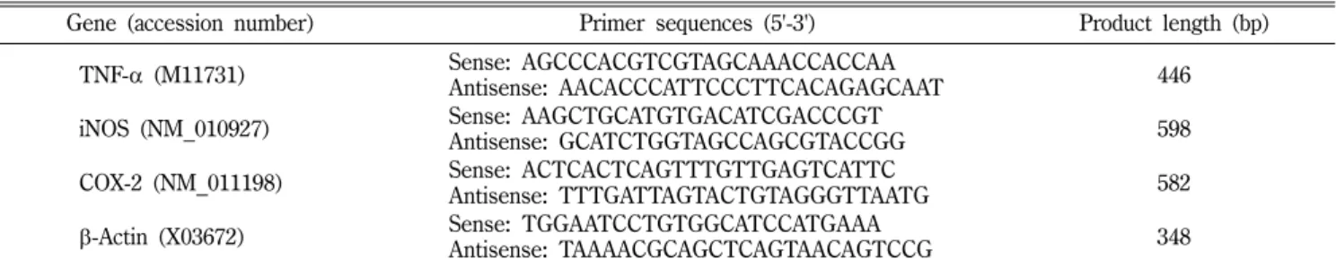
Fig. 1
는사염화탄소유도급성간독성모델에서치자가간의조직학적변화에미치는영향을살펴본것이다
.
그림에서보는 바와같이간조직의염색결과,
사염화탄소단독투여군(Fig. 1B)
에서는문맥주위염증
,
간세포괴사및Kupffer cell
비대가관찰되었으나치자
30 mg/kg
투여군(Fig. 1C)
에서이러한변화가 현저히완화되었다.
이는치자의혈중ALT
및AST
결과를일관성있게뒷받침하였으며
,
이로서치자가사염화탄소로유도된급성간독성에있어보호효과가있음을확인할수있었다
.
지질과산화물및glutathione정량
지질과산화는활성산소가세포막의불포화지방산을공격함으 로써일어나며
,
이로인하여세포막뿐아니라세포막으로둘러싸인소기관
,
즉미토콘드리아및소포체막등에도기능손상을 유발한다.
최근연구에따르면사염화탄소그자체는독성이강 하지않으나간장내일산소첨가효소계에의해활성화되어산 화적손상과세포막인지질내불포화지방산을공격하여간세 포의기능과구조를파괴시킨다고한다.
16)Table III
에서와같이 Table I −Characteristics of specific primers used for RT-PCR analysis
Gene (accession number) Primer sequences (5'-3') Product length (bp)
TNF-
α(M11731) Sense: AGCCCACGTCGTAGCAAACCACCAA
Antisense: AACACCCATTCCCTTCACAGAGCAAT 446
iNOS (NM_010927) Sense: AAGCTGCATGTGACATCGACCCGT
Antisense: GCATCTGGTAGCCAGCGTACCGG 598
COX-2 (NM_011198) Sense: ACTCACTCAGTTTGTTGAGTCATTC
Antisense: TTTGATTAGTACTGTAGGGTTAATG 582
β
-Actin (X03672) Sense: TGGAATCCTGTGGCATCCATGAAA
Antisense: TAAAACGCAGCTCAGTAACAGTCCG 348
Table II −
Effects of G. jasminoides on the serum aminotransferase activities in the CCl
4-induced mice
Group Dose (mg/kg) ALT (IU/ l ) AST (IU/
ℓ)
Control 0041.6±3.4 0030.2±5.5
CCl
4Vehicle 2115.6±235.0** 1629.6±127.4**
G. jasminoides 030 0903.0±80.0** 0662.6±63.1**
,++100 1012.7±76.5**
,++0861.1±85.4**
,++300 1112.5±53.9**
,+0979.5±115.0**
,++Silymarin 200 1365.0±176.5** 0832.2±94.7**
,++The results are presented as the mean±S.E.M. of 8 mice per group. ** Denotes significant differences ( P <0.01) versus control group;
+,++
denote significant differences ( P <0.05, P <0.01) versus CCl
4group.
사염화탄소단독투여군의
MDA
함량은대조군의약5.1
배로현저히증가되었으나
,
치자30 mg/kg
투여군에서사염화탄소단독투여군의
MDA
함량의약65.0%
정도로현저히억제되었다.
Glutathione
은세포의산화적손상에대해1
차적방어역할을하는중요한비단백성물질로서
,
세포가산화적손상을받으면GSH
가
GSSG
로산화됨으로써활성산소를무독화시킨다.
17)본실험에서사염화탄소단독투여후간조직에남아있는
GSH
의양은대조군의
82.9%
로감소하였으나,
치자30 mg/kg
투여는이러한 감소를억제하였다.
이는치자의간보호작용의일부가사염화 탄소로인한세포막의파괴를유발하는지질과산화물의생성억 제와간장내glutathione pool
을증가시킴으로써세포의산화적 손상에대한항산화방어기작을통해나타냄을알수있었다.
혈중TNF-α농도및유전자발현
간은인체주요염증기관
(inflammatory organ)
으로다양한간 독소노출후염증반응은일련의병리학적변화에관여한다.
간Fig. 1 −
Liver sections stained with hematoxylin and eosin. Mice were treated orally with vehicle or G. jasminoides (30 mg/
kg) 48 h, 24 h and 2 h before 6 h after CCl
4injection. (A):
Control group, showing normal hepatic architecture; (B):
CCl
4group, showing hepatocellular degeneration and necrosis with inflammatory infiltration; (C): G. jasminoides (30 mg/kg) + CCl
4group, showing mild hepatocellular necrosis and inflammatory infiltration. Original magnification
×400.
Table III −
Effects of G. jasminoides on the lipid peroxidation and hepatic glutathione content in the CCl
4-induced mice
Group Dose (mg/kg) MDA (nmol/mg protein) GSH (
µmol/g liver)
Control 0.34±0.01**
,++7.76±0.31
CCl
4Vehicle 1.76±0.14**
,++6.43±0.03**
G. jasminoides 30 1.16±0.06**
,++7.17±0.16*
,++The results are presented as the mean±S.E.M. of 8 mice per group. *
,** Denote significant differences ( P <0.05, P <0.01) versus control group;
++denotes significant differences ( P <0.01) versus CCl
4group.
Fig. 2 −
Effect of G. jasminoides (30 mg/kg) on the serum TNF-
αlevel of the CCl
4-treated mice. The values are reported as the means±S.E.M. of 8 mice per group. ** Denotes significant differences ( P <0.01) versus control group;
++denotes significant differences ( P <0.01) versus CCl
4group.
장내대식세포인
Kupffer cell
은간세포괴사혹은다양한간독 소에반응하여염증매개인자를유리하며이는사염화탄소로유 도된간손상의악화에도관여된다.
18)TNF-
α는조직손상시대식세포에서빠르게생성되는대표적인염증성사이토카인이다
.
사염화탄소노출후활성화된
Kupffer cell
에서TNF-
α는TNF-
α수용체와함께작용하여간조직괴사를일으킨다
.
19)본연구에서사염화탄소투여후혈중
TNF-
α농도는149.3±0.5 pg/ml
로대조군에비해현저히증가하였으나치자투여군에서
127.1±
1.3 pg/ml
로감소하였고,
이와유사하게유전자발현량도사염화탄소단독투여군에서대조군에비해약
3.8
배로증가하였으나 치자투여는이러한증가를현저히억제하였다(Figs. 2
및3).
iNOS및COX-2단백질과유전자발현
NO
는반응성이매우높은산화제로iNOS
의작용에의해L-
arginine
으로부터간장의실질세포와비실질세포에서생성된다.
정상시
NO
는세포내에낮은농도로존재하나염증반응시TNF-
α에의해서
iNOS
가유도되면장시간동안다량의NO
를생성하며
,
생성된NO
는혈관확장,
세포독성및조직손상등과같은생체유해작용을 나타낸다
.
20)또한iNOS
에의해 생성된NO
는superoxide anion
과반응하여좀더강력한산화물질인peroxy- nitrite
를생성하며,
세포내oxidant-sensitive
전사인자인NF-
κB
을활성화시켜다른염증매개체의생성을촉진하여염증반응 을 심화시킨다
.
21) 다수의 염증 억제 약물들의 작용기전은prostaglandin
합성억제이며,
이는COX-2
의생성및활성저해에의한것이다
. COX-2
는동물이나인간의염증반응부위에서발현되는효소로
, COX-2
에의한prostaglandin
의합성은염증반 응을매개한다.
연구보고에따르면염증반응에있어COX-2
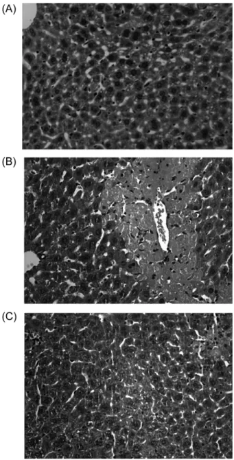
외Fig. 3 −
Effect of G. jasminoids (30 mg/kg) on the protein expression of iNOS and COX-2. Whole Hepatic proteins were extracted from mice 24 h after CCl
4injection. iNOS and COX-2 were detected by western blotting as described in Materials and Methods and
β-actin was used as a loading control. The results are presented as the mean±S.E.M. of 8 mice per group. *
,** Denote significant differences ( P <
0.05, P <0.01) versus control group;
+,++denote significant differences ( P <0.05, P <0.01) versus CCl
4group.
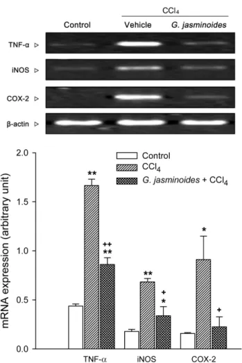
Fig. 4 −
Effect of G. jasminoides (30 mg/kg) on the mRNA expression of TNF-
α, iNOS and COX-2. The levels of TNF-
α, iNOS and COX-2 mRNA expression were measured by using RT- PCR as described in Materials and Methods. Also,
β-actin was used as a loading control. The result is presented as the mean±S.E.M. of 8 mice per group. *
,** Denote sig- nificant differences ( P <0.05, P <0.01) versus control group;
+,++
denote significant differences ( P <0.05, P <0.01) versus
CCl
4group.
생성은사염화탄소로유도된간독성의이차적효과에도관여한 다고한다
.
22)Figs. 3
및4
에서보는바와같이iNOS
및COX- 2
의단백질발현량은대조군에비해사염화탄소단독투여군에 서각각약4.4
배,
및3.3
배로현저히증가하였으나치자투여군에서이러한증가가현저히감소되었다
.
이와유사하게유전자발현량에있어서도
iNOS
및COX-2
의유전자발현량이사염화탄소단독투여군에서각각약
3.8
배및5.8
배로현저히증가하였으나치자투여군에서이러한증가가현저히감소되었다
.
이 는아마도치자가전사단계(transcriptional level)
에서사염화탄 소로유도된염증매개인자인iNOS
및COX-2
의생성을조절하여간보호작용을나타내는것으로여겨진다
.
결 론
치자의간보호작용과산화성스트레스및염증매개인자발현 억제에대한치자의역할을규명하기위하여사염화탄소를처치 한생쥐의간조직에서
ALT, AST,
지질과산화생성과염증매개 인자TNF-
α, iNOS
및COX-2
의혈중농도,
유전자및단백질발현량을관찰하였다
.
혈중
ALT
및AST
의수치는사염화탄소단독투여군에비해치자투여군에서더낮은활성도를나타내었고
,
지질과산화량은치자투여군에서감소하였고이와는반대로
GSH
는치자투여군에서증가하였다
.
치자투여군에서염증매개인자인TNF-
α 혈중농도및유전자발현량이사염화탄소단독투여군에비해 낮게나타났으며, iNOS
및COX-2
의단백질및유전자발현량역시사염화탄소단독투여군에비해현저히감소하였다
.
이상의결과들을종합하여볼때치자는사염화탄소로유도된 간독성모델에서산화성스트레스와염증매개인자발현억제를 통하여간보호작용을나타내는것으로여겨진다
.
감사의 말씀
본과제
(
결과물)
는교육과학기술부·지식경제부의출연금으로 수행한산학협력중심대학육성사업의연구결과입니다.
참고문헌
1) Di Pascoli, L., Lion, A., Milazzo, D. and Caregaro, L. : Acute liver damage in anorexia nervosa. Int. J. Eat. Disord.
36, 144 (2004).
2) Wang, G. X., Ben, C. E. and Ye, B. K. : Reparative effects of biphenyl dimethyl dicarboxylate on experimental liver injury in rats with histochemical and electronmicroscopy study. Zhong Xi Yi Jie He Za Zhi.
8, 158 (1988).
3) Bergamini, A., Bendandi, A., Maggi, G. and Chierego, G. :
Effect of lipotropic substances on the enzyme picture of liver tissue in fatty degeneration induced by carbon tetrachloride.
Boll. Soc. Ital. Biol. Sper.
31, 800 (1955).
4) Aterman, K. : Studies in fibrosis of the liver induced by carbon tetrachloride. III. Pantothenic acid and liver fibrosis. AMA Arch. Pathol.
57, 26 (1954).
5) Simler, M., Maurer, M. and Mandard, J. C. : Cancer of Liver on Cirrhosis Due to Carbon Tetrachloride. Strasb. Med.
15, 910 (1964).
6) Clawson, G. A. : Mechanisms of carbon tetrachloride hepatotoxicity. Pathol. Immunopathol. Res.
8, 104 (1989).
7) Ding, H., Huang, J. A., Tong, J., Yu, X. and Yu, J. P. : Influence of Kupffer cells on hepatic signal transduction as demonstrated by second messengers and nuclear transcription factors. World J. Gastroenterol.
9, 2519 (2003).
8) Paik, Y., Lee, C., Cho, M. and Hahn, T. : Physical stability of the blue pigments formed from geniposide of gardenia fruits:
effects of pH, temperature, and light. J. Agric. Food Chem.
49, 430 (2001).
9) Cheng, Y. Y., Chan, Y. S., Choang Tai, K. F. and Chang, H. M. : Effect of geniposide on acute jaundice in rats caused by ANIT poisoning. Zhongguo Yao Li Xue Bao.
7, 69 (1986).
10) Li, B. L., Chen, Y. H., Hu, R., Tang, J. J., Zhao, L. M. and Yuan, B. X. : Sedative, hypnotic and antiseizure effects of compound gardenia oil and jujube seed oil in mice. Nan Fang Yi Ke Da Xue Xue Bao.
28, 1636 (2008).
11) Yamazaki, M. and Chiba, K. : Genipin exhibits neurotrophic effects through a common signaling pathway in nitric oxide synthase-expressing cells. Eur. J. Pharmacol.
581, 255 (2008).
12) Auer, T., Khoschsorur, G. A., Rabl, H., Iberer, F., Petutschnigg, B., Wasler, A. and Tscheliessnigg, K. H. : Detection of lipid peroxidation products by malondialdehyde (MDA-TBA reaction) in organ transplantation. Transplant. Proc.
27, 2749 (1995).
13) Tietze, F. : Enzymic method for quantitative determination of nanogram amounts of total and oxidized glutathione:
applications to mammalian blood and other tissues. Anal.
Biochem.
27, 502 (1969).
14) Compton, S. J. and Jones, C. G. : Mechanism of dye response and interference in the Bradford protein assay. Anal. Biochem.
151
, 369 (1985).
15) Cherkasov, V. S. and Nemchenko, N. S. : Determination of the activity of blood serum alanine- and aspartate aminotransferases (ALT, AST) in various forms of hearing disorders. Zh Ushn Nos Gorl Bolezn. 23 (1978).
16) Weddle, C. C., Hornbrook, K. R. and McCay, P. B. : Lipid peroxidation and alteration of membrane lipids in isolated hepatocytes exposed to carbon tetrachloride. J. Biol. Chem.
251
, 4973 (1976).
17) Prokopenko, V. M., Partsalis, G. K., Pavlova, N. G., Burmistrov,
S. O. and Arutyunyan, A. V. : Glutathione-dependent system of antioxidant defense in the placenta in preterm delivery. Bull.
Exp. Biol. Med.
133, 442 (2002).
18) Curran, R. D., Billiar, T. R., Stuehr, D. J., Ochoa, J. B., Harbrecht, B. G., Flint, S. G. and Simmons, R. L. : Multiple cytokines are required to induce hepatocyte nitric oxide production and inhibit total protein synthesis. Ann. Surg.
212, 462 (1990).
19) Cubero, F. J. and Nieto, N. : Ethanol and arachidonic acid synergize to activate Kupffer cells and modulate the fibrogenic response via tumor necrosis factor alpha, reduced glutathione, and transforming growth factor beta-dependent mechanisms.
Hepatology
48, 2027 (2008).
20) Santos-Gomes, P. C., Seabra, R. M., Andrade, P. B. and Fernandes-Ferreira, M. : Determination of phenolic antioxidant compounds produced by calli and cell suspensions of sage (Salvia officinalis L.). J. Plant. Physiol.
160, 1025 (2003).
21) Yoshida, M., Korfhagen, T. R. and Whitsett, J. A. : Surfactant protein D regulates NF-kappa B and matrix metalloproteinase production in alveolar macrophages via oxidant-sensitive pathways. J. Immunol.
166, 7514 (2001).
22) Basu, S. : Oxidative injury induced cyclooxygenase activation in experimental hepatotoxicity. Biochem. Biophys. Res. Commun.
254