Korean J Vet Res(2018) 58(4) : 211~217 https://doi.org/10.14405/kjvr.2018.58.4.211
211
<원례보저>
하이드로젤 지지체 기반 3차원 환경에서 개 간엽줄기세포의 분화능 분석
구나연
†
·박미정†
·이지현*·변정수·정다운·조인수·차상호 농림축산검역본부 바이러스질병과(접수: 2018년 8월 31일, 수정: 2018년 12월 5일, 게재승인: 2018년 12월 10일)
Differentiation potential of canine mesenchymal stem cells on hydrogel scaffold-based three-dimensional environment
Na-Yeon Gu † , Mi Jeong Park † , Jienny Lee*, Jeong Su Byeon, Da-Un Jeong, In-Soo Cho, Sang-Ho Cha
Viral Disease Research Division, Animal and Plant Quarantine Agency, Gimcheon 39660, Korea(Received: August 31, 2018; Revised: December 5, 2018; Accepted: December 10, 2018)
Abstract: Mesenchymal stem cells (MSCs) are useful candidates for tissue engineering and cell therapy. Physiological cell environment not only connects cells to each other, but also connects cells to the extracellular matrix that provide mechanical support, thus exposing the entire cell surface and activating signaling pathways. Hydrogel is a polymeric material that swells in water and maintains a distinct 3-dimensional (3D) network structure by cross linking. In this study, we investigated the optimized cellular function for canine adipose tissue-derived MSCs (cAD-MSCs) using hydrogel. We observed that the expression levels of Ki67 and proliferating cell nuclear antigen, which are involved in cell proliferation and stemness, were increased in transwell-hydrogel (3D-TN) compared to the transwell-normal (TN). Also, transforming growth factor-β1 and SOX9, which are typical bone morphogenesis-inducing factors, were increased in 3D-TN compared to the TN. Collagen type II alpha 1, which is a chondrocyte-specific marker, was increased in 3D-TN compared to the TN. Osteocalcin, which is a osteocyte-specific marker, was increased in 3D- TN compared to the TN. Collectively, preconditioning cAD-MSCs via 3D culture systems can enhance inherent secretory properties that may improve the potency and efficacy of MSCs-based therapies for bone regeneration process.
Keywords: chondrogenesis, extracellular matrix, hydrogels, mesenchymal stem cells, osteogenesis
서 론
생체 내부는 3차원(3-dimensional)으로 구성되어 있고, 이 를 구성하는 세포는 또 다른 세포 및 주변 환경과 서로 교 류한다. In vitro에서 세포는 통상 평면(2차원; 2-dimensional) 으로 배양하게 되는데, 이는 체내 환경을 완벽하게 재현하지 못하는 단점을 가지지만 3차원 환경에서는 세포의 증식, 이 동, 분화 등에 중요한 역할을 하는 신호 전달이 용이하므로 체내와 유사한 환경의 재구성을 위해서는 3차원으로 세포를 배양하는 것이 더 이롭다 [2, 28]. 3차원 환경 연구는 성체 줄기세포를 포함한 다양한 세포에서 연구되고 있으며, 체내 와 유사한 환경을 위해 교반용 배양 용기(spinner culture flask), 친수성 고분자 구조물인 하이드로젤 (hydrogel), 다공
성 지지체 등 다양한 방법이 이용되고 있다 [30]. 3차원 배 양 용기와 코팅된 배양 용기는 세포의 응집을 유도하여 3차 원 구조 형성에 이용되고 하이드로젤과 다공성 지지체는 세 포가 하이드로젤과 다공성 지지체를 각각 세포외기질로 이 용하여 3차원 구조를 형성하는 데에 이용된다. 수의 분야에 서는 건염 [1]과 골 질환 [21] 등에 3차원 배양 연구가 진 행되고 있다.
3차원 환경 연구를 위해 흔히 사용되는 하이드로젤은 alginate, collagen, agarose 등의 천연 하이드로젤과 polye- thylene glycol 기반, poly(lactic-co-glycolic acid) 기반으로 하는 합성 하이드로젤로 나뉜다. 1954년 Wichterle와 Lím [32]이 최초로 하이드로젤을 합성하였고, 약물 전달 시스템 [14], 식품 첨가제 [6], 조직공학 및 재생의학 [10], 상처 처
*Corresponding author
Tel: +82-54-912-0809, Fax: +82-54-912-0812 E-mail: [email protected]
†
The first two authors contributed equally to this work.
치 [37] 등 다양한 분야에 활용되고 있고, 상처 처치 분야에 서는 제품으로도 판매되고 있다. 조직공학 및 재생의학 분야 에도 하이드로젤을 이용한 활발한 연구가 수행되고 있는데 [37], 조직공학 분야에서는 환자 자신의 조직이나 세포 일부 를 채취하여 생체 적합성 및 생체 기능성을 갖는 지지체 (scaffold)에 주입한 다음 인공적으로 체외에서 배양하고 체 내에 이식함으로써 실제 조직으로 또는 그와 유사한 조직으 로 복원하거나 대체하는 기술에 지지체로서 하이드로젤의 활 용이 주목받고 있다 [10].
성체줄기세포는 자가재생 및 면역관용 등 다양한 능력을 보유하므로, 골절 [9], 당뇨 [33], 심혈관 질환 [22], 알츠하 이머 [16] 등 많은 연구가 진행되고 있다. 반려동물에서도 다양한 분야에 활용되고 있는데, 대표적인 반려동물인 개의 골 관절염 [25], 골육종 [12] 간경변 [20], 신장 질환 [18], 아토피 피부염 [3] 등 다양한 질환의 치료를 위해 성체줄기 세포가 이용되고 있다.
이러한 성체줄기세포는 세포의 수명이 제한적이므로 생체 외에서 계대 배양 시 줄기세포로서의 능력을 점차 잃는다.
또한, 소규모 임상시험에서 증명되었던 성체줄기세포의 효과 가 대규모 임상시험에서는 효과가 동일하지 않았다고 보고 된 바 있고 [11, 27], 3차원 환경에서 배양한 세포와 2차원 환경에서 배양한 세포의 능력에 차이가 있다고 보고된 바 있 으므로 [23], 본 연구에서는 하이드로젤을 이용한 3차원 환
경에서의 개 유래 성체줄기세포의 특성 변화에 대해 연구하 고자 하였다.
재료 및 방법
지방 조직 유래 성체줄기세포 분리 및 배양
개의 복부 피하지방 조직으로부터 지방 조직을 일부 채취 하여 penicillin (300 U/mL; Gibco, USA)과 streptomycin (300 µg/mL; Gibco)이 포함된 Dulbecco’s phosphate-buffered saline (DPBS; Gibco)으로 2회 세척한 다음 가위를 이용하 여 조직을 잘게 자른 후, 0.1% collagenase type I (Gibco) 과 1% bovine serum albumin (BSA; bioWORLD, USA) 이 포함된 DPBS를 넣고 37oC에서 40분간 반응시켰다. Cell strainer (70–100 µm; BD, USA)를 사용하여 조직 부유물을 거르고 400 × g에서 5분간 원심분리하여 상층액을 제거하였 다. 원심분리된 pellet에 10% fetal bovine serum (Gibco), penicillin (100 U/mL), streptomycin (100 µg/mL)이 포함된 low glucose DMEM (Gibco) 배지를 넣고 잘 섞은 다음 세포 배양용 175 T-flask에 세포를 분주하여 5% CO2, 37oC 조건의 세포배양기에서 배양하였다. 다음 날 배지를 교 체하고 2일 간격으로 배지를 교체하였으며, 세포가 80% 밀 집하면 0.25% Trypsin-EDTA (Gibco)를 처리하여 다음 계 대로 세포를 배양하였다.
Table 1. List of primers used for quantitative real-time reverse transcriptase polymerase chain reaction (qRT-PCR)
Gene Primer sequence (5’-3’) PCR product size (bp) Annealing temperature (
oC)
CD29 F: CCTTGCACCAGTGAACAGAA
R: GATTGCATCAAAGCCACCTT 150 60
CD34 F: CACCCCAAGTACCATCAAGG
R: TCACACAGGACTTGGGTCAG 150 60
CD44 F: ACACAACCTCGGGTCCTATG
R: TCGACTATTGACAGCGATGC 113 60
CD45 F: CTCACGCACACAGGCTCGCA
R: CCCACCCACTGGCACTGCTG 159 60
CD90 F: CCACGAGAATGCTACCACCT
R: TGTGTATGTCCCCTCGTCCT 193 60
Ki-67 F: CCCACCTGTCCTGAAGAAAA
R: TGTGGTCACTTCCAGTTGGTT 88 60
PCNA F: AGTCACATCGGAGATGCTGTTGTA
R: AGCTGAACTGGCTCATTCATCTCTA 161 60
TGF-β1 F: CTGGAGTCGTGAGGCAGTG
R: GCAGTGTGTTATCTTTGCTGTCA 96 60
SOX9 F: GCTCGCAGTACGACTACACTGAC
R: GTTCATGTAGGTGAAGGTGGAG 101 60
COL2A1 F: GAAACTCTGCCACCCTGAATG
R: GCTCCACCAGTTCTTCTTGG 157 60
Osteocalcin F: CTGGTCCAGCAGATGCAAAG
R: GCTCGCATACTTCCCTCTTG 177 60
GAPDH F: CCATGTTCCAGGAGCGAGAT
R: TTCTCCATGGTGGTGAAGAC 97 60
F, forward; R, reverse; PCNA, proliferating cell nuclear antigen; TGF- β1, transforming growth factor-beta1; COL2A1, collagen type II alpha
1; GAPDH, glyceraldehyde 3-phosphate dehydrogenase.
정량적 중합효소연쇄반응(quantitative real-time reverse transcriptase polymerase chain reaction, qRT-PCR)을 이용한 유전자 분석
RNeasy Mini Kit (Qiagen, USA)를 이용하여 RNA를 추출 하고 NanoDrop 1000 Spectrophotometer (Thermo Scientific, USA)를 사용하여 RNA 상태와 농도를 측정한 다음, GoScript Reverse Transcriptase (Promega, USA)를 사용하여 cDNA를 합성하였다. qRT-PCR은 96-well plate에 LightCycler 480 SYBR Green I Master Mix (Roche Diagnostics, USA)를 사 용하여 pre-denaturation (95oC, 10분), annealing (60oC, 10초), elongation (72oC, 10초)을 45 cycle 반복하였다. 그리고, 2−ΔΔCT 계산법을 이용하여 melting curve를 분석하고 glyceraldehyde 3-phosphate dehydrogenase (GAPDH)를 reference gene으로 이용하여 qRT-PCR 결과를 분석하였다. qRT-PCR에 사용한 primer의 정보와 조건은 Table 1과 같다.
유세포 분석을 통한 세포 표면 특이 단백질 발현 검사 FACSCalibur flow cytometer (BD)를 이용하여 개 지방
조직 유래 성체줄기세포의 세포 표면 특이 단백질의 발현을 확인하였다. 0.25% Trypsin-EDTA를 처리하여 세포를 탈착 한 다음 DPBS로 2회 세척하여 5 × 10⁵개의 세포에 형광물 질이 결합된 mouse anti-dog CD34 (phycoerythrin [PE]
559369; BD Pharmingen, USA), mouse anti-dog CD44 (PE MAB5549; R&D System, USA), rat anti-dog CD90 (PE 12-5900; eBioscience, USA) 항체를 넣고 1시간 동안 반응시켰다. 반응 후 DPBS로 2회 세척하여 Cell Quest Pro (BD) 프로그램을 사용하여 결과를 분석하였다.
중배엽 분화 유도 및 염색
개 지방 조직 유래 성체줄기세포의 중배엽 분화능을 확인 하고자 adipogenesis, chondrogenesis, osteogenesis 유도용 배지를 사용하여 분화를 유도하였다(Table 2). 3~4일 주기로 분화용 맞춤 배지를 교체하고 분화능 확인을 위한 키트를 이 용하여 21일째에 분화 여부를 관찰하였다. Chondrogenesis 분화는 Alcian Blue stain kit (IHC World, USA), osteo- genesis 분화는 Alzarin Red stain kit (IHC World)를 이용
Table 2. Composition of differentiation media
Chondrogenesis Osteogenesis
Dexamethasone
Ascorbic acid 2-phosphate TGF- β1
100 nM 100 µM
510 ng/mLDexamethasone
Ascorbic acid 2-phosphate β-Glycerophosphate
100 nM
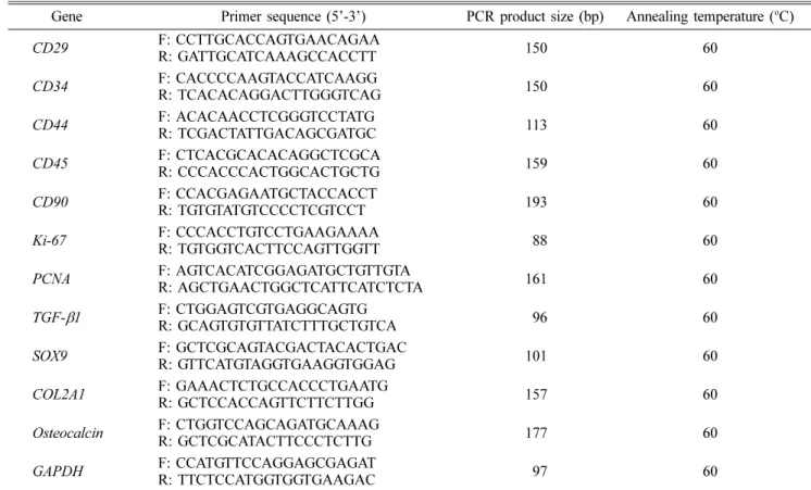
550 µM 510 mMFig. 1. Characterization of canine adipose tissue-derived mesenchymal stem cells (cAD-MSCs). (A) The cAD-MSCs obtained from canine adipose tissue were able to attach to the culture plates and expand in vitro. 40× (left), 100× (right). (B) Flow cytometry histograms show the expression of surface markers (CD34, CD44 and CD90) by cAD-MSCs populations compared with controls.
한 특이 세포 염색을 통해 성체줄기세포의 중배엽 분화능을 확인하였다.
하이드로젤을 이용한 3차원 환경 구성 및 세포 배양 하이드로젤(PuraMatrix; Corning, USA)을 이용하여 성체 줄기세포의 배양을 위한 3차원 환경을 구성하였다. 하이드로 젤의 준비는 제조사의 방법에 따랐으며, 6-well plate와 transwell (TN)에 0.25%의 하이드로젤을 각각 1.2 mL와 700 µL 넣은 뒤, 각 well에 배지를 넣어 액상의 하이드로젤 을 세포가 자랄 수 있도록 안정시켰다. 이후 TN에 5 × 10⁴ 개의 세포를 분주하고 5일간 배양하였으며, 대조군인 평면 배양에는 5일차에 세포가 90% 이상 밀집하는 1 × 10⁵개의 세포를 분주하여 5일간 배양하였다.
통계학적 분석
독립적인 3회의 실험을 수행하여 그 결과를 평균과 표준 편차로 표시하였으며, 통계는 SigmaPlot 12.0 (Systat Software, Germany)를 이용하여 분석하였다. 두 실험군 사이 의 유의성은 Student’s t-test 및 Mann-Whitney U test를 사용하여 검정하였고, 유의성 있는 결과(p < 0.05)를 판정하 였다.
결 과
개 지방 조직 유래 성체줄기세포 분리 및 특성 분석 개 지방 조직 유래 세포는 최초 분리 후 10일 이내에 colony를 형성하고 섬유아세포 모양으로 부착하여 안정적으
로 잘 자라는 것을 확인하였다(Fig. 1A). 성체줄기세포가 나 타내는 세포 표면 특이 단백질의 발현을 확인하고자 유세포 분석방법을 통해 해당 세포에서 CD34, CD44 및 CD90의 발현을 확인하였다. 그 결과, 성체줄기세포의 positive marker 인 CD44와 CD90의 발현을 확인하였으며, negative marker 인 CD34는 발현되지 않음을 확인하였다(Fig. 1B).
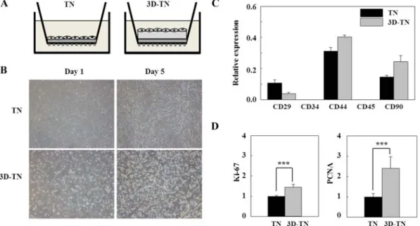
Fig. 3. Effects of TN and 3D-TN culture systems on cell phenotype in cAD-MSCs. (A) Schematic diagram of TN and 3D-TN culture systems. (B) Cellular morphology of TN and 3D-TN-cultured cells. 40×. mRNA expressions of (C) cell surface markers (CD29, CD34, CD44, CD45 and CD90), (D) Ki-67 and PCNA were confirmed by qRT-PCR and normalized to the GAPDH level.
GAPDH was used as a housekeeping control gene. Each data from three independent experiments was evaluated and expressed as the mean± SD (***p < 0.0001).
Fig. 2. Effects of 2-dimentional (2D) and transwell (TN) culture systems on cell phenotype in cAD-MSCs. (A) Schematic diagram of 2D culture and TN culture system. (B) mRNA expression of proliferation genes (Ki-67 and PCNA) were confirmed by qRT-PCR and normalized to the GAPDH level. GAPDH was used as a housekeeping control gene. Each data from three independent experiments was evaluated and expressed as the mean± SD (*p < 0.05, ***p < 0.0001).
개 지방 조직 유래 성체줄기세포의 평면 배양과 TN 배 양 비교
배양 환경에 따른 성체줄기세포의 성능 변화를 관찰하고 자 평면 배양과 TN 배양을 비교하였다. 세포 배양 5일 후 증식인자를 qRT-PCR로 확인한 결과, 증식하는 세포핵의 항 원인 proliferating cell nuclear antigen (PCNA)과 세포 증 식을 위한 핵단백질 Ki-67의 발현이 평면 배양보다 TN 배 양에서 유의적으로 높게 발현함을 확인하였다(Fig. 2B).
TN 배양과 하이드로젤을 이용한 3차원-TN(3D-TN) 배 양 비교
3차원 유사 환경에서의 성체줄기세포의 성능 변화를 관찰 하고자 TN 배양과 3D-TN 배양을 비교하였다(Fig. 3A).
TN 배양과 3D-TN 배양에서 성체줄기세포를 5일간 배양하 고 비교한 결과, TN 배양에서는 평면으로 세포가 자라지만 3D-TN 배양에서는 3차원 유사 형태로 서로 뭉치는 것이 관 찰되었다(Fig. 3B). 다음으로 TN과 3D-TN 배양 시 줄기세 포 표면 특이 단백질의 발현 변화를 비교한 결과, TN과 3D-TN 배양법 모두에서 성체줄기세포의 positive marker인 CD44와 CD90이 발현하였으며 negative marker인 CD34는 발현되지 않았다(Fig. 3C). 또한, PCNA와 Ki-67의 발현을 비교한 결과, PCNA와 Ki-67이 TN 배양보다 3D-TN 배양 에서 높게 발현함을 확인하였다(Fig. 3D).
연골 및 골분화능에 있어서 TN 배양과 3D-TN 배양 비교 위의 결과를 바탕으로 연골 및 골분화능에 있어서 TN과 3D-TN 배양을 비교하고자 연골세포와 골세포로의 분화를 확
인하는 특이적 세포 염색 및 분화 특이 단백질의 발현을 확 인하였다. 그 결과, TN 배양보다 3D-TN 배양에서 연골세포 특이 단백질인 collagen type II alpha 1 (COL2A1)과 연골 분화 유도인자인 SOX9의 발현이 현저히 높게 나타났으며, 골세포 특이 단백질인 osteocalcin (OC)과 골분화 유도인자 인 transforming growth factor-beta1 (TGF-β1)의 발현 또 한 TN 배양보다 3D-TN 배양에서 높게 발현함을 확인하였 다(Fig. 4).
고 찰
생체 내부는 세포와 세포외기질(extracellular matrix)로 구 성되는데, 이 세포외기질은 주로 동물의 구조적 지지 등을 담당하는 조직이며, 세포에 물리적 구조와 항상성 유지, 세 포 분화, 조직 형성 등에 필요한 다양한 생화학적 신호를 제 공한다. 이러한 세포외기질은 기본적으로 물, 단백질, 다당류 로 구성되고 각 조직은 조직 발생단계에서 다양한 세포(상피 세포, 섬유아세포, 지방세포, 내피세포 등)의 상호작용에 의 한 독특한 세포외기질 구성 비율을 가지게 되며 [13], 축종 마다 세포외기질의 조직 특이성은 다르다. 세포는 수용체를 이용하여 세포외기질에 부착하며, 이러한 수용체에는 integrin, discoidin domain 등이 있다 [19].
이러한 3차원 환경의 조직에서 세포를 분리하여 통상적으 로 2차원(평면) 환경에서 해당 세포를 배양하게 되는데, 이 과정에서 세포는 세포의 증식능, 분화능과 같은 정상적인 생 리학적 특성이 바뀌게 된다. 또한, 이러한 환경에서의 연속 적인 배양은 줄기세포를 더욱 빨리 노화시키고, 분화시키며, Fig. 4. Differentiation potentials of TN and 3D-TN culture systems in cAD-MSCs. (A) Using the specific staining kit, chondrocytes (Alcian Blue) and osteocytes (Alizarin Red) were positively stained in two groups. 100×. (B) The mRNA expressions of chondrocyte (COL2A1 and SOX9) and osteocyte (OC and TGF-β1) related genes were detected by qRT-PCR and normalized to the GAPDH level. GAPDH was used as a housekeeping control gene. Each data from three independent experiments was evaluated and expressed as the mean± SD (**p < 0.001, ***p < 0.0001).
다양한 능력을 잃어버리게 하는데 [29], 이러한 생리학적 특 성 변화는 줄기세포의 임상 적용에 있어서 큰 걸림돌이 될 수 있다. 생체 내부와 유사한 3차원 환경의 재구성을 위하 여 in vitro에서 다양한 방법이 응용되고 있으며, 하이드로젤 은 세포의 3차원 배양을 위한 지지체로 많은 연구에 활용되 고 있다 [30]. 하이드로젤은 펩타이드 기반으로 수분을 99%
함유하고 있다. 염분 첨가에 의해 나노 섬유를 만들어 3차 원 구조를 형성하는데 [35], 하이드로젤은 10 nm 크기의 나 노 섬유를 바탕으로 50~200 nm (pore size)의 구멍을 형성 하며, 하이드로젤 펩타이드의 서열은 RAD16-I, AcN–
RADARADARADARADA–CONH2로 세포의 부착 [34]과 이동 [24]을 향상한다고 알려져 있다.
세포의 증식과 관련하여 다양한 인자들이 관여하는데, Ki- 67은 휴지기의 세포에서는 발현되지 않으며 증식하는 모든 세포에서는 보편적으로 발현한다 [15]. PCNA 또한, 휴지기 세포에서는 발현되지 않고 증식하는 세포에서 발현이 증가 한다 [17]. Debnath 등 [8]은 사람 줄기세포를 대상으로 하 이드로젤 배양 시 줄기세포의 증식이 증가한다고 보고한 바 있다. 본 연구에서는 3차원 유사 환경하에서 개 지방 조직 유래 성체줄기세포의 증식능과 분화능을 향상시키고자 하였 다. 그 결과, 평면 배양에 비해 3차원 유사 환경인 3D-TN 배양에서 Ki-67과 PCNA의 발현이 현저하게 증가함을 확인 할 수 있었다.
이러한 줄기세포는 조직공학 및 재생의학의 재료로 다양 하게 사용되고 있다. Westhrin 등 [31]은 사람 줄기세포를 하이드로젤에서 배양 시 줄기세포의 골 분화능이 증가한다 고 보고하였고, Toh 등 [26]은 염소 줄기세포를 하이드로젤 에서 배양 시 줄기세포의 연골 분화능이 증가한다고 보고한 바 있다. 또, Zheng 등 [36]은 토끼 줄기세포를 하이드로젤 에서 배양 시 줄기세포의 연골 분화능이 증가한다고 보고하 였다. 줄기세포가 연골 및 골로 분화되기 위해서는 SOX9과 TGF-β1 등의 전사인자가 중요한 역할을 한다. SOX9은 연 골 형성 및 발달에 핵심적인 역할을 하는 전사인자로, 연골 의 주요 성분인 COL2A1과 aggrecan의 발현을 조절한다.
Cha 등 [4]은 bone morphogenic protein (BMP)-2와 SOX9 유전자를 encoding한 bicistronic vector의 동시 전달이 탈분 화된 연골세포의 정상연골 형성 특성을 향상시키고 연골 형 성을 개선할 수 있다고 보고하기도 하였다. 또한, TGF-β의 신호전달은 선택적 mitogen-activated protein kinases 및 Smad2/3 경로, 그리고 TGF-β와 parathyroid hormone, Wnt, BMP뿐만 아니라 fibroblast growth factor 신호 사이 의 협력을 통해 골 형성 전구세포를 모으는 것은 물론 골 형성 전구세포의 증식과 골세포로의 분화를 촉진시키며 [5], OC는 부분적으로 뼈의 미네랄 축적 조절을 담당하고 그 발 현은 미네랄화된 매트릭스 형성의 지표 역할을 한다고 알려 져 있다 [7]. 본 연구에서도 3차원 유사 환경하에서 개 지방 조직 유래 성체줄기세포의 연골 및 골 분화능을 조사하였다.
그 결과, 2차원 배양에 비해 3차원 유사 환경인 3D-TN 배 양에서 연골세포 특이 단백질인 COL2A1과 연골분화 유도
인자인 SOX9의 발현 증가와 골세포 특이 단백질인 OC와 골분화 유도인자인 TGF-β1의 발현이 현저하게 향상됨을 확 인하였다.
본 연구에서는 하이드로젤을 이용한 3차원 환경에서의 개 지방 조직 유래 성체줄기세포의 특성 변화에 대해 연구하였 으며, 이를 통해 연골 및 골 질환과 관련하여 개 성체줄기세 포의 줄기세포성을 유지하거나 향상시키는 데에 3차원 유사 환경이 유의성이 있음을 제안하고자 한다.
감사의 글
본 연구는 농림축산검역본부 연구사업(과제번호: B- 1543083-2018-20-01)의 지원으로 수행되었으며, 이에 감사드 립니다.