Bilateral Discoid Medial Menisci: Two Different Types in One Patient and Bony Changes on the Medial Tibial Plateau
Tae Seok Nam, M.D.
Department of Orthopedic Surgery, Haeundae Paik Hospital, Inje University College of Medicine, Busan, Korea
A discoid medial meniscus is a relatively rare pathology of the knee joint, and bilateral cases are extremely rare. We present one case of bilateral symptomatic discoid medial menisci. Arthroscopy revealed a complete type of discoid medial meniscus in one knee and an incomplete type in the other knee. Ours is a very rare case of bilateral discoid medial menisci with associated osseous changes in the tibia, and it is perhaps the first such reported case in the world. The patient was successfully treated by partial meniscectomy using routine arthroscopic procedures.
Key words: knee, meniscus, bilateral discoid medial menisci
Since the description of discoid medial meniscus by Cave and Sta- ples1) in 1941, several authors have published about this anomaly.2-7) The reported incidence rates range from 0.06% to 0.3%8); the bilateral cases are extremely rare.2-5) Only 18 cases of bilateral discoid medial meniscus have been reported. We experienced a unique case of bi- lateral discoid medial menisci with tears and osseous change in the adjacent proximal tibia.
CASE REPORT
A 23-year-old male soldier complained of pain and swelling on both knees on initial visit. He had a 13-month history of pain that initiat- ed first in the right, then in the left knee. He had pain while climbing stairs and squatting. The symptoms became worse with long distance walking and sports activities.
Examination of the knees showed no limitation of motion except pain in full flexion. He had medial joint line tenderness bilaterally.
Received August 2, 2010 Accepted December 8, 2010 Correspondence to: Tae Seok Nam, M.D.
Department of Orthopedic Surgery, Haeundae Paik Hospital, Inje University College of Medicine,1435, Jwa-dong, Haeundae-gu, Busan 612-862, Korea
TEL: +82-51-797-0990 FAX: +82-51-797-0991 E-mail: [email protected] This work was supported by Grant from Inje University, 2010.
Copyright © 2011 by The Korean Orthopaedic Association
“This is an Open Access article distributed under the terms of the Creative Commons Attribution Non-Commercial License (http://creativecommons.org/licenses/by-nc/3.0/) which permits unrestricted non-commercial use, distribution, and reproduction in any medium, provided the original work is properly cited.”
대한정형외과학회지:제 46권 제 2호 2011
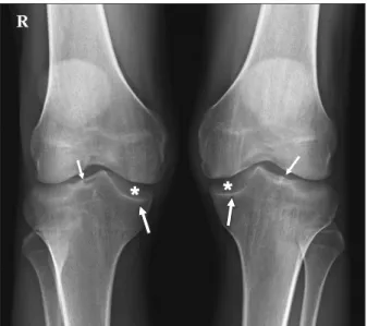
McMurray test was positive in both knees. Anteroposterior plain ra- diograph showed depression (cupping) of the both medial tibial pla- teau, widening of medial joint space, and hypoplasia of lateral tibial spine (Fig. 1).
MRI showed displaced torn discoid medial meniscus with hori- zontal tear and peripheral longitudinal tear on superior flap. Both superior flaps were displaced to the intercondylar notch. Ligamentous structures and lateral meniscus were intact (Fig. 2).
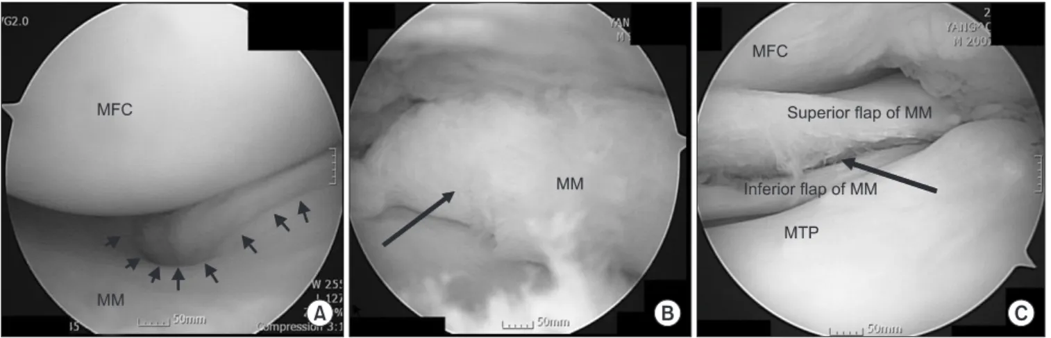
Arthroscopic examination of the right knee confirmed the pres- ence of the thickened complete type of discoid medial meniscus with horizontal tear on whole rim and fatty degeneration on posterior horn (Fig. 3A, B). On the left knee, similar tear pattern was shown except incomplete type of discoid medial meniscus (Fig. 3C). Degen- eration of tear site on meniscus was severe on whole rim, especially, posterior horn. So it seemed that healing potency on tear site was very low. Superior flap was resected and central discoid portion of the meniscus were partially meniscectomized. Anterior cruciate liga- ment was intact and had good stability (Fig. 4). Additionally, there were thick medial plica between patella and medial femoral condyle.
Medial femoral condyle in contact with medial plica showed cartilage degeneration. Excision of the pathological medial plica was per- formed.
In 3 months follow-up after the arthroscopic surgery, he had no
clinical examination.
DISCUSSION
Discoid medial menisci are rare and bilaterality of the cases was not known accurately.
Smillie9) in 1948 reported only 1 patient had bilateral involvement in 10 discoid medial menisci, in analysis of 8,040 medial meniscus.
in which 5 cases were bilateral in 10 patients with the discoid medial meniscus. Pinar et al.5) found 6 complete and 3 incomplete types of the bilateral discoid medial menisci have been reported.
In 2006, Kim and Seo3) reported a case of discoid medial meniscus with an incomplete type in one knee and a complete type in op- posite knee. These findings were similar in our case. But, in our case both knees were confirmed by arthroscopy, while only one knee was confirmed by arthroscopy in their case. In our case both knees had symptomatic meniscal tear. So, we performed arthroscopic proce- dures on both knees.
Following abnormal radiographic findings associated with dis- coid medial meniscus have been reported on anteroposterior plain radiographs; cupping of the medial tibial plateau,2,7) proximal tibial physis collapse,7) and widening of the medial joint space.2) However, only three cases had bilateral anomalies.2,5,7) Ours is the fourth case of bilateral discoid medial menisci with associated tibial osseous changes. Our case showed depression (cupping) of the both medial tibial plateau and widening of medial joint space. Additionally, our case showed hypoplasia of lateral tibial spine. In general, hypoplasia of lateral tibial spine have been associated with congenital absence of anterior cruciate ligament.10) But, our case presented good joint stability on clinical and arthroscopic examination. Whether this unique finding is limited in this case or not, more cases and study are needed.
Fatty degeneration on posterior horn of medial meniscus was shown on right knee. This finding was observed on degenerative meniscal Figure 1. Anteroposterior radiograph of both knee shows depression
(cupping) of medial tibial plateau (thick arrow), widening of medial joint space (asterisk), and hypoplasia of lateral tibial spine (narrow arrow).
Figure 2. Magnetic resonance images. (A) Right knee: coronal image shows discoid medial meniscus with horizontal tear (white arrow) and peripheral tear (asterisk) on superior flap. (B) Left knee: coronal image shows similar findings as of right knee.
tear in old age. So, we thought that fatty degeneration was caused by repeated trauma in the joint space.
In the normal knee, approximately 60% of the weight-bearing forces are transmitted through the medial compartment and approxi- mately 40% are transmitted through the lateral compartment. Even in severe valgus deformity (up to 30 degrees valgus), the medial plateau load never falls below 30%. This means importance of early inter- vention of tear of medial discoid meniscus. Torn meniscus is more harmful on articular cartilage of medial tibial plateau than lateral joint space in terms of the weight-bearing forces.
Several anomalies related to the discoid medial meniscus were reported; anomalous insertion of the anterior horn of the medial meniscus into the anterior cruciate ligament,4,10) discoid lateral menis- cus in the same knee, meniscal cyst10) and pathologic medial patella plica.5) In our case, pathologic medial patella plica was seen.
We report one case of bilateral discoid medial menisci with similar arthroscopic findings with complete type in one knee and incomplete type in opposite knee and bony changes in bilateral medial tibial pla- teau.
REFERENCES
1. Cave EF, Staples OS. Congenital discoid meniscus: a cause of internal derangement of the knee. Am J Surg. 1941;54:371-6.
2. Atay OA, Doral MN, Aydingöz U, Leblebicioglu G. Bilateral discoid medial menisci: association with bone changes in the tibia. Knee Surg Sports Traumatol Arthrosc. 2001;9:217-20.
3. Kim SJ, Seo YJ. Bilateral discoid medial menisci: Incomplete type in one knee and complete type in opposite knee. Knee.
2006;13:255-7.
4. Lee BI, Lee YS, Kwon SW, Choi SW, Cho KH, Kwon YJ. Bi- lateral symptomatic discoid medial meniscus: report of three cases. Knee Surg Sports Traumatol Arthrosc. 2007;15:739-43.
5. Pinar H, Akseki D, Karaoğlan O, Ozkan M, Uluç E. Bilateral discoid medial menisci. Arthroscopy. 2000;16:96-101.
6. Tachibana Y, Yamazaki Y, Ninomiya S. Discoid medial menis- cus. Arthroscopy. 2003;19:E12-8.
7. Weiner B, Rosenberg N. Discoid medial meniscus: associa- Figure 4. Arthroscopic finding of anterior cruciate ligament of right knee.
Figure 3. Arthroscopic findings. (A) Arthroscopic view shows complete discoid type of medial meniscus with peripheral tear (multiple arrows) of right knee. MFC; medial femoral condyle, MM; medial meniscus. (B) Arthroscopic view shows fatty degeneration on posterior horn (arrow) of medial meniscus on the right knee. MM; medial meniscus. (C) Arthroscopic view shows incomplete discoid type of medial meniscus with horizontal tear (arrow) of left knee. MFC; medial femoral condyle, MM; medial meniscus, MTP; medial tibial plateau.
Surg Am. 1974;56:171-3.
8. Kim SJ, Choi CH. Bilateral complete discoid medial menisci combined with anomalous insertion and cyst formation. Ar- throscopy. 1996;12:112-5.
Br. 1948;30B:671-82.
10. Thomas NP, Jackson AM, Aichroth PM. Congenital absence of the anterior cruciate ligament. A common component of knee dysplasia. J Bone Joint Surg Br. 1985;67:572-5.
양측성 원판형 내측 반월상 연골: 동일 환자에서 각각 다른 형태의 원판형 반월상 연골 및 내측 경골 고평부의 골성 변화를 동반한 증례
남태석
인제대학교 의과대학 해운대 백병원 정형외과학교실
원판형 내측 반월상 연골은 슬관절에 비교적 드문 경우이고, 양측성인 증례는 매우 드물다. 이에 증상있는 양측성 원판형 내측 반월 상 연골로 치료하였던 1예를 보고하고자 한다. 관절경 소견에서 한쪽 무릎은 완전형 내측 원판형 반월상 연골 소견을, 반대쪽은 불완 전형 내측 원판형 반월상 연골 소견을 보였다. 또한 본 증례의 경우 경골의 골성 변화를 동반한 양측성 원판형 내측 반월상 연골의 보 고된 세계에서도 드문 증례이다. 환자는 관절경적 술식으로 반월상 연골 부분절제술을 시행하여 성공적인 경과를 보였다.
색인단어: 슬관절, 반월상 연골, 양측성 원판형 내측 반월상 연골
접수일 2010년 8월 2일 게재확정일 2010년 12월 8일
교신저자 남태석, 부산시 해운대구 좌동 1435번지, 인제대학교 의과대학 해운대백병원 정형외과학교실 TEL 051-797-0990, FAX 051-797-0991, E-mail [email protected]
이 논문은 2010년도 인제대학교 학술연구 조성비 보조에 의한 것임.