ISSN 2234-3806 • eISSN 2234-3814
https://doi.org/10.3343/alm.2020.40.4.337 www.annlabmed.org 337
Ann Lab Med 2020;40:337-340
https://doi.org/10.3343/alm.2020.40.4.337
Letter to the Editor
Transfusion Medicine
First Korean Case of Partial D DBS-1
Sooin Choi , M.D.1,*, HongBi Yu , B.S.2,*, and Duck Cho , M.D.2,3
1Department of Laboratory Medicine, Soonchunhyang University Hospital Cheonan, Soonchunhyang University College of Medicine, Cheonan, Korea;
2Department of Health Sciences and Technology, Samsung Advanced Institute for Health Sciences and Technology, Sungkyunkwan University, Seoul, Korea;
3Department of Laboratory Medicine and Genetics, Samsung Medical Center, Sungkyunkwan University School of Medicine, Seoul, Korea
Dear Editor,
RHD and RHCE, encoding Rhesus proteins, are highly homolo- gous genes located adjacently on the same chromosome (chro- mosome 1). Therefore, hybrid RHD genes, in which some por- tions are substituted with the RHCE sequence can change the extracellular loop of the RhD antigen, leading to variable reactiv- ity to anti-D reagents [1].
Partial D phenotypes have historically been classified using epitope studies [2] and, recently, using genetic studies. DBS is a partial D phenotype characterized by c.[676G>C]+[697G>C]
(NM_016124.4) and has been named based on its positive re- activity with the D monoclonal antibodies (MoAbs) BS228 and BS233 (Biotest, Dreieich, Germany) [3]. Three DBS subtypes have been reported till date: DBS-0 [1], DBS-1 [3, 4], and DBS-2 [5]. Molecular studies have identified DBS-1 in an Arabian [3]
and a Japanese family [4]; however, it is unknown whether DBS-1 correlates with the same hybrid gene of a single evolutionary ori- gin in other, unrelated individuals from different ethnic groups.
To the best of our knowledge, this is the first report of a DBS-1 case in a Korean family. This study was approved by the Institu- tional Review Board of Samsung Medical Center, Seoul, Korea (SMC-2019-11-160), and written informed consent was obtained from the proband and all family members.
The proband was a Korean woman with fibrocystic breast chan-
ges, who was admitted to the Samsung Medical Center. D typ- ing using anti-D Bioclone (MAD2 clone; Ortho Clinical Diagnos- tics, Raritan, NJ, USA) was negative. Weak D testing using anti- D Bioclone and human IgG/IgM monoclonal anti-D (Millipore, Livingston, UK) yielded a result of grade 2+, while the result of partial D testing using D-screen (Diagast, Loos, France) was consistent with DBS-1. The phenotypes of the current case and previously reported DBS cases are summarized in Table 1.
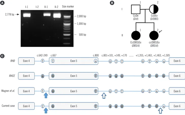
The RhCE phenotype was ccEe (anti-C, -c, -E, and -e anti- bodies were obtained from Bioclone, Ortho Clinical Diagnostics, Buckinghamshire, UK), and RHD genotyping was carried out according to a previously described method [6]. Exon 5 was not amplified by the primer sets used in this study, but the region from exon 4 to exon 6 was amplified and the PCR product was sequenced using RHCE exon 5-specific primers; the Rhesus box was also PCR-amplified (Fig. 1A). The proband harbored a hybrid RHD-cE(5)-D (DBS/d) allele involving the following amino acid changes: F223V, A226P, E233Q, V238M, V245L, G263R, and K267M. RHD genotypes of other family members are shown in Fig.1B. The breakpoints were confirmed between exon 4 and exon 6 by intron analysis [3, 4]. The 5´ breakpoint region was the same as that reported by Omi, et al. [4], whereas the 3´ break- point region was novel (Fig. 1C).
In various partial D phenotypes, such as DIIIa, DVa, DVI, DAR,
Received: August 1, 2019 Revision received: August 18, 2019 Accepted: November 12, 2019 Corresponding author: Duck Cho, M.D.
Department of Laboratory Medicine and Genetics, Samsung Medical Center, Sungkyunkwan University School of Medicine, 81 Irwon-ro, Gangnam-gu, Seoul 06351, Korea
Tel: +82-2-3410-2403, Fax: +82-2-3410-2719, E-mail: [email protected]
*These authors equally contributed to this study.
© Korean Society for Laboratory Medicine
This is an Open Access article distributed under the terms of the Creative Commons Attribution Non-Commercial License (https://creativecommons.org/licenses/by-nc/4.0) which permits unrestricted non-commercial use, distribution, and reproduction in any medium, provided the original work is properly cited.
1 / 1 CROSSMARK_logo_3_Test
2017-03-16 https://crossmark-cdn.crossref.org/widget/v2.0/logos/CROSSMARK_Color_square.svg
Choi S, et al.
First Korean Case of Partial D DBS-1
338 www.annlabmed.org https://doi.org/10.3343/alm.2020.40.4.337 DFR, DBT, and DBS, RHD exon 5 is substituted with a part of RHCE exon 5 [7]. Exon 5 is predicted to encode the fourth ex- tracellular loop of the D polypeptide. In DBS, the coexistence of two amino acid changes (A226P and E233Q) caused by c.676G>C and c.697G>C point variants is required for the characteristic phenotype [4]. A226P is observed in the fourth loop of antigen E [4] and is thought to have a considerable effect on D antigen density [1]. E233Q is also observed in the fourth loop as part of Dw (RH23) [8]. DBS-1 and -2, which share F223V, A226P, and E233Q, exhibited different reactivity to the MoAbs P3X249, P3X- 290, and P3X21211F1 (DBS-1, +/+/– and DBS-2, –/–/trace).
This difference might have been caused by the change in the extracellular amino acid, V238M, which is found in DBS-0 and DBS-1, but not in DBS-2. Other amino acid changes observed in DBS-1, including V245L, G263R, and K267M, are located in the intracellular or transmembrane regions; however, they might affect the RhD phenotype.
The genetic basis of the RhD blood group differs across races and ethnicities. For example, the RhD-negative phenotype mainly results from RHD deletion in Caucasians, whereas RHD altera- tions, such as RHD*D-CE(4-7)-D, are common in Africans [9].
Asians exhibit a high prevalence of c.1227G>A (NM_016124.4), known as “Asia type DEL.” These ethnicity-specific trends are used not only to diagnose RhD variants, but also for transfusion protocols [10]. The 3´ breakpoint observed in our case differed from that found in a Japanese family [4], suggesting that the DBS-1 cases have different genetic origins.
In conclusion, we reported the first case of DBS-1 in a Korean family. To understand the RhD characteristics specific to Korean ethnicity, further evaluation of RhD variants is required.
ACKNOWLEDGEMENTS
The authors wish to acknowledge the technical support of Ji Young Seo, M.T.
AUTHOR CONTRIBUTIONS
DC conceived the study; SC and HBY contributed to the inter- pretation of the results; all authors contributed to writing the man- uscript.
CONFLICTS OF INTEREST
The authors declare no conflicts of interest.
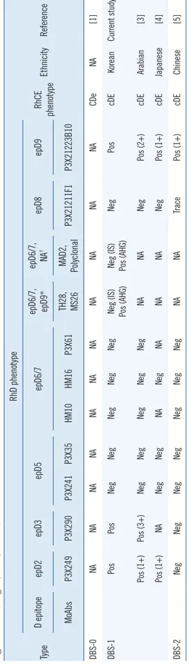
Table 1. Comparison of serologic characteristics based on analysis using MoAbs between previous DBS cases and the present DBS case. Partial D testing was performed us- ing D-screen (Diagast, Loos, France) Type
RhD phenotype RhCE phenotypeEthnicityReferenceD epitopeepD2epD3epD5epD6/7epD6/7, epD9*epD6/7, NA†epD8epD9 MoAbsP3X249P3X290P3X241P3X35HM10HM16P3X61TH28, MS26MAD2, PolyclonalP3X21211F1P3X21223B10 DBS-0NANANANANANANANANANANACDeNA [1] DBS-1PosPosNegNegNegNegNegNeg (IS) Pos (AHG)Neg (IS) Pos (AHG)NegPoscDEKoreanCurrent study Pos (1+)Pos (3+)NegNegNegNegNegNANANegPos (2+)cDEArabian [3] Pos (1+)NANegNegNANegNANANANegPos (1+)cDEJapanese [4] DBS-2NegNegNegNegNegNegNegNANATracePos (1+)cDEChinese [5] *The results using human IgG/IgM monoclonal anti-D (Millipore, Livingston, UK); †The results using anti-D Bioclone (MAD2 clone; Ortho Clinical Diagnostics, Raritan, NJ, USA). Abbreviations: ep, epitope; MoAbs, monoclonal antibodies; IS, immediate spin; AHG, antihuman globulin; Pos, positive; Neg, negative; NA, not available.
Choi S, et al.
First Korean Case of Partial D DBS-1
https://doi.org/10.3343/alm.2020.40.4.337 www.annlabmed.org 339
Fig. 1. Results of the genetic analysis of the proband and her family members. (A) Long-range PCR with primers located in non-Rhesus box sequences. A 2,778-bp fragment was amplified by PCR, indicating the presence of a hybrid RHD gene (lanes I-1, II-1, and II-2). (B) Pedigree, Rh phenotypes, and RHD genotypes of the Korean DBS-1 family. The genotypes and phenotypes of the DBS-1 family were de- termined using combined data from sequencing analysis, hybrid Rhesus box PCR, and serological analysis. Black circles indicate the DBS- 1 phenotype. The proband is indicated by a black arrow. Total RHD deletion is denoted as “d” in the genotype. (C) Part of the RHD nucleo- tide sequence in DBS reported by Wagner, et al. [3] and this case. In both cases, the 5´ breakpoint region was located between the RHD- specific c.642-249T and the first RHCE-specific nucleotide, c.667G (blue arrow). The 3´ breakpoint region, located between the last RHCE- specific nucleotide and the first RHD-specific nucleotide of intron 5, differed for each case; it was located between c.800 and c.801+101 in the case reported by Wagner, et al. [3] and between c.801+1463 and c.801+1505 in the current case (white arrow).
2,778 bp
I-1 I-2 II-1 II-2 Size marker
2,000 bp 1,000 bp
500 bp
A B
I
1
1
2
2 CcDe(D/d)
Cc(DBS)Ee (DBS/d)
CcDEe (D/DBS)
cc(DBS)Ee (DBS/d) II
C RHD
RHCE
Wagner et al.
Exon 4 Exon 5
Exon 5
Exon 5
Exon 5
Exon 6 c.642-249 c.667 c.800 c.801+101, +149, +170 ... +1,355, +1,461, +1,463, +1,505
Exon 6
Exon 6
Exon 6 Exon 4
Exon 4
Exon 4 Current case
RESEARCH FUNDING
None declared.
ORCID
Sooin Choi https://orcid.org/0000-0003-4746-4809 HongBi Yu https://orcid.org/0000-0002-2401-5958 Duck Cho https://orcid.org/0000-0001-6861-3282
REFERENCES
1. Avent ND, Finning KM, Liu W, Scott ML. Molecular biology of partial D phenotypes. Transfus Clin Biol 1996;3:511-6.
2. Flegel WA, Von Zabern I, Doescher A, Wagner FF, Vytisková J, Písačka M.
DCS-1, DCS-2, and DFV share amino acid substitutions at the extracel-
lular RhD protein vestibule. Transfusion 2008;48:25-33.
3. Wagner FF, Ernst M, Sonneborn HH, Flegel WA. A DV-like phenotype is obliterated by A226P in the partial D DBS. Transfusion 2001;41:1052-8.
4. Omi T, Takahashi J, Seno T, Tanaka M, Hirayama F, Matsuo M, et al.
Isolation, characterization, and family study of DTI, a novel partial D phenotype affecting the fourth external loop of D polypeptides. Transfu- sion 2002;42:481-9.
5. Ye L, Wang P, Gao H, Zhang J, Wang C, Li Q, et al. Partial D phenotypes and genotypes in the Chinese population. Transfusion 2012;52:241-6.
6. Fasano RM, Monaco A, Meier ER, Pary P, Lee-Stroka AH, Otridge J, et al. RH genotyping in a sickle cell disease patient contributing to hema- topoietic stem cell transplantation donor selection and management.
Blood 2010;116:2836-8.
7. Wagner F. The Human RhesusBase, version 2.4. http://www.rhesus- base.info (Updated on Dec 2018)
8. Omi T, Okuda H, Iwamoto S, Kajii E, Takahashi J, Tanaka M, et al. De- tection of Rh23 in the partial D phenotype associated with the DVa cat- egory. Transfusion 2000;40:256-7.
9. Wheeler MM, Lannert KW, Huston H, Fletcher SN, Harris S, Teramura G,
Choi S, et al.
First Korean Case of Partial D DBS-1
340 www.annlabmed.org https://doi.org/10.3343/alm.2020.40.4.337 et al. Genomic characterization of the RH locus detects complex and
novel structural variation in multi-ethnic cohorts. Genet Med 2019;21:
477.
10. Choi S, Chun S, Seo JY, Yang JH, Cho D. Planned transfusion of D-pos-
itive blood components in an Asia type DEL patient: proposed modifica- tion of the Korean National Guidelines for Blood Transfusion. Ann Lab Med 2019;39:102-4.