https://doi.org/10.4174/astr.2017.92.2.97 Annals of Surgical Treatment and Research
Inhibitory effect of sustained perivascular delivery of paclitaxel on neointimal hyperplasia in the jugular vein after open cutdown central venous catheter placement in rats
Seongyup Kim*, Younglim Kim1,*, Ji Woong Hwang2, Suk-Bae Moon1
Department of General Surgery, Wonju Severance Christian Hospital, Yonsei University Wonju College of Medicine, Wonju,
1Department of Surgery, Kangwon National University School of Medicine, Chuncheon, 2Department of Surgery, Eulji University Hospital, Daejeon, Korea
INTRODUCTION
Central venous catheter (CVC) placement in pediatric patients is performed for a variety of clinical indications. Although the percutaneous approach to CVC placement has become the standard of care, the more invasive cutdown technique is occasionally required, particularly in premature infants [1].
Sequelae of the cutdown method for CVC placement include
local venous deformity, such as stenosis or stricture [2,3]. These complications can prevent repeated CVC insertion through the same venous access site, and may eventually lead to exhaustion of readily accessible central veins.
We previously conducted a qualitative analysis of the serial histological changes in the unligated external jugular vein (EJV) wall in rats after open cutdown, including their interactions with the pericatheter sleeve. Our study demonstrated that Purpose: Inhibitory effect of paclitaxel on neointimal hyperplasia after open cutdown has not been elucidated.
Methods: For the control group (n = 16), silicone 2.7-Fr catheters were placed via the right external jugular vein with the cutdown method. For the treatment group (n = 16), a mixture of 0.65 mg of paclitaxel and 1 mL of fibrin glue was infiltrated around the exposed vein after cutdown. After scheduled intervals (1, 2, 4, and 8 weeks), the vein segment was harvested and morphometric analysis was performed on cross-sections.
Results: Proliferation of smooth muscle cell (SMC) was strongly suppressed in the treatment group, and the ratio of neointima to vein wall was significantly reduced in the treatment group (8 weeks; 0.63 ± 0.08 vs. 0.2 ± 0.08, P < 0.05).
Luminal patency was significantly more preserved in the treatment group, and the luminal area was significantly wider in the paclitaxel-treated group compared to the control group (8 weeks; 1.91 ± 0.43 mm2 vs. 5.1 ± 0.43 mm2, P < 0.05). Mean SMC counts measured at 1 and 2 weeks after cutdown were significantly lower in the treatment group (2 weeks; 115 ± 22 vs. 62 ± 22). Paclitaxel was undetectable in systemic circulation (<10 ng/mL).
Conclusion: Sustained perivascular delivery of paclitaxel with fibrin glue was effective in inhibiting neointimal hyperplasia in rat jugular vein after open cutdown.
[Ann Surg Treat Res 2017;92(2):97-104]
Key Words: Paclitaxel, Neointima, Central venous catheters, Fibrin, Jugular veins
Received August 3, 2016, Reviewed September 27, 2016, Accepted September 28, 2016
Corresponding Author: Suk-Bae Moon
Department of Surgery, Kangwon National University Hospital, 156 Baengnyeong-ro, Chuncheon 24289, Korea
Tel: +82-33-258-9209, Fax: +82-33-258-2169 E-mail: [email protected]
*Seongyup Kim and Younglim Kim contributed equally to this study as co- first authors.
Copyright ⓒ 2017, the Korean Surgical Society
cc Annals of Surgical Treatment and Research is an Open Access Journal. All articles are distributed under the terms of the Creative Commons Attribution Non- Commercial License (http://creativecommons.org/licenses/by-nc/4.0/) which permits unrestricted non-commercial use, distribution, and reproduction in any medium, provided the original work is properly cited.
circumferential neointimal hyperplasia in the vein and conse
quent luminal narrowing were caused by smooth muscle cell (SMC) activation, proliferation, and infiltration into the peri
catheter sleeve [4]. Having examined the histopathological changes that lead to neointimal hyperplasia, investigation of a method to prevent this irreversible complication in a similar subject group was warranted.
Paclitaxel is an antiproliferative agent that has been demon
strated to inhibit arterial neointimal hyperplasia in various ani
mal models. For example, paclitaxelcoated vascular grafts were shown to suppress neointimal hyperplasia in arteriovenous hemodialysis grafts in porcine models [57]. In human studies, paclitaxeleluting stents in coronary and infrainguinal lower extremity arterial interventions have demonstrated positive clinical results in terms of instent restenosis and target lesion revascularization, presumably by suppressing neointimal hyperplasia [811]. However, the impact of paclitaxel on venous neointimal hyperplasia has not been specifically addressed. In the setting of indwelling CVCs, minimizing venous neointimal hyperplasia and the resultant vascular deformity could offer the potential to preserve vital access sites for future central venous access. Thus, the purpose of this study was to evaluate the potentially protective effect of paclitaxel on EJVs in rats following open cutdown CVC placement. We performed quan
titative histopathological analysis of the veins to test our hypothesis that sustained perivascular application of paclitaxel would inhibit the previously reported circumferential venous neointimal hyperplasia by suppressing SMC activation and proliferation, without associated systemic toxicity.
METHODS
Animals
This study was approved by the Kangwon National University Institutional Animal Care and Use Committee. Humane care was applied in compliance with the “Guide for the Care and Use of Laboratory Animals” (National Institute of Health, publication No. 8523, revised 1996). Eightweekold, pathogen
free male SpragueDawley rats with initial weight of 250 g were purchased for the study (DBL Co., Eumseong, Korea). Diet was not restricted at any point in the investigation.
Subject groups
A total of 32 rats underwent CVC placement using the cutdown method. The control group (n = 16) was defined as rats that only underwent cutdown CVC placement, while the treatment group (n = 16) comprised rats that received paclitaxel after cutdown. Overall, the materials and methods utilized in this study were identical to those used in our previous investi
gation [4], with the exception of paclitaxel application in the treatment group of the current study.
Catheters
Sterile, 2.7Fr, singlelumen, beveledtip silicone catheters were used (Broviac, Bard Access System Inc., Salt Lake City, UT, USA) for both the control and treatment groups. The catheters were flushed with heparin solution (100 IU/mL) before the surgical procedure.
Preparation of paclitaxel mixture
A mixture of 0.65 mg of paclitaxel (Genexol, Samyang Genex Inc., Daejeon, Korea) and 1 mL of fibrin glue prepared according to the manufacturer’s instructions (Greenplast, Greencross Co., Seoul, Korea) was prepared shortly before application to rats in the treatment group.
Surgical procedure
Prior to the operation, each rat was anesthetized with a single intraperitoneal dose of tiletamine/zolazepam (Zoletil, Virbac Laboratories, Carros, France), 20 mg/kg. A single subcutaneous dose of atropine (Daihan Pharm Co., Seoul, Korea), 0.04 mg/
kg, was administered to decrease tracheal secretions. Finally, cefmetazole (Daewoong Co., Seoul, Korea), 50 mg/kg, was administered as a single intraperitoneal dose for antibiotic prophylaxis.
A vertical right paramedian incision was made after identi
fying the pulsating right EJV. Subsequently, the platysma mus cle was divided to expose the right EJV, which was then mobilized from the junction of the facial and maxillary veins to its junction with the right subclavian vein. After securing the proximal and distal right EJV with fine vascular loops, a 20gauge needle was used for venipuncture of the anterior wall of the EJV. Next, the beveledtip catheter was inserted through the venipuncture and advanced to a depth of 4 cm, positioning the tip at the inferior aspect of the superior vena cava. Closure of the venipuncture was not required. After a bolus of heparin solution (100 IU/mL) was injected into the catheter to prevent catheter thrombosis, the catheter end was occluded with a titanium clip (Surgiclip; Covidien, North Haven, CT, USA). For the treatment group, the paclitaxel mixture was infiltrated around the exposed vein with 24gauge hypodermic needle. For both control and treatment groups, the platysma layers were approximated with 60 absorbable sutures (Vicryl, Ethicon, Somerville, NJ, USA), while the skin was closed with a skin stapler.
The catheters were left in situ for different durations prior to histopathological assessment in order to investigate the changes in the veins over time. Catheters were removed at the following intervals: 1 week (n = 4), 2 weeks (n = 4), 4 weeks (n = 4), and 8 weeks (n = 4) postoperatively.
Tissue preparation for staining and morphometric analysis
Once the appropriate interval for each indwelling catheter had elapsed, the animals were anesthetized in the same man
ner as for the surgical procedure. A long, midline incision was made from the chin to the xiphoid process, exposing the right carotid artery and left EJV, which were then cannulated with 26gauge catheters. Heparin (1,000 IU/kg) was administered into the left EJV, followed by a bolus of 1,000mg/kg anesthetic solution for euthanasia.
Next, 100 mL of 10% buffered formalin solution was infused at a physiologic pressure of 100 mmHg through the right carotid artery cannula (inflow) and drained by the left EJV can nula (outflow). After in situ fixation, 10 mL of 10% gelatin solution (SigmaAldrich, St. Louis, MO, USA) heated to 36oC was infused into the veins via the left EJV cannula to prevent luminal collapse. The rats were then cooled for 40 minutes in a
refrigerator set to 4oC.
After cooling, the entire length of the right EJV, superior vena cava, heart, and surrounding tissues were excised en bloc through a median sternotomy and immersed in formalin over
night. Following immersion fixation, we removed a 0.5cm
thick segment of the EJV centered at the catheter entry site and the junction with the subclavian vein. The venous segments were embedded in cassettes, and stained with hematoxylin and eosin (H&E). Immunohistochemical staining included SMC
specific antiαactin to confirm the presence of SMCs.
Morphometric analysis
All morphometric analyses were performed using a light microscope and ToupView software (ver 3.7, ToupTek Photo
nics Co., Zheijang, China). Neointimal hyperplasia index (NHI), patent luminal area, and SMC count measured on represen
tative crosssections were compared between control and
A B
C D
300 m 300 m
300 m 300 m
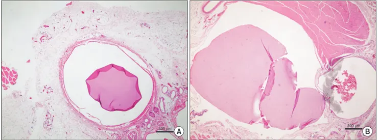
Fig. 1. Delineation of neointimal hyperplasia (arrow) at 1 and 2 weeks postoperatively (PO) following cutdown central venous catheter placement against whole vein wall (arrowhead). Neointimal hyperplasia was consistently thicker in the control group than in the treatment group (H&E, ×40). (A) Control group, 1 week PO, (B) paclitaxel treatment group, 1 week PO, (C) control group, 2 weeks PO, and (D) paclitaxel treatment group, 2 weeks PO.
treatment groups. NHI was calculated as the quotient of the area of neointimal hyperplasia divided by the total area of the vein wall on the H&Estained crosssections. Patent luminal area was measured from H&Estained cross sections as the difference of catheter area subtracted from the total luminal area, reported in mm2.
Using highpower fields (×600), the number of SMC nuclei was counted in four locations in each αactinstained cross
section, with each location separated by approximately one
fourth of the total vein circumference. The mean counts were reported, and compared between control and treatment groups.
Detection of paclitaxel in systemic circulation
Blood samples were obtained from the left EJV at the time of animal euthanasia. Plasma concentration of paclitaxel in the blood samples was measured using highperformance liquid chromatography (HPLC). Of note, a threshold plasma level of 10 ng/dL has been demonstrated as the lower limit for detection of paclitaxel by HPLC [12]. Detailed description of the mechanism of HPLC is beyond the scope of this study.
Statistical analysis
All the parameters were measured as continuous variables and expressed as mean±standard deviation. Statistical analyses were performed using IBM SPSS ver. 18.0 (IBM Co., Armonk, NY, USA). Continuous variables were compared using the Mann
Whitney Utest between the control groups and treatment groups, and significance was assigned at a Pvalue of <0.05.
RESULTS
None of the subjects died or exhibited clinical signs of infec
tion during the perioperative or observational periods.
Neointimal hyperplasia index
Neointimal hyperplasia was observed in both the control and treatment groups. The greatest measured thickness of neointimal hyperplasia was consistently thicker in the control group (Fig. 1). NHI was significantly lower in the treatment group than in the control group at each measured interval in the observation period (1 week, 0.11 ± 0.04 vs. 0.34 ± 0.04; 2 weeks, 0.26 ± 0.12 vs. 0.53 ± 0.09; 4 weeks, 0.16 ± 0.07 vs. 0.54
± 0.11; 8 weeks, 0.2 ± 0.08 vs. 0.63 ± 0.08; Fig. 2). In contrast to the treatment group, the control group demonstrated an in
crease in NHI over the course of the observation period.
Patent luminal area
Patent luminal area at final observation (8 weeks) was sig
ni fi cantly larger in the treatment group than in the control group (Fig. 3). Furthermore, the measured luminal area in
Neointimalhyperplasiaindex
1
Weeks 0
Control Treatment 0.8
0.7 0.6 0.5 0.4 0.3 0.2 0.1
P = 0.000
P = 0.001 P = 0.000 P = 0.000
2 4 8
Fig. 2. Neointimal hyperplasia index was significantly lower in the treatment group.
300 m 300 m
A B
Fig. 3. (A, B) Measurement of luminal area at 8 weeks postoperatively (H&E ×40). Measured patent luminal area was larger in the treatment group than in the control group.
the treatment group was larger at all measured intervals and enlarged over the course of the observation period (1 week, 1.73
± 0.59 vs. 0.84 ± 0.11; 2 weeks, 1.84 ± 0.54 vs. 0.91 ± 0.13; 4 weeks, 3.04 ± 0.79 vs. 2.14 ± 0.32; 8 weeks, 5.08 ± 0.43 vs. 1.91
± 0.43; Fig. 4).
SMC count
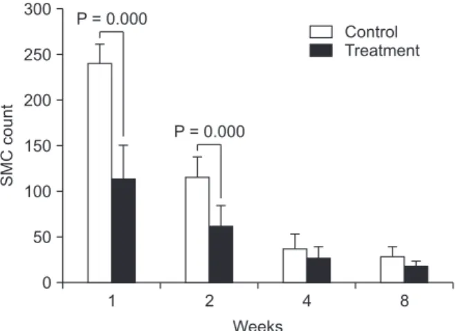
Mean SMC counts measured at 1 week and 2 weeks after cutdown CVC placement were significantly lower in the treat
ment group than in the control group (1 week, 113 ± 36 vs. 240
± 21; 2 weeks, 62 ± 22 vs. 115 ± 22; Fig. 5). However, counts measured at 4 weeks and 8 weeks demonstrated no statistically significant difference between the treatment and control groups (4 weeks, 27 ± 12 vs. 37 ± 16; 8 weeks, 18 ± 5 vs. 29 ± 10).
SMC counts decreased over the course of observation in both groups (Fig. 6).
Patentluminalarea(mm)2
Control Treatment
Weeks 0
6 5 4 3 2 1
1
P = 0.002 P = 0.013
2
P = 0.026
4
P = 0.000
8
Fig. 4. Patent luminal area was significantly wider in the treat ment group than in the control group at each measured inter val in the observation period.
20 m 20 m
20 m 20 m
A B
C D
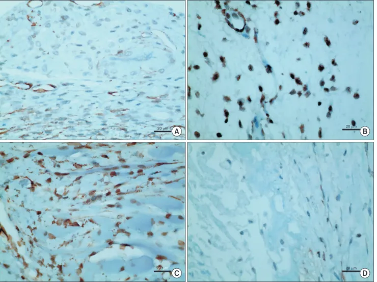
Fig. 5. Smooth muscle cell (SMC) counts in the early postoperative phases (1 and 2 weeks) measured on highpowered fields (×600) with immunohistochemical staining. SMC counts were reduced in the treatment group compared to those in the control group. (A) Control group, 1 week PO, (B) paclitaxel treatment group, 1 week PO, (C) control group, 2 weeks PO, and (D) paclitaxel treatment group, of 2 weeks PO.
Plasma paclitaxel concentration
The concentration of paclitaxel in venous samples from the treatment group was below the detection limit of HPLC in all subjects.
DISCUSSION
The aim of our study was to evaluate the preventive effect of sustained, perivascular delivery of paclitaxel on venous neointimal hyperplasia in rats following CVC placement using the cutdown method. Compared to controls, our treatment group exhibited significantly decreased NHI, lower SMC counts, and widened luminal area throughout the observation period, confirming paclitaxel’s inhibition of venous neointimal hyper
plasia and attendant luminal preservation. This study is the first to report the positive effect of paclitaxel on the prevention of neointimal hyperplasia in veins, and invites further investi
gation to supplement our observations.
In our previous study, proliferation of SMCs was first identi
fied 1 week after open cutdown CVC placement, stabilizing by 4week postprocedure [4]. As shown in SMC counts in current study, SMC counts were significantly reduced in the treatment group at early postoperative period (1 and 2 weeks) when SMC proliferation was active, whereas the counts were similar between groups after postoperative 4 weeks when SMC proliferation was stabilized. Therefore, adequate treatment should include sustained delivery of paclitaxel for a duration that covers the time period for ongoing neointimal hyperplasia.
Due to the substantial toxicity associated with systemic therapy, vascular applications of paclitaxel have favored local over systemic routes of delivery. For example, paclitaxelcoated and paclitaxeleluting devices that capitalize on the local inhibitory effects on neointimal hyperplasia have been employed to augment a variety of endovascular interventions in arteries.
Yet, for open surgical procedures such as venous cutdowns, direct, perivascular application of paclitaxel may represent the only reasonable route to deliver the medicine without associated systemic toxicity. Drugeluting bioabsorbable vas
cular wrap showed promising results in surgicallyplaced dialysis grafts in sheep [13]. Furthermore, Kwon et al. [14] and Park et al. [15] reported that perivascular delivery of antipro
liferative agents such as rapamycin or paclitaxel with F127 pluronic gel successfully inhibited neointimal hyperplasia following carotid artery injury in rats. Our study, on the other hand, employed fibrin glue as a vehicle to deliver paclitaxel over an extended period of time. Fibrin glue is composed of supraphysiological levels of key components of the coagulation cascade, including fibrinogen, thrombin, calcium, and apro
tinin. Fibrin's efficacy as a tissue sealant and hemostatic agent has fostered its widespread usage by a variety surgical spe
cialties [16]. Additionally, fibrin glue has yielded promising results as a tissueadherent carrier for the local delivery and slow release of medications, including antibiotics, growth factors, and chemotherapeutic agents [16]. Although the in vivo pharmacokinetics of paclitaxel release from fibrin glue have not yet been defined, our observed efficacy in inhibiting neointimal hyperplasia in the absence of evidence of local toxicity such as medial wall thinning or tissue blistering suggests that fibrin glue may represent a useful alternative for sustained perivas
cular drug delivery. Future studies are required to elucidate the in vivo pharmacokinetics of paclitaxel delivery via fibrin glue, as well as to assess the relative perivascular and transmural concentrations of paclitaxel over time.
The typical doses of paclitaxel administered as a chemother
apeutic agent for adult malignant tumors (135 or 175 mg/m2) [17] are much larger than those reported for prevention of neointimal hyperplasia in various clinical and experimental settings [5,6,18,19]. It has generally been accepted that these extremely low doses of paclitaxel for preventing neointi
mal hyperplasia cannot generate sufficient systemic drug concentration to produce clinical effects. Masaki et al. [20]
observed effective inhibition of neointimal hyperplasia in arteriovenous hemodialysis grafts in dogs using a thermo
sensitive, biodegradable copolymer for sustained local delivery.
In the study, 0.26–0.65 mg of paclitaxel was mixed with biodegradable polymer and applied to perivascular space.
Paclitaxel concentrations from all peripheral venous blood sam
ples were below the detection limit (10 ng/mL) of the enzyme
linked immunosorbent assay. By contrast, limited data has been present regarding the use of paclitaxel in pediatric patients, and Hurwitz et al. [21] showed that paclitaxel was well tolerated in children with recurrent or progressive brain tumors, ages 4 months to 19 years, at a dosage of 350 mg/m2 every 3 weeks. Our study used doses of 0.65 mg of paclitaxel, with all measured paclitaxel concentrations in plasma from 1 to 8 weeks
SMCcount
Control Treatment
Weeks 0
300 250 200 150 100 50
1 P = 0.000
P = 0.000
2 4 8
Fig. 6. Mean smooth muscle cell (SMC) counts were signifi
cantly lower in the treatment group than in the control group at 1 and 2 weeks postoperatively.
postoperatively also falling below the detection limit. Since the dosages employed in the present study were approximately 1,000 fold lower than those administered for chemotherapy in study of Hurwitz et al. [21], it was expected that paclitaxel would not be detected in the systemic circulation.
In conclusion, sustained perivascular delivery of paclitaxel in a mixture with fibrin glue effectively prevented neointimal hyperplasia and subsequent venous narrowing after open cutdown CVC placement in rats while maintaining systemic paclitaxel concentrations below the detectable limit.
CONFLICTS OF INTEREST
No potential conflict of interest relevant to this article was reported.
ACKNOWLEDGEMENTS
This study was supported by a 2014 Research Grant from Kangwon National University (grant number: 120140657).
1. Hong SM, Lee HS, Moon SB. Central ve
nous cutdown in neonates: feasibility as a bedside procedure without general anesthesia. J Pediatr Surg 2013;48:17226.
2. Kim MJ, Chang HK, Lee MS, Han SJ, Oh JT. Internal jugular vein deformities after central venous catheterisation in neo
nates: evaluation by Doppler ultra sound. J Paediatr Child Health 2010;46:1548.
3. Willetts IE, Ayodeji M, Ramsden WH, Squire R. Venous patency after open cen
tralvenous cannulation. Pediatr Surg Int 2000;16:4113.
4. Kim S, Kim Y, Moon SB. Histological changes of the unligated vein wall adja
cent to the central venous catheter after open cutdown in rats. J Pediatr Surg 2015;
50:192832.
5. Dake MD, Van Alstine WG, Zhou Q, Ragheb AO. Polymerfree paclitaxelcoated Zilver PTX Stentsevaluation of pharma
coki netics and comparative safety in por
cine arteries. J Vasc Interv Radiol 2011;22:
60310.
6. Lee BH, Nam HY, Kwon T, Kim SJ, Kwon GY, Jeon HJ, et al. Paclitaxelcoated ex
panded polytetrafluoroethylene hae mo
dialysis grafts inhibit neointimal hyper
plasia in porcine model of graft stenosis.
Nephrol Dial Transplant 2006;21:24328.
7. Baek I, Hwang J, Park J, Kim H, Park JS, Kim DJ. Paclitaxel coating on the terminal portion of hemodialysis grafts effectively sup presses neointimal hyperplasia in a porcine model. J Vasc Surg 2015;61:1575
82.e1.
8. Nakano Y, Ishikawa T, Mutoh M. Long
term angiographic outcomes of sirolimus
and paclitaxeleluting stent placement in diabetes, long lesions, and small vessels.
Cardiovasc Interv Ther 2015;30:32737.
9. Laird JR, Hong M. Drugeluting stents in the superficial femoral artery: The Long and Winding Road. Circulation 2016;133:
14357.
10. De Luca G, Wirianta J, Lee JH, Kaiser C, Di Lorenzo E, Suryapranata H. Sirolimus
eluting versus paclitaxeleluting stent in pri mary angioplasty: a pooled patient
level metaanalysis of randomized trials. J Thromb Thrombolysis 2014;38:35563.
11. Hur SH, Cho YK, Nam CW, Kim H, Han SW, Kim YN, et al. Comparison of long
term outcomes following sirolimuselu
ting stent vs paclitaxeleluting stent im
plan ta tion in patients with long cal ci fied co ro nary lesions. Clin Cardiol 2009;32:
6338.
12. Yonemoto H, Ogino S, Nakashima MN, Wada M, Nakashima K. Determination of paclitaxel in human and rat blood sam ples after administration of low dose pa cli taxel by HPLCUV detection. Biomed Chromatogr 2007;21:3107.
13. Kohler TR, Toleikis PM, Gravett DM, Avelar RL. Inhibition of neointimal hyper
plasia in a sheep model of dialysis access failure with the bioabsorbable Vascular Wrap paclitaxeleluting mesh. J Vasc Surg 2007;45:102937.
14. Kwon JS, Park SS, Kim YG, Son JH, Lee YS, Kim KS, et al. Perivascular delivery of paclitaxel with F127 pluronic gel inhi bits neo intimal hyperplasia in a rat caro tid ar tery injury model. Korean Circ J 2005;
35:2217.
15. Park D, Kim SM, Min SI, Ha J, Kim IG, Min SK. Inhibition of intimal hyperplasia by local perivascular application of rapa
mycin and imatinib mesilate after caro tid balloon injury. J Korean Surg Soc 2013;85:
296301.
16. Spotnitz WD. Commercial fibrin sealants in surgical care. Am J Surg 2001;182(2 Suppl):8S14S.
17. McEvoy GK. American hospital formulary service drug information, Bethesda (MD):
American Society of HealthSystem Phar
ma cists; 2000.
18. Dake MD, Ansel GM, Jaff MR, Ohki T, Saxon RR, Smouse HB, et al. Paclitaxel
eluting stents show superiority to balloon angioplasty and bare metal stents in fe mo ropopliteal disease: twelvemonth Zilver PTX randomized study results. Circ Cardiovasc Interv 2011;4:495504.
19. FernandezParra R, Laborda A, Lahuerta C, Lostale F, Aramayona J, de Blas I, et al. Pharmacokinetic study of paclitaxel con centration after drugeluting balloon angio plasty in the iliac artery of healthy and atherosclerotic rabbit models. J Vasc Interv Radiol 2015;26:13807.e1.
20. Masaki T, Rathi R, Zentner G, Leypoldt JK, Mohammad SF, Burns GL, et al. Inhibition
REFERENCES
of neointimal hyperplasia in vascular grafts by sustained perivascular delivery of paclitaxel. Kidney Int 2004;66:20619.
21. Hurwitz CA, Strauss LC, Kepner J, Kretschmar C, Harris MB, Friedman H, et al. Paclitaxel for the treatment of pro
gre ssive or recurrent childhood brain tu
mors: a pediatric oncology phase II study.
J Pediatr Hematol Oncol 2001;23:27781.