DOI: 10.4174/jkss.2011.80.5.348
ORIGINAL ARTICLE
Journal of the Korean Surgical Society
JKSS
pISSN 2233-7903ㆍeISSN 2093-0488
Received September 24, 2010, Accepted November 17, 2010 Correspondence to: Young-Wook Kim
Division of Vascular Surgery, Department of Surgery, Samsung Medical Center, Sungkyunkwan University School of Medicine, 50 Irwon-dong, Gangnam-gu, Seoul 135-710, Korea
Tel: +82-2-3410-3461, Fax: +82-2-3410-0040, E-mail: [email protected]
cc Journal of the Korean Surgical Society is an Open Access Journal. All articles are distributed under the terms of the Creative Commons Attribution Non-Commercial License (http://creativecommons.org/licenses/by-nc/3.0/) which permits unrestricted non-commercial use, distribution, and reproduction in any medium, provided the original work is properly cited.
Measurement of carotid artery stenosis: correlation
analysis between B-mode ultrasonography and contrast arteriography
Kyo Won Lee, Yang Jin Park, Young-Nam Rho, Dong-Ik Kim, Young-Wook Kim
Division of Vascular Surgery, Department of Surgery, Samsung Medical Center, Sungkyunkwan University School of Medicine, Seoul, Korea
Purpose: To evaluate the efficacy of B-mode ultrasonography (US) in measurement of carotid stenosis% (CS%). Methods:
One hundred and thirth-three carotid arteries in 96 patients who underwent both carotid US and carotid arteriography (CA) were included in this retrospective study. To measure CS% on US, a cross sectional view of the most stenotic segment of the internal carotid artery was captured and residual diameter and original diameter of that segment were measured with elec- tronic caliper on the same plane and in the same direction. To measure CS% on an angiogram, we used European Carotid Surgery Trial (ECST) and the North American Symptomatic Carotid Endarterectomy Trial (NASCET) methods. Pearson’s correlation analysis and linear regression analysis were used to determine the correlation between CS% on an US and angiogram. Results: Pearson’s correlation coefficient (R) between CS% measured in US and CA were 0.853 (ECST method, P
< 0.001) and 0.828 (NASCET method, P < 0.001). Accuracies of B-mode US were 93.2%, 88.0%, and 81.2% for estimating CS% by ECST method and 86.5%, 82.7%, and 82% for estimating CS% by NASCET method. Conclusion: CS% measured in B-mode US was simpler and showed a strong positive correlation with that measured on an arteriogram either ECST or NASCET method.
Key Words: Carotid stenosis, Ultrasonography, Accuracy, Arteriography
INTRODUCTION
According to the current guidelines, the indications for treatment of carotid artery stenosis are based on the pres- ence of clinical symptoms and the degree of carotid steno- sis% (CS%) [1]. For decades, trans-femoral contrast an- giography has been used as the gold standard for meas- urement of the CS%. However, transfemoral carotid arte-
riography (CA) cannot be used as a routine diagnostic pro- cedure because of its invasiveness, potential risk of cere- bral infarction due to carotid artery embolism and the side effects associated with the contrast agent. To avoid an in- vasiveness of transfemoral CA, computed tomography angiography or magnetic resonance angiography were al- so used in the diagnosis of carotid stenosis.
CA provides only luminogram showing features of ar-
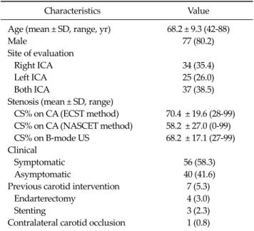
Table 1. Patient characteristics
Characteristics Value
Age (mean ± SD, range, yr) 68.2 ± 9.3 (42-88)
Male 77 (80.2)
Site of evaluation
Right ICA 34 (35.4)
Left ICA 25 (26.0)
Both ICA 37 (38.5)
Stenosis (mean ± SD, range)
CS% on CA (ECST method) 70.4 ± 19.6 (28-99) CS% on CA (NASCET method) 58.2 ± 27.0 (0-99) CS% on B-mode US 68.2 ± 17.1 (27-99) Clinical
Symptomatic 56 (58.3)
Asymptomatic 40 (41.6)
Previous carotid intervention 7 (5.3)
Endarterectomy 4 (3.0)
Stenting 3 (2.3)
Contralateral carotid occlusion 1 (0.8)
Values are presented as mean ± SD (range) or no. of patients (%).
CS%, carotid stenosis%; CA, carotid angiography; ECST, European Carotid Surgery Trial; NASCET, North American Symptomatic Carotid Endarterectomy Trial; US, ultrasonography; ICA, internal carotid artery.
terial lumen and narrowing or dilatation of the artery. But it cannot show lesion morphology in the arterial wall and outside of the arterial wall. On the contrary, US shows pla- que morphology in the arterial wall on a B-mode image and the physiological characteristics of blood flow includ- ing flow velocity and flow direction. Because of its non- invasiveness, US has become more frequently used for the diagnosis of carotid artery disease. However, CS% meas- ured on a B-mode US has not been accepted as an in- dependent diagnostic test in measurement of the CS%.
According to Society of Radiologists in Ultrasound Consensus Conference [2], plaque estimate (diameter re- duction) on grey-scale or color Doppler US image was used as a primary parameter of criteria for the diagnosis of CS. But that image parameter was just classified into less than 50% and ≥50% not as an independent criterion for measurement of carotid stenosis.
In current practice, duplex US machine is equipped col- or-coded Doppler sonography, power Doppler and B- mode US image with higher resolution. Considering the advanced equipment and technique of US machine, we can assume that a more accurate measurement of vessel lumen is available with US than before.
In this study, we attempted to determine correlation be- tween CS% measured on B-mode US and CA.
METHODS
The clinical protocol for this study was approved by the Institutional Review Board. The database of carotid US and arteriogram in 101 patients who underwent both ex- aminations for an assessment of carotid artery stenosis at a tertiary referral center in Seoul, Korea over 3 years from October 2006 to June 2009 were retrospectively reviewed.
Among them, 133 carotid arteries (37 bilateral, 59 unilat- eral) in 96 patients were included for this correlation analysis. One severely calcified artery and five arteries with missing data were excluded. Table 1 shows the clin- ical and demographic features of the enrolled patients.
Measurement of CS% on B-mode US
All US examinations were performed by experienced
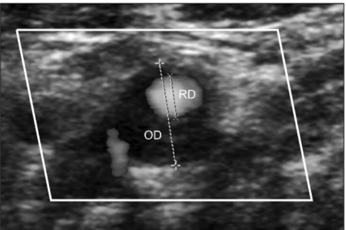
registered vascular technologists (RVTs) under the in- spection of vascular surgeon and one of the RVTs re- viewed the results of examinations. To measure CS% on a B-mode US or color Doppler (Logiq 9, GE Medical Systems, Milwaukee, WI, USA; iU22, Philips Ultrasound, Bothell, WA, USA), the most stenotic segment of the ICA was captured using 9-3 MHz linear transducer on a longi- tudinal image and by measurement of peak systolic veloc- ity (PSV) at the Doppler angle of insonation 60°. After cap- turing a transverse scan of the most stenotic segment of ICA on a B-mode US or color Doppler, the original diame- ter (OD) and residual diameter (RD) were measured using electronic calipers. The RD was defined as the shortest di- ameter of the residual lumen at the most stenotic segment of ICA and OD was defined as the measured diameter from the outer media to the outer media of the diseased ar- tery on the same plane and at same direction with the RD (Fig. 1). The CS% on B-mode US was calculated using the following equation: CS% = (1 − [RD/OD]) x 100%. This method is used in our institution according to textbooks and previous studies [3,4].
Table 2. Severity of carotid artery stenosis
CS% B-mode US ECST NASCET
<50 20 (15.0) 19 (14.3) 47 (35.3)
50-69 40 (30.1) 38 (28.6) 32 (24.1) 70-99 73 (54.9) 76 (57.1) 54 (40.1) Values are presented as number (%).
CS%, carotid stenosis%; US, ultrasonography; ECST, European Carotid Surgery Trial; NASCET, North American Symptomatic Carotid Endarterectomy Trial.
Fig. 1. Calculation of carotid stenosis% on a transverse scan color flow ultrasonography image of the internal carotid artery: residual diameter (RD) indicates the shortest diameter of residual lumen of the most stenotic segment of the internal carotid artery and original diameter (OD) indicates a diameter from the outer media-to-outer media in a same plane and a same direction with RD.
Measurement of CS% on CA
CA was indicated only when the patients are planned for intervention such as, stent insertion and/or balloon di- latation and performed with a trans-femoral catheter on a biplane angiographic system: Allura Xper FD 20/10 sys- tem (Phillips Healthcare, Andover, MA, USA) and Altis zee system (Siemens Medical System, Forchheim, Ger- many). The CA images were reviewed by two inves- tigators who were blinded to the results of the B-mode US exam. There was no significant interobserver variability.
The CS% on CA was measured at the most stenotic seg- ment of the internal carotid artery (ICA) according to North American Symptomatic Carotid Endarterectomy Trial (NASCET) and European Carotid Surgery Trial (ECST) methods using electronic calipers on a picture ar- chiving and communication system image. Between ante- rior-posterior and the lateral views of the carotid artery stenosis, the more stenotic one was selected in measure- ment of CS%.
Statistical analysis
To determine a correlation between CSs% on a B-mode US and angiogram, Pearson’s correlation analysis and lin- ear regression analysis was conducted. The correlation analysis was performed using CS% on an angiogram measured by NASCET and ECST methods. Receiver oper-
ator characteristic curves were generated for sensitivity, specificity, positive predictive value, negative predictive value, and correlation coefficients of CS% on a B-mode US was estimated in subclass of CS% of 50 to 99%, 60 to 99%, and 70 to 99% on arteriogram. The PASW ver. 17.0 (SPSS Inc., Chicago, IL, USA) was used for all statistical calculations.
RESULTS
Table 2 demonstrates the distribution of carotid artery stenosis according to the CS% on B-mode US and arteriogram. The distribution of CS% on B-mode US was more similar to that of ECST method than that of NASCET method.
Correlation between CS% on B-mode US and arte- riogram by ECST method
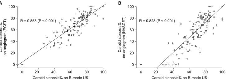
Correlation between CS% on B-mode US and CA (by ECST method) is shown in the scatter plot diagram (Fig.
2A). The Pearson’s correlation coefficient (R) between them was 0.853 (P < 0.001). The following equations were derived by the linear regression analysis:
CS% on CA (by the ECST method) = 0.974 x CS% on B-mode US + 4.075
On a subgroup analysis according to CS%, accuracies of B-mode US were 93.2%, 88.0%, and 81.2% in a subgroup with CS% on B-mode >50%, >60%, and >65%, respectively. The sensitivity, specificity, positive and neg- ative predictive values are listed in Table 3.
Fig. 2. Scatter diagrams of the carotid stenosis% (CS%) measured ultrasonography (US) in reference to CS% measured on carotid angiogram.
(A) Angiographic CS% measured by European Carotid Surgery Trial method. (B) Angiographic CS% measured by North American Symptomatic Carotid Endarterectomy Trial method.
Table 3. Accuracy of B-mode ultrasonography in measurement of CS% according to the angiographic CS% calculated by the ECST method CS% on
CA
CS% on
US Sensitivity Specificity PPV NPV Accuracy AUC P-value
50-99% >45% 98.3 57.9 93.3 84.6 92.5 0.967 <0.001
>50% 95.6 79.0 96.5 75.0 93.2
>55% 89.5 79.0 96.2 55.6 88.0
60-99% >55% 93.6 53.9 83.0 77.8 82.0 0.928 <0.001
>60% 92.6 76.9 90.6 81.1 88.0
>65% 85.1 84.6 93.0 70.2 85.0
70-99% >60% 96.1 60.7 77.1 91.9 81.0 0.907 <0.001
>65% 89.6 69.6 80.2 83.0 81.2
>70% 80.5 80.4 84.9 75.0 80.5
Values are presented as %.
CS%, carotid stenosis%; ECST, European Carotid Surgery Trial; CA, carotid angiogram; US, ultrasonography; PPV, positive predictive value; NPV, negative predictive value; AUC, area under curve.
Correlation between CS% on B-mode US and arte- riogram by NASCET method
The correlation between CS% on B-mode US and CA (by NASCET method) is shown in the scatter plot diagram (Fig. 2B). The Pearson’s correlation coefficient (R) between them was 0.828 (P < 0.001). The following equations were derived by the linear regression analysis:
CS% on CA (by the NASCET method) = 1.304 x CS% on B-mode US - 30.711
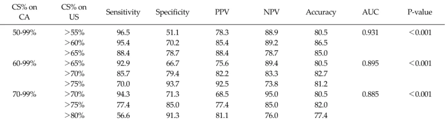
On a subgroup analysis according to CS%, accuracies of
B-mode US were 86.5%, 82.7%, and 82.0% in a subgroup with CS% on B-mode >60%, >70%, and >75%, respectively. The sensitivity, specificity, positive and neg- ative predictive values are shown in Table 4.
DISCUSSION
In this study, we found a strong positive correlation be- tween CS% measured on B-mode US and an arteriogram which was calculated by either ECST or NASCET method.
The efficacy of carotid endarterectomy for symptomatic
Table 4. Accuracy of B-mode ultrasonography inmeasurement of CS% according to the angiographic CS% calculated by the NASCET method
CS% on CA
CS% on
US Sensitivity Specificity PPV NPV Accuracy AUC P-value
50-99% >55% 96.5 51.1 78.3 88.9 80.5 0.931 <0.001
>60% 95.4 70.2 85.4 89.2 86.5
>65% 88.4 78.7 88.4 78.7 85.0
60-99% >65% 92.9 66.7 75.6 89.4 80.5 0.895 <0.001
>70% 85.7 79.4 82.2 83.3 82.7
>75% 70.0 93.7 92.5 73.8 81.2
70-99% >70% 94.3 71.3 68.5 95.0 80.5 0.885 <0.001
>75% 77.4 85.0 77.4 85.0 82.0
>80% 56.6 91.3 81.1 76.0 77.4
Values are presented as %.
CS%, carotid stenosis%; NASCET, North American Symptomatic Carotid Endarterectomy Trial; CA, carotid angiogram; US, ultrasonography; PPV, positive predictive value; NPV, negative predictive value; AUC, area under curve.
and asymptomatic patients with critical stenosis of carotid artery (>50% by NASCET method; >70% by ECST meth- od; >60% by Asymptomatic Carotid Artherosclerosis Study [ACAS] criteria) has already been proven by many prospective randomized trials including NASCET, ECST, and the ACAS [5,6]. In ACAS trial and NASCET, arterio- graphic measurements were used to determine the degree of carotid stenosis, which was calculated by comparing the residual lumen at the site of maximal stenosis to the lu- men of the nondiseased distal ICA.
Although angiographic stenosis of carotid artery was used as a gold standard to give the degree of carotid steno- sis in prior trials, each method of calculation is different and has its own drawbacks. In ECST, they used an antici- pated outer wall of the carotid bulb as a denominator in calculation of CS% while distal normal ICA was used as a denominator in NASCET and ACAS.
The ECST Method can give the best estimate of plaque thickness which is relevant for the ensuing risk of embo- lism and the true extent of stenosis. However, angiog- raphy enables only a rough and indirect estimate of the lo- cal degree of a stenosis because, unlike B-mode US, it does not depict the original vessel diameter. The NASCET method is frequently used in the United States and is reli- able but tends to underestimate the degree of stenosis.
Some practice guideline recommended not to use NASCET method in patients with a near-occlusive sten- otic lesion or with a reduced diameter at the distal internal
carotid artery beyond the stenotic lesion [2,7].
In performing duplex US for patients with carotid ar- tery disease, the criteria of CS% can be different from cen- ter to center. The main diagnostic parameters of CS% on duplex US is based on velocity criteria. Accordingly, meas- urement errors due to inappropriate positioning of the Doppler gate or accounting for the Doppler angle can lead to measurement error in diagnosis of the CS% during a du- plex US of the carotid artery.
US image of the carotid plaque has not been commonly used for classifying CS% but for determine the presence, location and characteristics of the plaque so far. As de- scribed above, diagnostic criteria from Society of Radiologists used ICA PSV, plaque estimate (diameter re- duction) on gray-scale or color Doppler US as primary pa- rameter of criteria and ICA/ common carotid artery veloc- ity and ICA/end-diastolic velocity as additional parame- ters [2]. But they suggested only 2 categories of plaque esti- mate <50% and ≥50% as parameter of CS<50% and CS
≥50%, respectively. No more detailed parameter of pla- que estimate was suggested to subclassify CS≥50% [8].
With an evolution of US equipment and examination techniques, it has been available to delineate arterial wall layers and measure the thickness of the layer on a US image. In current practice, duplex US has become an initial diagnostic modality of choice in carotid artery disease.
However, the duplex US has some limitations with regard to the technical, hemodynamic and anatomic factors.
Furthermore, the results of the duplex US are exam- iner-dependent. Variability between laboratories and be- tween examiners using the same equipment have been previously documented [8-10]. Anatomic factors such as tortuosity or kinking of the carotid artery, hemodynamic factors such as an occlusion of the contralateral carotid ar- tery, the presence of tandem lesion or previous carotid in- tervention [11-14], and systemic factors such as change of blood pressure or the cardiac output can also influence ve- locity mesurement of the duplex US [15,16].
The measurements CS% using B-mode US is free from those limitations however, it also has limitations include possibility of angle-dependant measurement bias, diffi- culties in case with too high carotid bifurcation, severely calcified arterial lesions, echo-lucent lesions, or patients with near-occlusive stenotic lesions. A near-occlusive sten- otic lesion can be interpreted as a total occlusion. During the measurement of CS% with B-mode US, eccentric pla- que configuration can also lead to a measurement error.
However, compared to velocity measurement using du- plex US, measurement of the CS% on a hard copy of B-mode US is simple in its technique and examination re- sults are less affected by such technical factors. Another advantage of US over the CA is its ability to assess carotid plaque morphology [17-19].
Some authors have reported the efficacy of CS% meas- ured on B-mode US [2,15,16,20-23]. Among them, some authors reported increased accuracy of B-mode US of car- otid stenosis when it is interpreted in conjunction with flow velocity criteria [20,23]. MacKenzie et al. [16] de- scribed that the accuracy of CS% on B-mode US was sim- ilar to that of velocity criteria in detecting CS% of 50 to 99%, 60 to 99%, and 70 to 99% on TFCA. According to them, the B-mode US image provided the best overall ac- curacy rates in subgroups of CS of 50 to 99%, 60 to 99%, and 70 to 99% were 85.3% (CS on B-mode US >65%), 82.2% (CS on B-mode US >70%, and 87.0% (CS on B-mode US >78%), respectively. These results are similar to our re- sults in this study. Sprouse et al. [15] suggested that the CS% on B-mode US can be a reliable predictor of carotid stenosis independent of the velocity criteria. According to them, CS% calculated using distal non-stenotic ICA diam- eter as a denominator and RD of the most stenotic segment
of ICA as a numerator (similar to NASCET method) also could be a reliable reliable predictor of carotid stenosis in addition to bulb diameter reduction (similar to ECST method).
Though opponents of B-mode US in the measurement of CS% reported the above-described limitations of US, Sprouse et al. [15] reported that B-mode US assessment was available in 91% of patients. They expected that B-mode US measurement of CS% will be available on more than 95% of patients as examiner’s experience and US equipment continues to improve. It was hard for us to determine technical availability of B-mode US examina- tion of the carotid artery in this retrospective study.
In conclusion, although we take some drawbacks of B-mode US in measurement of CS%, we found that US measurement of carotid stenosis was simpler compared to velocity measurement, available in most patients and its results showed a strong positive correlation with CS%
measured on transfemoral CA either ECST or NASCET methods (R = 0.853, P < 0.001 and R = 0. 828, P < 0.001, re- spectively).
CONFLICTS OF INTEREST
No potential conflict of interest relevant to this article was reported.
REFERENCES
1. Hobson RW 2nd, Mackey WC, Ascher E, Murad MH, Calligaro KD, Comerota AJ, et al. Management of athero- sclerotic carotid artery disease: clinical practice guidelines of the Society for Vascular Surgery. J Vasc Surg 2008;
48:480-6.
2. Grant EG, Benson CB, Moneta GL, Alexandrov AV, Baker JD, Bluth EI, et al. Carotid artery stenosis: gray-scale and Doppler US diagnosis--Society of Radiologists in Ultra- sound Consensus Conference. Radiology 2003;229: 340-6.
3. Lemne C, Jogestrand T, de Faire U. Carotid intima-media thickness and plaque in borderline hypertension. Stroke 1995;26:34-9.
4. Wendelhag I, Gustavsson T, Suurküla M, Berglund G, Wikstrand J. Ultrasound measurement of wall thickness in the carotid artery: fundamental principles and description
of a computerized analysing system. Clin Physiol 1991;
11:565-77.
5. Endarterectomy for asymptomatic carotid artery stenosis.
Executive Committee for the Asymptomatic Carotid Atherosclerosis Study. JAMA 1995;273:1421-8.
6. Beneficial effect of carotid endarterectomy in symptomatic patients with high-grade carotid stenosis. North American Symptomatic Carotid Endarterectomy Trial Collaborators.
N Engl J Med 1991;325:445-53.
7. Higashida RT, Meyers PM, Phatouros CC, Connors JJ 3rd, Barr JD, Sacks D, et al. Reporting standards for carotid ar- tery angioplasty and stent placement. J Vasc Interv Radiol 2004;15:421-2.
8. Corriveau MM, Johnston KW. Interobserver variability of carotid Doppler peak velocity measurements among tech- nologists in an ICAVL-accredited vascular laboratory. J Vasc Surg 2004;39:735-41.
9. Kuntz KM, Polak JF, Whittemore AD, Skillman JJ, Kent KC. Duplex ultrasound criteria for the identification of car- otid stenosis should be laboratory specific. Stroke 1997;28:597-602.
10. Mead GE, Lewis SC, Wardlaw JM. Variability in Doppler ultrasound influences referral of patients for carotid surgery. Eur J Ultrasound 2000;12:137-43.
11. AbuRahma AF, Robinson PA, Strickler DL, Alberts S, Young L. Proposed new duplex classification for threshold stenoses used in various symptomatic and asymptomatic carotid endarterectomy trials. Ann Vasc Surg 1998;12:
349-58.
12. Hood DB, Mattos MA, Mansour A, Ramsey DE, Hodgson KJ, Barkmeier LD, et al. Prospective evaluation of new du- plex criteria to identify 70% internal carotid artery stenosis.
J Vasc Surg 1996;23:254-61.
13. Moneta GL, Edwards JM, Papanicolaou G, Hatsukami T, Taylor LM Jr, Strandness DE Jr, et al. Screening for asymp- tomatic internal carotid artery stenosis: duplex criteria for discriminating 60% to 99% stenosis. J Vasc Surg 1995;21:
989-94.
14. Moneta GL, Edwards JM, Chitwood RW, Taylor LM Jr, Lee
RW, Cummings CA, et al. Correlation of North American Symptomatic Carotid Endarterectomy Trial (NASCET) an- giographic definition of 70% to 99% internal carotid artery stenosis with duplex scanning. J Vasc Surg 1993;17:152-7.
15. Sprouse LR 2nd, Meier GH, Parent FN, Demasi RJ, Lesar CJ, Nelms C, et al. Are we undertreating carotid stenoses diagnosed by ultrasound alone? Vasc Endovascular Surg 2005;39:143-51.
16. MacKenzie KS, French-Sherry E, Burns K, Pooley T, Bassiouny HS. B-mode ultrasound measurement of carotid bifurcation stenoses: is it reliable? Vasc Endovascular Surg 2002;36:123-35.
17. Handa N, Matsumoto M, Maeda H, Hougaku H, Ogawa S, Fukunaga R, et al. Ultrasonic evaluation of early carotid atherosclerosis. Stroke 1990;21:1567-72.
18. O'Donnell TF Jr, Erdoes L, Mackey WC, McCullough J, Shepard A, Heggerick P, et al. Correlation of B-mode ultra- sound imaging and arteriography with pathologic find- ings at carotid endarterectomy. Arch Surg 1985;120:443-9.
19. Wolverson MK, Bashiti HM, Peterson GJ. Ultrasonic tissue characterization of atheromatous plaques using a high res- olution real time scanner. Ultrasound Med Biol 1983;9:
599-609.
20. Rotstein AH, Gibson RN, King PM. Direct B-mode NASCET-style stenosis measurement and Doppler ultra- sound as parameters for assessment of internal carotid ar- tery stenosis. Australas Radiol 2002;46:52-6.
21. Jmor S, El-Atrozy T, Griffin M, Tegos T, Dhanjil S, Nicolaides A. Grading internal carotid artery stenosis us- ing B-mode ultrasound (in vivo study). Eur J Vasc Endovasc Surg 1999;18:315-22.
22. Golledge J, Ellis M, Sabharwal T, Sikdar T, Davies AH, Greenhalgh RM. Selection of patients for carotid endarterectomy. J Vasc Surg 1999;30:122-30.
23. Beebe HG, Salles-Cunha SX, Scissons RP, Dosick SM, Whalen RC, Gale SS, et al. Carotid arterial ultrasound scan imaging: a direct approach to stenosis measurement. J Vasc Surg 1999;29:838-44.