ISSN 2234-3806 • eISSN 2234-3814
http://dx.doi.org/10.3343/alm.2016.36.6.590
Soluble Suppression of Tumorigenicity 2 and Echocardiography in Sepsis
Hyun Suk Yang, M.D.1, Mina Hur, M.D.1, Hanah Kim, M.D.2, Laura Magrini, M.D.3, Rossella Marino, M.D.3, and Salvatore Di Somma, M.D.3; on behalf of GREAT Network
Departments of Cardiovascular Medicine1 and Laboratory Medicine2, Konkuk University School of Medicine, Seoul, Korea; Department of Medical-Surgery Sciences and Translational Medicine3, University La Sapienza Rome, Sant’Andrea Hospital, Rome, Italy
Soluble suppression of tumorigenicity 2 (sST2) has emerged as a biomarker of cardiac stretch or remodeling, and has demonstrated a role in acutely decompensated heart fail- ure. However, its role in sepsis-induced cardiac dysfunction is still unknown. We explored whether sST2 serum concentration reflects either systolic or diastolic dysfunction as mea- sured by Doppler echocardiography. In a total of 127 patients with sepsis, correlations be- tween sST2 and blood pressure, left ventricular (LV) ejection fraction, LV diastolic filling (ratio of early transmitral flow velocity to early diastolic mitral annulus velocity), and resting pulmonary arterial pressure were evaluated. Correlations between sST2 and other sepsis biomarkers (high-sensitivity C-reactive protein [hs-CRP] and procalcitonin) were also ex- amined. sST2 showed a moderate correlation with mean arterial pressure (r =-0.3499) but no correlation with LV ejection fraction, diastolic filling, or resting pulmonary hyperten- sion. It showed moderate correlations with hs-CRP and procalcitonin (r =0.2608 and r=0.3829, respectively). sST2 might have a role as a biomarker of shock or inflammation, but it cannot reflect echocardiographic findings of LV ejection fraction or diastolic filling in sepsis.
Key Words: sST2, Sepsis, Echocardiography, Left ventricular ejection fraction, Diastolic fill- ing
Received: January 11, 2016 Revision received: May 22, 2016 Accepted: June 29, 2016 Corresponding author: Mina Hur Department of Laboratory Medicine, Konkuk University School of Medicine, Konkuk University Medical Center, 120-1 Neungdong-ro, Gwangjin-gu, Seoul 05030, Korea
Tel: +82-2-2030-5581 Fax: +82-2-2636-6764 E-mail: [email protected] Co-corresponding author:
Salvatore Di Somma
Department of Medical-Surgery Sciences and Translational Medicine, University La Sapienza Rome, Sant’Andrea Hospital, Via di Grottarossa 1035/1039 00189 Rome, Italy
Tel: +39-0633775581 Fax: +39-0633775890
E-mail: [email protected]
© The Korean Society for Laboratory Medicine This is an Open Access article distributed under the terms of the Creative Commons Attribution Non-Commercial License (http://creativecom- mons.org/licenses/by-nc/4.0) which permits unrestricted non-commercial use, distribution, and reproduction in any medium, provided the original work is properly cited.
Sepsis is life-threatening organ dysfunction from a dysregulated host response to infection [1]. Among sepsis-induced complica- tions, the development of hemodynamic instability or cardiac dysfunction is a major risk factor of in-hospital mortality. For early detection of sepsis-induced cardiac dysfunction (SICD), immediate echocardiography is crucial but may be delayed ow- ing to inability of bedside skilled staff or high cost. For early di- agnosis and decision-making in this critical setting, biomarkers like high-sensitivity C-reactive protein (hs-CRP) and procalcito-
nin (PCT) have been suggested to indicate septic processes [2].
A multi-marker or complementary approach with other biomark- ers related to SICD might be helpful to determine the appropri- ate therapy for cardiovascular support.
Among the emerging cardiac inflammatory biomarkers, solu- ble suppression of tumorigenicity 2 (sST2), a member of the Toll-like/interleukin (IL)-1 receptor superfamily, seems promis- ing. Clinical studies revealed the diagnostic value of sST2 for acute myocardial infarction, acute heart failure, sepsis, and
trauma [3-5]. Recently, we demonstrated the prognostic role of sST2 in sepsis and its association with the cardiovascular sub- score of sepsis-related organ failure assessment (SOFA) scores [6]. However, the cardiac subset of SOFA elements consist of only mean arterial pressure and use of a vasopressor, which are too limited to reflect SICD alone [7]. In this study, we hypothe- sized that SICD would result in enough mechanical stress to re- lease sST2, and sST2 may reflect systolic or diastolic dysfunc- tion as measured by Doppler echocardiography in sepsis pa- tients. We explored whether sST2 correlates with echocardio- graphic findings and other sepsis biomarkers or not.
A retrospective study was conducted in two tertiary referral hospitals: Sant’Andrea Hospital (SAH), Rome, Italy and Konkuk University Medical Center (KUMC), Seoul, Korea. The hospital echocardiographic database including medical/surgical inten- sive care units, general wards, and emergency department was searched for the period from November 1, 2013 to May 31, 2014 (SAH) and November 1, 2013 to August 31, 2014 (KUMC). This study was conducted separately from our previ- ous study [6] that has the overlapping study period, using a dif- ferent study protocol. This study protocol involved reviewing medical records on clinical, laboratory, and echocardiographic data. The protocol met the criteria of the Declaration of Helsinki and was approved by the Institutional Review Board of each hospital. Informed consent was obtained from each patient in SAH for the biomarker measurement and review of medical re- cords; in KUMC, it was exempted for this retrospective study.
Among the eligible adult patients (≥18 yr) who had available echocardiographic data, only the patients diagnosed as having sepsis and with relevant laboratory data were included. Al- though echocardiography is not a routine practice for every sep- tic patient, the decision to perform echocardiography was made by the attending clinicians when the patient had hemodynamic instability, dyspnea, and/or abnormal radiographic findings on a chest X-ray. Each patient was considered eligible only when it was his or her first time in the hospital due to sepsis. Because the patients were searched not by the whole hospital database but by the echocardiographic database, information on how many patients were diagnosed as having sepsis during the study period was not available for this study. The following ex- clusion criteria were applied in the following order: patients’ age
<18 yr; absence of routine laboratory data for PCT and/or hs- CRP that were measured concomitantly with echocardiography (on the same day or within 24 hr); and absence of concomi- tantly obtained frozen samples, acute coronary syndrome, and documented history of symptomatic heart failure.
Recently, the diagnosis of sepsis was redefined according to the most up-to-date consensus definition [1]. Among the eligible 136 patients, nine patients were excluded owing to acute coro- nary syndrome (N =2), documented history of symptomatic heart failure (N=4), and the new sepsis definition (N=3) (Sup- plemental Data Fig. S1). We enrolled 127 patients (110 patients from KUMC and 17 patients from SAH), consisting of 95 pa- tients with sepsis (74.8%) and 32 patients with septic shock (25.2%) (Table 1). The median age was 71 yr (range, 58-80 yr), and 55% of patients were male. No statistical difference was observed between the patients from KUMC and SAH, in terms of age, sex, use of vasopressor, mean arterial pressure, left ven- tricular (LV) ejection fraction (EF), or sST2 concentrations. Their underlying medical conditions were: pneumonia (N =47), he- matologic immunocompromised state (N=19), urinary tract in- fection (N=15), hepatobiliary (N=14), cerebral (N=12), solid organ malignancy (N=8), bone or spine (N=6), gastrointestinal (N=5), and others (N=1, mucormycosis).
Serum PCT and hs-CRP concentrations were measured ac- cording to the manufacturers’ instructions [1]. In KUH, PCT was measured by an Elecsys BRAHMS PCT electrochemilumines- cence assay (BRAHMS, Hennigsdorf, Germany) in a Roche Co- bas system (Roche Diagnostics, Basel, Switzerland), and in SAH PCT was measured by using a BRAHMS PCT sensitive Kryptor immunofluorescent assay system (BRAHMS). hs-CRP was measured by using the BN II Nephelometer (Dade Behring Ins, Newark, DE, USA). Frozen sera stored at -70°C were thawed once for sST2 measurement. Briefly, sST2 concentra- tions were measured in duplicate by using an enzyme-linked immunosorbent assay, the Presage ST2 assay (Critical Diagnos- tics, San Diego, CA, USA) [8]. The maximum total CV (cumula- tive calculation) of sST2, PCT, and hs-CRP assays in both labo- ratories were 3%, 3.2%, and 3.3%, respectively.
The transthoracic Doppler echocardiogram records on the Car- diology PACS system (INFINITT healthcare, Seoul, Korea) were reviewed. LV dimensions were measured by M-mode, tracing both end-diastolic and end-systolic period. LV systolic function was evaluated with EF by two-dimensional images using the modified Simpson’s method [9]. LV diastolic function was as- sessed with the ratio of pulsed-wave Doppler-derived early trans- mitral inflow velocity (E velocity) and mitral septal annular tissue Doppler-derived early diastolic velocity (e’ velocity) (E/e’ ratio) [10]. Resting pulmonary arterial systolic pressure (PASP) was as- sessed via the tricuspid regurgitation jet peak velocity using the simplified Bernoulli equation with an assumption of right atrial pressure [11]. Two experienced cardiologists reviewed the im-
ages for apical ballooning, LV outflow dynamic obstruction, or right ventricular dysfunction [12]. For discrepant opinions be- tween them, a consensus was drawn throughout their discussion.
We performed statistical analysis using dBSTAT (DBSTAT Ver- sion 5. Chuncheon, Korea). Data were expressed as a median (interquartile range) for continuous variables and as a number (percentage) for categorical variables. We compared clinical, laboratory, and echocardiographic data in patients with or with- out sepsis-induced LV systolic dysfunction (EF <50%). Correla- tion between sST2 and cardiovascular or inflammatory markers was assessed by using Pearson’s correlation coefficient (95%
confidence interval). Mann-Whitney U test was used for contin- uous variables, and Fisher’s exact test for categorical variables.
Obtained P values were not adjusted for multiple comparisons and are therefore descriptive only.
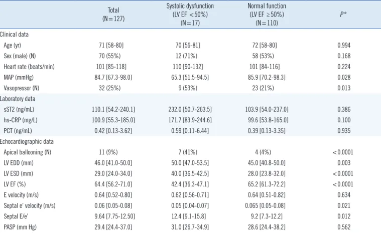
Sepsis-induced LV systolic dysfunction was observed in 17 patients (13.4%) (Table 1). Except for E velocity and PASP, the other echocardiographic data showed significant differences
between the patients with and without LV systolic dysfunction.
Although median sST2, hs-CRP, and PCT concentrations tended Table 1. Comparison between sepsis-induced cardiac dysfunction and normal function
Total (N=127)
Systolic dysfunction (LV EF <50%)
(N=17)
Normal function (LV EF ≥50%)
(N=110) P*
Clinical data
Age (yr) 71 [58-80] 70 [56-81] 72 [58-80] 0.994
Sex (male) (N) 70 (55%) 12 (71%) 58 (53%) 0.168
Heart rate (beats/min) 101 [85-118] 110 [90-132] 101 [84-116] 0.224
MAP (mmHg) 84.7 [67.3-98.0] 65.3 [51.5-94.5] 85.9 [70.2-98.3] 0.028
Vasopressor (N) 32 (25%) 9 (53%) 23 (21%) 0.013
Laboratory data
sST2 (ng/mL) 110.1 [54.2-240.1] 232.0 [50.7-263.5] 103.9 [54.0-237.0] 0.386
hs-CRP (mg/L) 100.9 [55.3-185.0] 171.7 [83.9-244.6] 99.6 [53.8-165.0] 0.100
PCT (ng/mL) 0.42 [0.13-3.62] 0.59 [0.11-6.44] 0.39 [0.13-3.35] 0.935
Echocardiographic data
Apical ballooning (N) 11 (9%) 7 (41%) 4 (4%) <0.0001
LV EDD (mm) 46.0 [41.0-50.0] 50.0 [47.0-53.5] 45.0 [40.8-50.0] 0.003
LV ESD (mm) 29.0 [24.0-34.0] 40.0 [36.5-42.5] 28.0 [23.8-32.0] <0.0001
LV EF (%) 64.4 [56.2-71.0] 42.4 [36.3-47.1] 65.2 [61.3-72.2] <0.0001
E velocity (m/s) 0.64 [0.52-0.80] 0.62 [0.56-0.71] 0.64 [0.51-0.82] 0.634
Septal e’ velocity (m/s) 0.06 [0.05-0.08] 0.05 [0.04-0.07] 0.065 [0.05-0.08] 0.021
Septal E/e’ 9.64 [7.75-12.50] 12.4 [9.1-15.8] 9.2 [7.3-12.2] 0.012
PASP (mm Hg) 29.4 [24.4-37.0] 31.0 [26.7-34.9] 28.6 [24.4-38.2] 0.562
Data are expressed as number (percentage) or median [interquartile range].
*Chi-square test (sex), Fisher’s exact test (vasopressor and apical ballooning), or Mann-Whitney U test was applied to compare two groups.
Abbreviations: MAP, mean arterial pressure; sST2, soluble suppression of tumorigenicity 2; hs-CRP, high-sensitivity C-reactive protein; PCT, procalcitonin; LV, left ventricular; EF, ejection fraction; EDD, end-diastolic dimension; ESD, end-systolic dimension; E velocity, early transmural flow velocity; e’ velocity, early di- astolic mitral annulus velocity; E/e’, ratio of early transmitral flow velocity to early diastolic mitral annulus velocity; PASP, pulmonary arterial systolic pressure.
Table 2. Correlation of sST2 with cardiac functional parameters and inflammatory markers
Pearson’s correlation coefficient (r)
95%
confidence interval P sST2 vs Cardiac functional parameters
MAP (mm Hg) -0.3499 -0.4940--0.1871 <0.0001 Heart rate (beats/min) 0.2382 0.0668-0.3960 0.007
LV EF (%) -0.0267 -0.2000-0.1482 0.766
Septal E/e’ 0.0248 -0.1599-0.2077 0.478
PASP (mm Hg) -0.0398 -0.2154-0.1382 0.662
sST2 vs Inflammatory markers
hs-CRP (mg/L) 0.2608 0.0907-0.4161 0.0031
PCT (ng/dL) 0.3829 0.2156-0.5283 <0.0001
Abbreviations: see Table 1.
to be higher in the systolic dysfunction group, unadjusted P val- ues did not indicate a meaningful difference. sST2 concentra- tion correlated moderately with hs-CRP and PCT concentra- tions; it also correlated moderately with the hemodynamic pa- rameters of blood pressure and heart rate. No correlation was observed between sST2 concentration and LV EF, septal E/e’, or PASP (Table 2).
The heart is one of the most frequently affected organs in sepsis; up to 60% of patients with septic shock develop LV EF
<45% during the first three days [13]. The prevalence or de- gree of LV dysfunction depends on the timing and severity of sepsis. In this study, depressed LV systolic function was ob- served only in 13.4% of patients, possibly owing to the relatively small portion of patients with septic shock (25.2%). In addition, the examination timing within 24 hr after blood collection for PCT and hs-CRP measurement (approximately the time point when the diagnosis of sepsis was established) may include the compensatory hyperdynamic contractile period. During early phases of sepsis, the LV EF is high ( >55%) with insufficient cardiac preload due to high vascular permeability and low vas- cular tone; however, during the later phase with fluid loading, EF decreases [14]. Therefore, serial echocardiography Doppler monitoring is warranted.
To the best of our knowledge, this is the first study to compare sST2 concentration with concomitant Doppler echocardio- graphic findings in septic patients. We speculated that sepsis- induced acute LV systolic or diastolic dysfunction, with de- creased LV EF or elevated LV filling pressure, would be enough mechanical stress to release sST2. However, our findings did not support this hypothesis, similarly to the recent report that revealed no correlation between inflammatory cytokines and myocardial dysfunction in septic shock [15]. Sepsis itself may trigger sST2 release as an inflammatory mediator, covering up any sST2 upregulation from myocardial mechanical strain. The median sST2 concentration was 110 [54.2-240.1] ng/mL, and most patients (118/127 patients, 92.9%) exhibited higher sST2 concentrations than the reference value ( ≤35 ng/mL) [8].
These results are consistent with our previous study on patients with stress cardiomyopathy in a non-cardiac medical intensive care unit; initial sST2 concentration did not predict stress car- diomyopathy, but the 48-hr follow-up sST2 concentration (per- sistently or newly increased sST2) did [16]. A limitation of our study is that we only performed a single sST2 measurement. A short-term follow-up of sST2 concentration is necessary to de- lineate its cardiac-specific role. More specifically, an algorithm combining sST2 and echocardiography may be proposed: sST2
and echocardiography can be evaluated in the early period of sepsis (within 24 hr of diagnosis) and followed up after 48 hr of the initial evaluation.
Although we excluded the patients with documented history of symptomatic heart failure, it is possible that some patients had undiagnosed/asymptomatic heart failure or new onset of symptomatic failure. This is another limitation of our study.
Moreover, we could not include other cardiac biomarkers in our retrospective analysis. Although cardiac troponins and natri- uretic peptides have been studied in relation to echocardio- graphic findings, they did not show satisfactory results [17, 18].
Absence of comparison data with the other cardiac biomarkers may be another limitation of this study.
It is unclear whether elevated sST2 in acute heart failure is produced in cardiac myocytes or elsewhere. Intuitively, sST2 protein is upregulated by myocardial stress in cultured myo- cytes [19], but other studies did not show elevated myocardial sST2 expression during heart failure [20]. In our sepsis popula- tion, sST2 is mainly a marker of inflammation, and even in heart failure, the major source of circulating sST2 is likely not the heart. Therefore, we speculate the possibility of non-myocardial or systemic vascular production of sST2 is triggered by altered myocardial load or low blood pressure in sepsis (Table 2).
In conclusion, sST2 might have a role as a biomarker of shock or inflammation in sepsis. A single measurement of sST2, however, cannot reflect sepsis-induced LV systolic or diastolic function evaluated by Doppler echocardiography. Serial mea- surement of sST2 along with echocardiography Doppler moni- toring would be necessary to better delineate the role of sST2 as a marker of SICD.
Authors’ Disclosures of Potential Conflicts of Interest
No potential conflicts of interest relevant to this article were re- ported.
Acknowledgments
This paper was supported by Konkuk University in 2016.
REFERENCES
1. Singer M, Deutschman CS, Seymour CW, Shankar-Hari M, Annane D, Bauer M, et al. The third international consensus definitions for sepsis and septic shock (Sepsis-3). JAMA 2016;315:801-10.
2. Dellinger RP, Levy MM, Rhodes A, Annane D, Gerlach H, Opal SM, et
al. Surviving sepsis campaign: international guidelines for management of severe sepsis and septic shock: 2012. Crit Care Med 2013;41:580- 637.
3. Weir RA, Miller AM, Murphy GE, Clements S, Steedman T, Connell JM, et al. Serum soluble ST2: a potential novel mediator in left ventricular and infarct remodeling after acute myocardial infarction. J Am Coll Car- diol 2010;55:243-50.
4. Januzzi JL, Mebazaa A, Di Somma S. ST2 and prognosis in acutely de- compensated heart failure: the International ST2 Consensus Panel. Am J Cardiol 2015;115(7 Suppl):26B-31B.
5. Brunner M, Krenn C, Roth G, Moser B, Dworschak M, Jensen-Jarolim E, et al. Increased levels of soluble ST2 protein and IgG1 production in patients with sepsis and trauma. Intensive Care Med 2004;30:1468-73.
6. Hur M, Kim H, Kim HJ, Yang HS, Magrini L, Marino R, et al. Soluble ST2 has a prognostic role in patients with suspected sepsis. Ann Lab Med 2015;35:570-7.
7. Vincent JL, Moreno R, Takala J, Willatts S, De Mendonça A, Bruining H, et al. The SOFA (Sepsis-related Organ Failure Assessment) score to de- scribe organ dysfunction/failure. On behalf of the Working Group on Sepsis-Related Problems of the European Society of Intensive Care Medicine. Intensive Care Med 1996;22:707-10.
8. Mueller T and Dieplinger B. The Presage(®) ST2 Assay: analytical con- siderations and clinical applications for a high-sensitivity assay for mea- surement of soluble ST2. Expert Rev Mol Diagn 2013;13:13-30.
9. Lang RM, Bierig M, Devereux RB, Flachskampf FA, Foster E, Pellikka PA, et al. Recommendations for chamber quantification: a report from the American Society of Echocardiography’s Guidelines and Standards Committee and the Chamber Quantification Writing Group, developed in conjunction with the European Association of Echocardiography, a branch of the European Society of Cardiology. J Am Soc Echocardiogr 2005;18:1440-63.
10. Nagueh SF, Mikati I, Kopelen HA, Middleton KJ, Quiñones MA, Zoghbi WA. Doppler estimation of left ventricular filling pressure in sinus tachy- cardia. A new application of tissue doppler imaging. Circulation 1998;
98:1644-50.
11. Lima CO, Sahn DJ, Valdes-Cruz LM, Goldberg SJ, Barron JV, Allen HD, et al. Noninvasive prediction of transvalvular pressure gradient in pa- tients with pulmonary stenosis by quantitative two-dimensional echocar- diographic Doppler studies. Circulation 1983;67:866-71.
12. Y-Hassan S, Settergren M, Henareh L. Sepsis-induced myocardial de- pression and takotsubo syndrome. Acute Card Care 2014;16:102-9.
13. Vieillard-Baron A, Caille V, Charron C, Belliard G, Page B, Jardin F. Ac- tual incidence of global left ventricular hypokinesia in adult septic shock. Crit Care Med 2008;36:1701-6.
14. Rudiger A and Singer M. Mechanisms of sepsis-induced cardiac dys- function. Crit Care Med 2007;35:1599-608.
15. Landesberg G, Levin PD, Gilon D, Goodman S, Georgieva M, Weissman C, et al. Myocardial dysfunction in severe sepsis and septic shock: no correlation with inflammatory cytokines in real-life clinical setting. Chest 2015;148:93-102.
16. Yang HS, Kim HJ, Shim HJ, Kim SJ, Hur M, Di Somma S, et al. Soluble ST2 and troponin I combination: Useful biomarker for predicting devel- opment of stress cardiomyopathy in patients admitted to the medical intensive care unit. Heart Lung 2015;44:282-8.
17. Miller WL, Hartman KA, Burritt MF, Grill DE, Jaffe AS. Profiles of serial changes in cardiac troponin T concentrations and outcome in ambula- tory patients with chronic heart failure. J Am Coll Cardiol 2009;54:
1715-21.
18. Logeart D, Saudubray C, Beyne P, Thabut G, Ennezat PV, Chavelas C, et al. Comparative value of Doppler echocardiography and B-type natri- uretic peptide assay in the etiologic diagnosis of acute dyspnea. J Am Coll Cardiol 2002;40:1794-800.
19. Weinberg EO, Shimpo M, De Keulenaer GW, MacGillivray C, Tominaga S, Solomon SD, et al. Expression and regulation of ST2, an interleukin-1 receptor family member, in cardiomyocytes and myocardial infarction.
Circulation 2002;106:2961-6.
20. Bartunek J, Delrue L, Van Durme F, Muller O, Casselman F, De Wiest B, et al. Nonmyocardial production of ST2 protein in human hypertrophy and failure is related to diastolic load. J Am Coll Cardiol 2008;52:2166- 74.